Continually Updated Clinical Reference
 
 
  All Sources     eMedicine     Medscape     Drug Reference     MEDLINE
 
eMedicine - Neonatal Brachial Plexus Palsies : Article by

Quick Find
Authors & Editors
Introduction
Clinical
Differentials
Workup
Treatment
Medication
Follow-up
Miscellaneous
Multimedia
References

Related Articles
Traumatic Brachial Plexopathy


Patient Education
Click here for patient education.


Author: Jennifer Semel-Concepcion, MD, Director, Assistant Professor of Physical Medicine and Rehabilitation, Department of Physical Medicine and Rehabilitation, St Charles Hospital and Rehabilitation Center; Chair, State University of New York at Stony Brook School of Medicine

Jennifer Semel-Concepcion is a member of the following medical societies: American Academy of Pediatrics, American Academy of Physical Medicine and Rehabilitation, and American Medical Association

Coauthor(s): Hany Nasr, MBBCh, Staff Physician, Department of Physical Medicine and Rehabilitation, State University of New York at Stony Brook; Anne Conway, PT, Clinical Coordinator, Department of Physical Therapy, Children's National Medical Center of Washinton DC

Editors: Teresa L Massagli, MD, Residency Director, Professor, Department of Rehabilitation Medicine and Pediatrics, University of Washington School of Medicine; Francisco Talavera, PharmD, PhD, Senior Pharmacy Editor, eMedicine; Kat Kolaski, MD, Assistant Professor, Departments of Orthopedics and Pediatrics, Wake Forest University School of Medicine; Kelly L Allen, MD, Consulting Staff, Department of Physical Medicine and Rehabilitation, Lourdes Regional Rehabilitation Center, Our Lady of Lourdes Medical Center; Robert H Meier III, MD, Director, Amputee Services of America, Presbyterian St Luke's Hospital; Consulting Staff, North Valley Rehabilitation Hospital, Kindred Hospital, North Suburban Hospital

Author and Editor Disclosure

Synonyms and related keywords: brachial plexus injury, obstetric brachial plexus palsy, obstetrical brachial plexus palsy, brachial plexus palsy, brachial plexus birth palsy, birth brachial plexus palsy, traumatic peripheral nervous system injury, congenital brachial plexus palsy, Erb's palsy, Klumpke's palsy, brachial plexopathy, Duchenne-Erb's palsy, Erb palsy, Klumpke palsy, brachial plexopathy, Duchenne-Erb palsy

Background

The first known description of neonatal brachial plexus palsy (BPP) dates from 1779 when Smellie reported the case of an infant with bilateral arm weakness that resolved spontaneously within a few days after birth. In the 1870s, both Duchenne and Erb described cases of upper trunk nerve injury, attributing the findings to traction on the upper trunk, now called Duchenne-Erb palsy. In 1885, Klumpke described injury of C8-T1 nerve roots and the nearby stellate ganglion that now bears her name.

Many cases of BPP are transient, with the child recovering full function in the first week of life. A smaller percentage of children continue to have weakness leading to long-term disability from the injury. The mainstay of treatment for these children is physical and/or occupational therapy in concert with a regular home exercise program. A select few patients may benefit from surgical intervention in the early stages to improve innervation of the affected muscles. Others benefit from tendon transfers performed later to improve shoulder and, sometimes, elbow function. Numerous other nonsurgical treatments, including electrical stimulation and botulinum toxin injections, also may prove effective in the treatment of children with BPP. In view of the variability in presentation, treatment options, and outcome measures, a multidisciplinary approach to the care of the infant with BPP is recommended.

Pathophysiology

To understand the clinical presentation and provide anticipatory guidance for families affected by BPP, the clinician must first know basic anatomy. As seen in Image 2, the brachial plexus consists of nerves (the ventral rami) from C5-T1. 
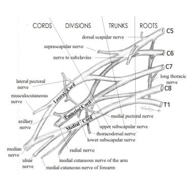
C5 and C6 join to form the upper trunk, C7 travels alone as the middle trunk, and C8-T1 join as the lower trunk. Each trunk divides into anterior and posterior divisions to create the cords, which then subdivide further into branches that supply the muscles of the arm. Injuries of the brachial plexus may be mild, with only temporary sequelae, or devastating, leaving the child with a flaccid insensate arm.

Severity depends upon the number of nerves involved and the degree to which each level is injured. The basic types of brachial plexus palsies include the following:

  • Duchenne-Erb palsy affects nerves arising from C5 and C6.
  • Upper-middle trunk brachial plexus palsy involves nerve fibers from C5, C6, and C7 levels.
  • Klumpke palsy results in deficits at levels C8 and T1, although many clinicians agree that pure C8-T1 injuries do not occur in infants and may be indicative of spinal cord injury (SCI).
  • Total BPP affects nerves at all levels (C5-T1).
  • Bilateral BPP demonstrates bilateral involvement.

When defining severity of a peripheral nerve injury, differentiation between neurapraxic, axonotmetic, and neurotmetic lesions is helpful.

  • Purely neurapraxic lesions do not affect the axon itself. These lesions generally are reversible and do not leave sequelae.
  • Axonotmetic lesions involve disruption of both the myelin sheath and the axon, leading to degeneration of the axon distal to the injury. The connective tissue across the lesion remains intact. These injuries improve gradually over 4-6 months, depending on the level of the lesion.
  • Neurotmetic lesions are the most severe and destroy not only the axon and myelin, but also the supporting structures across a nerve. As the proximal end of the nerve attempts to regenerate without this supportive connective tissue, a neuroma may develop. The extent of improvement in the patient's condition depends upon the ultimate number of nerve fibers that reconnect distal to the neuroma. Muscle atrophy from a neurotmetic lesion begins 3-6 months after injury and by 1.5-2 years is irreversible.

Although the traditional mechanism of injury is lateral neck flexion, the upper rootlets (C5-C7) are 25% as likely to be avulsed as the lower roots (C7-T1). The upper roots (C5-C6), however, are far more likely to be ruptured (88%) because of the anatomy of the transverse processes and the degree of flexibility at that level.

The clinician must also distinguish neonatal BPP from traumatic BPP in older children and adults. The damage in neonates usually results from slow traction injuries, unlike the high-energy shearing type of trauma seen in older individuals. Not only are the latter injuries often more severe, but, with similar injuries, the infants show a better functional outcome. This clinical observation is confirmed by Vredeveld et al who studied 14 infants and 19 adults with surgical evidence of complete avulsion of the C5-C6 roots or upper trunk. Electromyelogram (EMG) showed normal recruitment of biceps and deltoid in the infants and complete denervation in the older individuals. When C7 also was torn, the infants demonstrated complete denervation. Vredeveld et al attribute this observation to neonatal C7 innervation of the biceps and deltoid that is lost subsequently if C5-C6 roots are functional.

Frequency

United States

0.5-4.4 cases per 1000 full-term births

International

Studies in France and Saudi Arabia suggest an incidence of 1.09-1.19 cases per 1000 live births.

Mortality/Morbidity

  • Incidence of permanent impairment is 3-25%.
  • Rate of recovery in the first few weeks is a good indicator of final outcome. Complete recovery is unlikely if no improvement is noted in the first 2 weeks of life.

Race

No studies were found to support a link between race and the risk of BPP.

Sex

Eng et al examined 191 infants with BPP. Nearly half of them (49%) were male, and 51% were female.

Age

Neonatal BPP is noted at birth.


History

When an infant is born with a BPP, the condition generally is apparent from birth. In a common scenario, the baby weighs over 4 kilograms and is the product of a difficult delivery to a multiparous woman, requiring use of vacuum extraction or forceps. Upon delivery, which may involve anterior shoulder dystocia, the arm hangs loosely at the child's side. Respiratory depression may indicate an associated phrenic nerve palsy.

Physical

  • Newborn findings
    • The infant with complete BPP (C5-T1) typically lies in the nursery with the arm held limply at his/her side. Deep tendon reflexes (DTRs) in the affected arm are absent, and the Moro response is asymmetric with no active abduction of the ipsilateral arm.
    • In children with total arm involvement, careful examination of the child's eye often demonstrates Horner syndrome (ie, miosis, ptosis, anhidrosis), suggesting injury to the stellate ganglion.
    • Children with intrinsic hand weakness associated with BPP generally have Horner syndrome and vice versa.
    • Respiratory status should be evaluated since the phrenic nerve can be injured simultaneously.
    • The infant with an upper plexus palsy (C5-C7) keeps the arm adducted and internally rotated with the elbow extended, forearm pronated, wrist flexed, and the hand in a fist. In the first hours of life, the hand also may appear flaccid, but strength soon returns.
    • The right side is injured more commonly, comprising 51% of cases. Left BPP occurs in 45% of patients and bilateral injuries in 4%.
    • The infant with a nerve injury to the lower plexus (C8-T1) holds the arm supinated with the elbow bent and wrist extended.
    • Sensation should be assessed closely, with the clinician noting any sensory loss in corresponding dermatomes.
    • Reflexes, typically absent in the affected limb, should be evaluated. This examination is particularly important in distinguishing BPP from hemiparesis. Reflexes do not typically return except in the mildest injuries.
    • In the newborn nursery, it is essential that the physician carefully inspect the size of the hand and arm, the bulk of the pectoralis major muscle, as well as palmar dermatoglyphics and limb range of motion (ROM), looking for clues into the time when the injury occurred. On occasion, injuries occur prior to onset of labor. These children already may have a smaller limb with asymmetric palmar creases, pectoralis muscle atrophy, and/or joint contractures at the time of delivery.
  • Associated injuries
    • The pediatrician must perform a careful examination of the infant with a brachial plexus palsy to look for associated injuries.
    • The most common associated injuries (not causative) include the following:
      • Clavicular and humeral fractures
      • Torticollis
      • Cephalohematoma
      • Facial nerve palsy
      • Diaphragmatic paralysis
  • Findings in older children
    • The root level(s) and severity of injury ultimately determine the clinical picture and, in part, the outcome as a child ages.
    • The older child with BPP involving the upper trunk typically has difficulty with active shoulder abduction, forward flexion, symmetric elbow flexion, and forearm supination.
    • With shoulder abduction, the medial edge of the scapula often can be seen protruding above the shoulder line, a manifestation referred to as Putti sign.
    • The reduction in shoulder abduction is due in part to weakness of the deltoid and in part to the lack of external rotation, which is needed for the greater trochanter to slide past the coracoacromial arch.
    • The term trumpet sign describes the child's typical pattern of bringing objects to the mouth (ie, shoulder abduction accompanied by elbow flexion).
    • Posterior subluxation of the humeral head can develop as the internal rotators of the shoulder overpower the weaker external rotators and become contracted.
    • Mild shortening and atrophy of the limb are observed.
    • Biting of the fingernails and hands, to the point of tissue damage, is not infrequent (4.7%) in children with BPP and is more prevalent in children with total BPP.
    • The child should be re-evaluated on a regular basis to ensure that scoliosis does not develop from muscle imbalance and asymmetric motor patterns.

Causes

For many years, blame has been placed on the obstetrician when a neonate is diagnosed with BPP. The assumption has been that the method of delivery and traction applied to the head and neck during the birthing process caused the injury as the shoulder crossed the pubic arch. This theory was supported by the fact that BPP is exceedingly rare (ie, in <1% of all BPP) with cesarean section deliveries.

A retrospective study by Jennett et al (reiterated by Allen et al in 2005) questions this assumption and notes that there are 2 separate populations of children with BPP, those with shoulder dystocia and those without. Jennett et al found that 22 of the 39 children with BPP did not have documented shoulder dystocia. Unlike the traditional risk factors listed above, these infants had an average birthweight of 2.5-3.5 kg and were born more often to young nulliparous women.

Gherman et al propose that the brachial plexus in many cases has been stretched in utero or in the descent of the fetus and may not represent a traction injury associated with the final stages of delivery. They reviewed birth records of 9071 children delivered vaginally to determine the extent of association between shoulder dystocia and BPP. A total of 40 cases of BPP were noted (17 cases without shoulder dystocia and 23 cases with associated shoulder dystocia).

When shoulder dystocia occurred, the risk of BPP was 18.3-32%. According to Gherman the characteristics of the injury in children with BPP were different in the presence and absence of shoulder dystocia. When dystocia is present, the affected shoulder usually is anterior (81%), but, in children with BPP and no shoulder dystocia, the injured shoulder often is posterior (68%). Children who did not have shoulder dystocia but sustained BPP tended to be slightly smaller than unaffected children, exhibited an associated clavicular fracture, and were subject to a less favorable outcome.

In 2003, Raio et al identified an increased incidence of brachial plexus injury among fetuses weighting more than 5000 g (2.86% vs 0.85% in fetuses weighting 4500-4599 g), especially fetuses that developed shoulder dystocia. The authors suggested that when the estimated birth weight exceeded 4500 g, women should be informed of the increased risk of perinatal morbidity (including brachial plexus palsy) prior to making a decision on the mode of delivery.

In 2002, the American College of Obstetricians and Gynecologists recommended cesarean delivery for fetuses with an estimated weight of 5000 g or more to reduce the prevalence of shoulder dystocia. The cesarean delivery rate would be negligibly affected, but this would reduce the shoulder dystocia rate, which, in this category of births, is otherwise 20%.

  • Most neonatal BPP occurs in the birthing process. Risk factors for this type of injury, also referred to as obstetrical brachial plexus palsy (OBPP), include the following:
    • Large birth weight (average vertex BPP 3.8-5.0 kg; average breech BPP 1.8-3.7 kg; average unaffected 2.8-4.5 kg)
    • Breech presentation
    • Maternal diabetes
    • Multiparity
    • Second stage of labor that lasts more than 60 minutes
    • Assisted delivery (eg, use of mid/low forceps, vacuum extraction)
    • Previous child with OBPP
    • Intrauterine torticollis
    • Shoulder dystocia
  • Other less common causes of neonatal BPP include the following:
    • Neoplasm (eg, neuromas, rhabdoid tumors)
    • Intrauterine compression
    • Humeral osteomyelitis
    • Hemangioma
    • Exostosis of the first rib


Traumatic Brachial Plexopathy

Other Problems to be Considered

Preplexus lesions are a manifestation of the effects of tearing of a rootlet, root, or spinal nerve that feeds the brachial plexus and may demonstrate the same clinical findings. Electrodiagnostic testing, however, can distinguish these lesions from those in BPP.

Cervical SCI may be involved. Bowel and bladder function should be assessed carefully. MRI of the spine should be performed in any child with bilateral BPP to rule out an associated SCI.

Patients with hemiparesis should demonstrate presence of DTRs, absence of apparent abnormalities in EMG findings, and an exaggerated, not depressed, Moro reflex.

Patients with hypotonia of central origin should have preserved DTRs and absence of findings on EMG.

Amyoplasia congenita (a form of arthrogryposis) can be distinguished from BPP by rigidity of the joint and skin dimpling.

Children who have sustained humeral fracture demonstrate pseudoparalysis secondary to pain.

Anterior horn cell injury is unusual, but children with congenital varicella or congenital cervical spinal atrophy can present with a weak or flaccid arm accompanied by reduced sensation.


Lab Studies

  • Lab studies generally are not necessary for the diagnosis of BPP.

Imaging Studies

  • Until the advent of MRI, CT myelography was the standard method for evaluating the integrity of the brachial plexus, and it remains arguably the most sensitive radiographic study to detect nerve root injuries. A water-soluble dye is injected intrathecally, and CT images of the area in question are obtained. The main drawbacks of the procedure are the radiation exposure, the need for sedation, significant false-positive rate, and the lack of information on the distal brachial plexus. Some medical centers have abandoned the use of CT myelography because direct observation during surgical exploration did not always correlate with CT myelographic findings.
  • High-resolution MRI is the best imaging study available for evaluating neonatal BPP. MRI requires no radiation exposure, is noninvasive, and provides more detail than CT myelography. This test is most useful preoperatively to show the extent of trauma, including pseudomeningocele, and the presence of roots in the neural foramen.
  • While of little use in providing information on the anatomy of the brachial plexus, plain radiographs can be helpful in diagnosing hemidiaphragm paralysis from phrenic nerve involvement and fractures of the clavicle or humerus. Axillary radiographs also should be performed in children who show progressive loss of external rotation to rule out posterior shoulder dislocation.

Other Tests

  • Electrodiagnostic studies are used as an extension of the physical examination and can provide data on both the severity and timing of the injury. The initial study usually is performed 2-3 weeks after injury, when signs of denervation are seen in children with moderate or severe injuries. Some authors feel that EMG provides useful information to track the reinnervation process and guide in surgical decision-making. Others feel that the EMG does not provide prognostic information.
  • The examination typically includes study of latencies of musculocutaneous and axillary nerves in Erb palsy. In complete injuries, motor and sensory nerve conduction studies (NCS) of median, ulnar, and, on occasion, radial nerves are performed. Sensory NCS are useful in discerning an avulsion injury; if the sensory nerve potential is intact in the context of a clinically insensate arm, an unfavorable prognosis is suggested. If respiratory distress was noted at birth, ipsilateral phrenic nerve conduction also is tested. Needle EMG is performed on muscles innervated by the affected nerve. In Erb palsy, these muscles include the supraspinatus, deltoid, infraspinatus, triceps, and biceps; in cases of total BPP, the muscles tested include those above and also the dorsal interossei and opponens pollicis.


Rehabilitation Program

Physical Therapy

The rehabilitation of children with BPP must begin in infancy to achieve optimal functional returns. For the first 2 weeks, the child may have some pain in the affected shoulder and limb, either from the injury or from an associated clavicular or humeral fracture. The arm can be fixed across the child's chest by pinning of his or her clothing to provide more comfort. Recently, some authors have discouraged this pinning in favor of immediate institution of gentle ROM exercises. Parents should be instructed in techniques for dressing the child to avoid further traction on the arm. Often a wrist extension splint is necessary to maintain proper wrist alignment and reduce the risk of progressive contractures.

  • Therapy is the cornerstone in the management of the symptoms of a child with BPP. The role of the treating physician is to guide the program and make critical decisions regarding the need for further medical or surgical intervention. As the child gets older, bimanual activities (eg, swimming, basketball, wheelbarrow walking, climbing) should be encouraged. A comprehensive therapy program, designed and implemented by a pediatric physical therapist, is essential both for children whose cases are being managed conservatively and for those requiring surgical interventions.
  • A pediatric physical or occupational therapist's role is 2-fold. The first responsibility of the therapist is to provide ongoing therapeutic treatment and parental instruction. By the very nature of therapy, the therapist's second function is to provide precise and ongoing assessment of the infant's functional status and recovery, to assist the physician in determining future medical and surgical considerations, and to assess the efficacy of these interventions.
  • When dealing with infants and young children, the pediatric therapist should evaluate the child based on normal development and age appropriate skills. The therapist's initial evaluation of an infant with BPP should include specific details about passive and active ROM, strength of each muscle or muscle groups, posture of the affected limb compared with the other extremity, sensibility, and overall function.
  • Formal goniometry should be employed to measure active and passive ROM. Standardized strength testing, although difficult in young children, is necessary for objective documentation of recovery. Physical therapists at the Hospital for Sick Children of Toronto have devised a simple observation tool that evaluates active joint movement against gravity. Based on observations of movement, a clinical grade is assigned to quantify the patient's status, and progress can be tracked over time. Comparison of the movement patterns of the affected and unaffected arm also is useful. Testing of sensation, posture, and functional activity is performed through clinical observation.
  • A comprehensive therapy program should consist of ROM exercises, facilitation of active movement, strengthening, promotion of sensory awareness, and provision of instructions for home activities. Overall goals should focus on minimizing bony deformities and joint contractures associated with BPP, while optimizing functional outcomes.

  • For normal movement to occur, it is crucial that soft tissue mobility, joint motion, and proper joint kinematics be present. Shortening and tightness of the soft tissues and joints occurs when normal motion is restricted in any way due to muscle imbalance. These deficits ultimately may lead to contracture and further joint deformity. Movements most commonly restricted in BPP include shoulder flexion, abduction and external rotation, and elbow extension. Severe contractures should be avoidable with consistent therapeutic exercise including passive and active stretching, flexibility activities, myofascial release techniques, and joint mobilization.
  • Specific guidelines and special consideration should be given to the shoulder and elbow in any program of ROM exercises. In children with BPP and persistent peripheral neurologic deficits, internal rotation and adduction contractures develop because of muscular imbalance around the glenohumeral joint.
  • Over time, these contractures can lead to progressive bony deformity and shoulder dislocation. Early and consistent stretching of internal rotators should minimize the risk of this problem. External rotation, performed with the shoulder adducted alongside the chest and the elbow flexed to 90°, provides maximum stretch of internal rotators (specifically, the subscapularis) and the anterior shoulder capsule. The scapula should be stabilized while stretching shoulder girdle muscles to maintain mobility and preserve some scapulohumeral rhythm. Early development of flexion contractures at the elbow is common and can be exacerbated by radial head dislocation caused by forced supination. Aggressive forearm supination, therefore, should be avoided.
  • Active mobility and strengthening initially are facilitated through age appropriate developmental activities. As the child gets older, standard strengthening exercises are used, and specific functional skills are introduced. Specific muscle groups can be targeted for strengthening through functional movement. Compensatory and substitute movements should be avoided, as they may perpetuate weak muscles and deformity.
  • Static and dynamic splinting of the arm is useful to reduce contractures, prevent further deformity, and, in some cases, assist movement. Commonly prescribed splints include resting hand and wrist splints, elbow extension splints, dynamic elbow flexion and supinator splints. Careful selection and timing of splint use is essential to optimize the desired effect.
  • Taping techniques may be used by the therapist to control scapular instability, and hence promote improved shoulder mobility.
  • Sensory awareness activities are useful to enhance active motor performance, as well as to minimize neglect of the affected limb. Use of infant massage and drawing visual attention to the affected arm can be incorporated easily into play and daily activities. Weight-bearing activities with the affected arm in all positions not only provide necessary proprioceptive input but also can contribute to skeletal growth.
  • Instructing parents and family in a home exercise program is instrumental in effective management of BPP cases. A comprehensive program including stretching exercises, safe handling and early positioning techniques, developmental and strengthening activities, and sensory awareness should be developed and updated as needed. In older children with persistent disability, the focus on home instruction shifts to independence with self-stretching and strengthening exercises and instruction on strategies to achieve specific life skills. The focus of therapy often is directed to more recreational activities, such as swimming or basketball.

Occupational Therapy

See Physical Therapy section above.

Recreational Therapy

Bimanual recreational activities, such as swimming, basketball, wheelbarrow walking, and climbing, should be encouraged.

Medical Issues/Complications

  • Aggressive forearm supination can lead to radial head dislocation. Unlike nursemaid's elbow, radial head dislocation does not relocate easily in children with BPP and can lead to a permanent elbow flexion contracture.
  • A small but significant percentage of children mutilate their fingers and hands as toddlers. Parents should be warned of this possibility, and they should take care to avoid cutaneous infection.
  • Without regular stretching, the child with residual weakness from BPP is at risk for progressive contractures, posterior shoulder dislocation, and agnosia of the affected limb.
  • Scoliosis can develop from muscle imbalance and asymmetric motor patterns.

Surgical Intervention

  • Early surgery/neurosurgical intervention
    • By the early 1900s, surgeons began performing exploratory surgery on children with BPP. In 1925, when Sever's series of 1100 cases failed to show significant functional benefit, interest in neurosurgical intervention faded. With the advent of new microsurgical techniques, renewed interest arose in the mid 1980s, and now many centers across the US, Canada, and Europe are performing these procedures for neurologic intervention in patients with BPP.
    • Debate continues among experts in the field on the timing and indications for neurosurgical intervention. Physicians who believe that spontaneous recovery occurs gradually over the first few years and early surgical intervention may be unwarranted in many cases are on one side. Physicians who feel that surgical intervention is most effective when performed when the patient is young, in some cases as young as 2 months, and that a delay in surgery results in less favorable outcome are on the other. Additionally, there is controversy about whether the EMG provides useful prognostic information to select appropriate surgical candidates (see Other Tests). Unfortunately, the lack of uniform outcome measures and large controlled studies have prevented this debate from being put to rest. Many authors do agree that early surgery should be considered in children who have injuries affecting the entire brachial plexus (ie, C5-T1).
    • Two neurosurgical options (ie, neurolysis vs excision of the neuroma and nerve graft reconstruction) exist.
    • Neurolysis involves removal of scar tissue while taking care to avoid damaging the underlying nerve fibers. This procedure is performed most often when nerve grafting is necessary for treatment of more extensive brachial plexus lesions. Generally, intraoperative nerve stimulation is performed to see the extent of transmission across a neuroma. Differences of opinion exist on the criteria for neurolysis. Some surgeons perform neurolysis if there is conduction across the neuroma and appropriate distal muscle contractions while others resort to nerve grafting when the amplitude of the motor unit action potential drops 50% or more as it crosses the neuroma.
    • Nerve graft reconstruction involves taking a donor nerve, usually sural, and transposing it to the area of the excised neuroma. The nerve is reversed and attached (with fibrin glue or suture) proximally to a donor spinal nerve, in most cases, and then to the nerve fibers distal to the excised neuroma. The arm usually is immobilized for 1 month postoperatively to allow the graft to begin healing. Subsequently, gentle ROM exercises are resumed. When clinical improvement occurs, it usually is noted by 3-9 months after the operation. Nerve transfer (neurotization) is required in cases where there is not a sufficient donor nerve, such as with avulsion or intraforaminal rupture.
    • A number of studies have examined outcomes of surgical intervention in patients with BPP.
      • Gilbert et al performed nerve grafts on 178 children with BPP between 1978 and 1986 and reported that their results were superior to cases with spontaneous recovery. They quote 0% spontaneous recovery of C5-C6 and C5-C7 injuries at 5 years compared to 80% and 45% respectively in the surgical group. As a result of their experience, they recommend surgical exploration in patients with total BPP and associated Horner or Erb palsy who do not demonstrate contraction of the biceps by 3 months. These findings are supported by other authors (Chuang, 2005) and more recent articles by the same author (Haerle, 2004).
      • Gilbert et al also emphasize the importance of achieving a functional hand after brachial plexus repair. In infants who have extensive paralysis of the hand, a surgical repair of the lower roots at the expense of the upper roots is recommended.
      • Laurent et al performed surgery on 50 infants (aged 2-6 mo in 44 cases and 7-24 mo in 6 cases). Neurolysis was performed if conduction across the neuroma was greater than 50%. In total, end-to-end repairs were performed 60 times and neurolysis 41 times. One year after surgery, 95% of the children with upper BPP had an improvement of at least 1 grade (using the traditional muscle strength scale 0-5) in 2 different muscle groups. Furthermore, 81% regained antigravity strength in the biceps at 1 year, and 95% regained biceps antigravity strength by 18 months. For the total BPP group, 64% of the combined muscles showed 1 grade of improvement by 9 months, and, after 1 year, only the deltoid and biceps muscles still were improving. At 18 months postoperation, 87% of the children had antigravity use of biceps and deltoid muscles. No child lost function as a result of the operation.

        Laurent et al concluded that without surgical repair, these children would not have achieved antigravity shoulder function. Bodensteiner et al comment in an editorial that the claims of superior outcome are not based on substantive data, and, when comparing the surgical outcome quoted, it is not significantly better than previously published statistics of natural recovery.

      • Clarke et al performed neurolysis on 16 infants, 9 with Erb palsy and 7 with total BPP. The average age at the time of surgery was 10 months. They found that, postoperatively, the Erb palsy group had stronger shoulder movement, elbow flexion, forearm supination, and wrist extension. Clinically useful improvements were seen at the shoulder and elbow. The total plexus palsy group also demonstrated some improvement in strength of shoulder movements and elbow flexion with additional improvement in finger and thumb extension, but no significant improvement in useful function was identified. The study concluded that neurolysis improves muscle grade and function in Erb palsy patients but not in those with total plexus palsy. The authors felt that nerve grafting might offer better functional improvement in patients with total plexus palsy. One criticism of this and several other studies has been the lack of appropriate nonoperative controls.
      • In 2000, Strombeck et al published the first retrospective series comparing outcomes for children treated surgically and nonsurgically. They analyzed 247 children with BPP of varying severity at age 5 years and compared those who had undergone surgery with those who had opted for conservative treatment. The groups were matched and assessed for active ROM of each upper limb joint, tactile sensibility, grip strength, and fine motor skills with the pick up test. The group that had undergone surgery demonstrated more shoulder movement at age 5 years, but, otherwise, the groups had similar outcomes. Children who underwent surgical intervention before or after 6 months demonstrated similar outcomes.
      • The authors discouraged using deltoid or biceps activity at 3 months as the criterion for surgical intervention and came to the conclusion that children with little or no deltoid and biceps activity at 6-9 months were more appropriate candidates.
      • In 2003, McNeely and Drake reviewed all relevant articles from 1966-2002 with the goal of establishing evidence-based recommendations for the surgical management of brachial plexus injuries. Twenty-three articles were reviewed, and they found that while surgery may be a valid treatment option, no compelling evidence showed a benefit for surgery over conservative management in birth-related brachial plexus injuries.
      • In 2003, Grossman et al reported on the outcome of combined surgeries to the brachial plexus and the shoulder girdle in children aged 11-29 months. While 3 patients required additional surgery before follow-up, of the 22 cases all demonstrated an improvement of at least 2 grades on the modified Gilbert scale.
      • Recent research has focused on the use of neurotrophic factors following brachial plexus injuries (Aszmann, 2002; Aszmann, 2004). Studies in laboratory animals have shown that when neurotrophic factors are administered following a brachial plexus injury, the motor neuron survival is significantly enhanced when compared with untreated controls. They conclude that this may be a useful treatment in severe brachial plexopathies, particularly when used in conjunction with reconstructive neurosurgical techniques.
  • Late surgery/orthopedic and plastic surgery
    • Late surgery for BPP most often involves tendon transfers and/or osteotomies. Tendon transfers are performed most often to improve the flexibility and active movement of the shoulder. Release, followed by transfer of the preserved internal rotators (ie, subscapularis, teres major, pectoralis major, latissimus dorsi) to the weaker shoulder abductors and external rotators is most common. Unless the shoulder joint is dislocated, tendon transfers often are delayed until age 4 years to allow for motor recovery. The glenohumeral joint begins taking its permanent form by age 3 years. Thus, if the shoulder is displaced, surgical intervention is expedited in order to promote normal glenoid development. If elbow strength does not permit flexion past 90° with gravity present, surgical tendon transfers may be considered. The most common transfers include (1) triceps to biceps and (2) pectoralis major or latissimus dorsi to biceps.
    • Osteotomies generally are reserved for children with BPP who present at a later age, once bony changes are seen at the glenohumeral joint. In these cases, a humeral external rotation osteotomy can improve function.
    • Outcomes of muscle transfer procedures are discussed in several sources. Hoffer et al performed muscle transfers on 8 children (average age 28 months) with posterior shoulder dislocation. The latissimus dorsi and teres major muscles were released and transferred to the rotator cuff. At 3-year follow-up examinations, all children showed improved muscle strength in shoulder abduction and external rotation. Mean active shoulder abduction improved from a baseline of 84° preoperatively to 164° postoperatively. Passive external rotation improved by 62° and radiographs showed reduction of the shoulder dislocation.
    • Chuang et al describe one technique to improve the flexibility of the shoulder in BPP. As a result of cross-innervation, the existing muscle imbalance is exaggerated. The authors of this study have proposed several muscle transfers for the shoulder, specifically, a release of internal rotators (ie, teres major, pectoralis major), followed by transferring teres major to infraspinatus and reinserting clavicular ends of pectoralis major laterally to augment weak muscles. In the Chuang study of 29 patients, the average age at surgical intervention was 8.5 years. The average improvement in shoulder abduction was from 77° and was 48° in external rotation.
    • Supination and pronation deformities may be suitable for surgical intervention, such as biceps rerouting and pronator teres lengthening respectively. Price et al report good results in 20 of 21 patients who underwent these tendon transfers.
    • Waters et al studied 48 patients prospectively with neonatal BPP to determine and compare the outcomes of humeral derotation osteotomies with tendon transfers. These children had sequelae of internal rotation contracture, external rotation weakness, and shoulder dysfunction.

      CT scan or MRI of the shoulder was performed to delineate the glenohumeral relationship. External rotational humeral osteotomies were performed on older children (average age 8.4 years) with severe glenohumeral deformity, while younger children (average age 4.9 years) with less glenohumeral pathology underwent tendon transfers (pectoralis major lengthening, latissimus and teres major transfer to the rotator cuff). In both groups, the combined Mallet score increased significantly from 9.5 to 15.1 in the osteotomy group, and 9.5 to 15.6 in the transfer group.

    • Price et al recommend latissimus dorsi and/or teres major transfers to the posterior aspect of the upper humerus to assist in shoulder external rotation and abduction. In the absence of joint deformity, surgery is recommended at age 6 years or older, as the best results are achieved when the child is able to participate actively in intensive postoperative rehabilitation.
    • In 2002, Terzis et al suggested that surgical correction to improve scapular stabilization may be of some functional and cosmetic benefit. In a series of 26, patients they performed a transfer of the contralateral trapezius muscle and/or rhomboids to anchor the affected scapula. In severe cases, the contralateral latissimus dorsi was also used. They found improved scapular stability and gains in active shoulder flexion, abduction, and external rotation.

Consultations

If multidisciplinary evaluation is not available, consultations from orthopedics, neurosurgery, and plastic surgery should be obtained for evaluation of the patient's suitability for surgical intervention.

Other Treatment

Assessment tools

Mallet classification

See Image 3. The Mallet classification is arguably the most widely used tool to measure recovery after brachial plexus injury or subsequent surgical repair. It primarily reflects the integrity of muscles innervated by the upper brachial plexus. The arm is tested in 5 different natural movements: abduction, external rotation, hand-behind-head, hand-to-back, and hand-to-mouth. Scores can be affected not only by strength, but by joint contracture, bony deformity, and neglect of the affected limb.

Grades II-IV are depicted in Image 3. Grade I denotes no active motion and grade V reflects normal movement (equal to the contralateral limb if unaffected). Hence aggregate scores range from 5-25.

Active Movement Scale

The Active Movement scale was created by the Hospital for Sick Children in Toronto to assess motor function in infants with brachial plexus injuries. An infant is scored on 15 separate movements based on observational analysis. A muscle grade score of 0 (no contraction) to 7 (full motion) is assigned based on motion elicited. Fifteen movements are evaluated from the affected shoulder to the hand: shoulder abduction, adduction, external rotation, flexion, and internal rotation; elbow flexion and extension; forearm supination and pronation; wrist flexion and extension; finger extension and flexion; and thumb flexion and extension. Recent studies (Bae, 2003; Curtis, 2002) denote good interrater reliability with this tool.

Gilbert shoulder classification

  • Grade 0 is a complete flail shoulder.
  • Grade 1 (poor) is abduction equal to 45°, with no active external rotation.
  • Grade 2 (fair) is abduction less than 90°, with no external rotation.
  • Grade 3 (satisfactory) is abduction equal to 90°, with weak external rotation.
  • Grade 4 (good) is abduction of less than 120°, with incomplete external rotation.
  • Grade 5 (excellent) is abduction greater than 120°, with active external rotation.

Pediatric Outcomes Data Collection Instrument

The Pediatric Outcomes Data Collection Instrument is an established tool that measures upper extremity function, transfers and basic mobility, sports and physical function, comfort and pain, and happiness with physical condition.

In 2005, Huffman et al reported on the administration of the test to 23 children with BPP who were candidates for shoulder surgery. They found that clear differences existed between these children and age-matched controls in (1) upper extremity function, (2) sports, and (3) global function. They concluded that the Pediatric Outcomes Data Collection Instrument may have further application as a tool to measure baseline function and postoperative functional gains for children with BPP.

Other treatment

Neuromuscular electrical stimulation

Neuromuscular electrical stimulation (NMES) is used widely for children with BPP. NMES is a modality in which muscles are stimulated by pulsating alternating currents. The 2 main forms used are threshold and functional electrical stimulation (FES). The former can begin when the patient is young and involves application of low-frequency currents to the muscle. This technique has been reported to increase blood flow and, possibly, muscle bulk, but has not been studied rigorously. FES involves stimulation with a higher frequency current, causing the muscle to contract and the arm to move.

The stimulator needs to be titrated with assistance from the child to allow for sufficient muscle contraction while avoiding pain. Many children can cooperate sufficiently with this procedure by age 3 years, and the technique is helpful in prompting weak muscles to contract in functional situations. NMES has been reported in the literature as useful for facilitating muscle contraction and is used widely to minimize atrophy of affected muscles. No large studies have been published on use of NMES with BPP, and its effect on reinnervation is not clear.

Botulinum toxin A therapy

Botulinum toxin A therapy is being used by several facilities to improve the flexibility of shoulder internal rotators. The usefulness of this intervention still is being studied.


No studies were found to support the use of medications in the treatment of neonatal BPP.


Further Outpatient Care

Complications

  • Children with BPP are at risk for developing complications such as progressive contractures, bony deformities, scoliosis, posterior shoulder dislocation, and agnosia of the affected limb.

Prognosis

  • Statistics on children who attain complete recovery after BPP vary widely, with reports ranging from 4-93%. This discrepancy is due, at least in part, to the time of evaluation. Many children present to the newborn nursery with temporary weakness (neurapraxia) that resolves prior to discharge, and, thus, is unaccounted for in most of these studies.
  • Each child with more severe injuries presents with a slightly different degree of injury and responds differently to growth and therapeutic interventions. Experience in treating children teaches that children who present at 2 weeks of age with no signs of recovery generally are subject to development of sequelae, including mild scapular winging, inability to supinate the forearm fully, limitation in shoulder abduction, and forward flexion.
  • Peripheral nerves remyelinate at a rate of 1 mm/day. Thus, if the nerve is not transected, recovery can be expected by 4-5 months in Erb palsy, 6-7 months for an upper-middle trunk palsy, and 14 months for a total BPP. Some authors believe that infants who do not show signs of spontaneous recovery by 3-5 months usually are left with residual deficits if managed conservatively. Papazian et al add that unfavorable functional outcome is related more often to aberrant reinnervation than to lack of reinnervation. Aberrant reinnervation is especially common in brachial plexus lesions secondary to the close proximity of the nerves involved.
  • As one might expect, findings consistent with a more extensive initial injury (Horner syndrome and total BPP) portend a less favorable prognosis. The converse also is true; children with isolated upper trunk lesions generally have a better prognosis. The presence or absence of phrenic nerve involvement does not appear to have prognostic value in BPP.
  • Yilmaz et al compared MRI, electrophysiologic studies, and muscle strength scoring in 13 infants with BPP to determine which indicator provided the most accurate prognosis of the outcome at 1 year. They found that scoring of muscle strength (eg, elbow flexion; wrist, finger, and thumb extension) was the most reliable measure, with 94.8% confidence at 3 months. Electrophysiologic studies, while helpful in identifying patients with unfavorable prognosis, occasionally are overoptimistic (in 1 of 13 cases). MRI findings of pseudomeningoceles were seen in 2 of 5 patients with an unfavorable prognosis and 2 of 8 with good prognosis.
  • Michelow and Clarke et al studied 66 children with BPP retrospectively. They found that elbow flexion capacity at 3 months correlated well with good recovery of the arm at 1 year; however, predictions based on the presence or absence of elbow flexion at 3 months were incorrect in 12.8% of cases. When elbow flexion was combined with other physical findings (eg, improved extension of the elbow, wrist, thumb, finger), the prediction was incorrect in only 5.2% of cases.
  • Eng et al point out that most patients treated conservatively exhibit return of biceps function. Biceps strength at 3-4 months, therefore, should not be the sole selection criterion for neurosurgical intervention. Many children did not show reinnervation of the biceps until 4-6 months of age, the forearm until 7-8 months, and the hand muscles (when affected) until 12-14 months. EMG signs of reinnervation are apparent 3-4 weeks before clinical recovery is seen. Motor function continues to improve until age 2.5 years with conservative management alone.
  • Eng et al classified these sequelae as mild, moderate, and severe. Mild impairment was defined as minimal winging of the scapula, shoulder abduction to 90° or more, minimal limitation of shoulder rotation and elbow supination, normal hand function, and normal sweat and sensation. Moderate impairment included moderate winging of the scapula, shoulder abduction less than 90° with substitution by other muscles, elbow flexion contracture, no supination, weak wrist and finger extensors, good hand intrinsic muscles, and some loss of sweat and sensation. Severe impairment was defined as marked winging of the scapula, shoulder abduction less than 45°, severe elbow flexion contracture, no supination, poor or no hand function, severe loss of sweat and sensation, or agnosia of the limb. Outcomes were followed in 149 children whose cases were managed conservatively. A total of 4% recovered completely, 62% developed mild sequelae, 19% had moderate symptoms, and 15% sustained
    severe impairment.
  • In 1999, Waters performed a retrospective analysis of 94 children with BPP, comparing outcomes following neurosurgical repair, tendon transfers, osteotomy of the humerus, and conservative management. Sixty-six infants with BPP presented in the first 3 months of life; 6 underwent microsurgical repair after biceps showed no clinical improvement in biceps contraction and antigravity strength at 6 months. Twenty-seven patients were referred after 6 months secondary to persistent deficits; latissimus and teres major transfer was performed in 9 and a humeral derotation osteotomy in 7 for weakness in external rotation or tightness of the internal rotators.

    The patients who underwent microsurgical repair had more favorable outcomes (based on the Mallet classification) than those who did not have biceps function by 5 months, but not better than those who had return of biceps function by 4 months. The Mallet scores, 4 in each category on average, after tendon transfers and osteotomy were equal to those of children who spontaneously regained biceps function in the first 3 months of life. Despite the small sample size, he concluded that microsurgical repair leads to improved function in children without clinical improvement of the biceps by 6 months. If biceps function by 3 months has been used as the main criterion for neurosurgical intervention, 39 additional patients would have qualified.

  • In 2004, Smith et al concluded a 13-year prospective study that looked at the long-term outcome of patients with absent biceps function at 3 months. Of the 170 patients studied, 28 had absent biceps function at 3 months. Twenty-seven of the 28 had at least antigravity biceps function at the time of follow-up. They concluded that patients with C5-C6 injury and absent biceps function at age 3 months often have good long-term shoulder function without brachial plexus surgery.

Patient Education


Medical/Legal Pitfalls

  • Inform parents of the risk of radial head dislocation with forceful supination of the forearm.
  • Examine the patient and document the findings of clinical examination at each visit. Note comparative size, muscle bulk, length, motor strength, and sensory examination of the affected limb.
  • At birth note dermatoglyphics, any restriction in passive motion of the affected limb, and associated findings, such as clavicular or humeral fracture.


Media file 1:  The brachial plexus.
Click to see larger pictureClick to see detailView Full Size Image
Media type:  Image

Media file 2:  The brachial plexus.
Click to see larger pictureClick to see detailView Full Size Image
Media type:  Image

Media file 3:  Mallet classification.
Click to see larger pictureClick to see detailView Full Size Image
Media type:  Image


  • Al-Qattan MM. Self-mutilation in children with obstetric brachial plexus palsy. J Hand Surg [Br]. Oct 1999;24(5):547-9. [Medline].
  • Al-Qattan MM, Clarke HM, Curtis CG. The prognostic value of concurrent phrenic nerve palsy in newborn children with Erb''s palsy. J Hand Surg [Br]. Apr 1998;23(2):225. [Medline].
  • Alfonso I, Alfonso DT, Papazian O. Focal upper extremity neuropathy in neonates. Semin Pediatr Neurol. Mar 2000;7(1):4-14. [Medline].
  • Allen RH, Gurewitsch ED. Temporary Erb-Duchenne palsy without shoulder dystocia or traction to the fetal head. Obstet Gynecol. May 2005;105(5 Pt 2):1210-2.
  • Alsunnari S, Berger H, Sermer M, et al. Obstetric outcome of extreme macrosomia. J Obstet Gynaecol Can. Apr 2005;27(4):323-8.
  • Aszmann OC, Korak KJ, Kropf N, et al. Simultaneous GDNF and BDNF application leads to increased motoneuron survival and improved functional outcome in an experimental model for obstetric brachial plexus lesions. Plast Reconstr Surg. Sep 15 2002;110(4):1066-72. [Medline].
  • Aszmann OC, Winkler T, Korak K, et al. The influence of GDNF on the timecourse and extent of motoneuron loss in the cervical spinal cord after brachial plexus injury in the neonate. Neurol Res. Mar 2004;26(2):211-7. [Medline].
  • Bae DS, Waters PM, Zurakowski D. Reliability of three classification systems measuring active motion in brachial plexus birth palsy. J Bone Joint Surg Am. Sep 2003;85-A(9):1733-8. [Medline].
  • Bahm J. [Secondary procedures in obstetric brachial plexus lesions]. Handchir Mikrochir Plast Chir. Feb 2004;36(1):37-46.
  • Bar J, Dvir A, Hod M, et al. Brachial plexus injury and obstetrical risk factors. Int J Gynaecol Obstet. Apr 2001;73(1):21-5. [Medline].
  • Belzberg AJ, Dorsi MJ, Storm PB, Moriarity JL. Surgical repair of brachial plexus injury: a multinational survey of experienced peripheral nerve surgeons. J Neurosurg. Sep 2004;101(3):365-76. [Medline].
  • Birch R, Ahad N, Kono H, Smith S. Repair of obstetric brachial plexus palsy: results in 100 children. J Bone Joint Surg Br. Aug 2005;87(8):1089-95.
  • Birchansky S, Altman N. Imaging the brachial plexus and peripheral nerves in infants and children. Semin Pediatr Neurol. Mar 2000;7(1):15-25. [Medline].
  • Capek L, Clarke HM, Curtis CG. Neuroma-in-continuity resection: early outcome in obstetrical brachial plexus palsy. Plast Reconstr Surg. Oct 1998;102(5):1555-62; discussion 1563-4. [Medline].
  • Christoffersson M, Rydhstroem H. Shoulder dystocia and brachial plexus injury: a population-based study. Gynecol Obstet Invest. 2002;53(1):42-7. [Medline].
  • Christoffersson M, Kannisto P, Rydhstroem H, et al. Shoulder dystocia and brachial plexus injury: a case-control study. Acta Obstet Gynecol Scand. Feb 2003;82(2):147-51. [Medline].
  • Chuang DC, Ma HS, Wei FC. A new strategy of muscle transposition for treatment of shoulder deformity caused by obstetric brachial plexus palsy. Plast Reconstr Surg. Mar 1998;101(3):686-94. [Medline].
  • Chuang DC, Mardini S, Ma HS. Surgical strategy for infant obstetrical brachial plexus palsy: experiences at Chang Gung Memorial Hospital. Plast Reconstr Surg. Jul 2005;116(1):132-42; discussion 143-4.
  • Clarke HM, Al-Qattan MM, Curtis CG, Zuker RM. Obstetrical brachial plexus palsy: results following neurolysis of conducting neuromas-in-continuity. Plast Reconstr Surg. Apr 1996;97(5):974-82; discussion 983-4. [Medline].
  • Curtis C, Stephens D, Clarke HM, Andrews D. The active movement scale: an evaluative tool for infants with obstetrical brachial plexus palsy. J Hand Surg [Am]. May 2002;27(3):470-8. [Medline].
  • Dubuisson A, Kline DG. Indications for peripheral nerve and brachial plexus surgery. Neurol Clin. Nov 1992;10(4):935-51. [Medline].
  • Duchenne GBA. De L'Electrisation Localise et de Son Application a La Pathologie et La Therapeutique. JB Balliere. 1872;357-62.
  • Eng GD, Binder H, Getson P, O''Donnell R. Obstetrical brachial plexus palsy (OBPP) outcome with conservative management. Muscle Nerve. Jul 1996;19(7):884-91. [Medline].
  • Eng GD, Koch B, Smokvina MD. Brachial plexus palsy in neonates and children. Arch Phys Med Rehabil. Oct 1978;59(10):458-64. [Medline].
  • Erb WS. Ueber Eine Eigenthumliche Lokalisation Von Lahmengen im Plexus Brachialis. Verhandl D Naturhist-Med. 1874;2:130-137.
  • Gherman RB, Ouzounian JG, Miller DA, et al. Spontaneous vaginal delivery: a risk factor for Erb''s palsy?. Am J Obstet Gynecol. Mar 1998;178(3):423-7. [Medline].
  • Gilbert A, Razaboni R, Amar-Khodja S. Indications and results of brachial plexus surgery in obstetrical palsy. Orthop Clin North Am. Jan 1988;19(1):91-105. [Medline].
  • Gilbert WM, Nesbitt TS, Danielsen B. Associated factors in 1611 cases of brachial plexus injury. Obstet Gynecol. Apr 1999;93(4):536-40. [Medline].
  • Gosk J, Rutowski R. [Analysis of risk factors for perinatal brachial plexus palsy]. Ginekol Pol. Apr 2005;76(4):270-6. [Medline].
  • Grossman JA. Early operative intervention for birth injuries to the brachial plexus. Semin Pediatr Neurol. Mar 2000;7(1):36-43. [Medline].
  • Grossman JA, Price AE, Tidwell MA, et al. Outcome after later combined brachial plexus and shoulder surgery after birth trauma. J Bone Joint Surg Br. Nov 2003;85(8):1166-8. [Medline].
  • Grossman JA, DiTaranto P, Price A, et al. Multidisciplinary management of brachial plexus birth injuries: 2004. The Miami experience. Semin Plast Surg. 2004;18(4):319-26.
  • Grossman JA, DiTaranto P, Price A, et al. Multidisciplinary management of brachial plexus birth injuries: 2004. The Miami experience. Semin Plast Surg. 2004;18(4):319-26.
  • Haerle M, Gilbert A. Management of complete obstetric brachial plexus lesions. J Pediatr Orthop. Mar-Apr 2004;24(2):194-200. [Medline].
  • Hoffer MM, Phipps GJ. Closed reduction and tendon transfer for treatment of dislocation of the glenohumeral joint secondary to brachial plexus birth palsy. J Bone Joint Surg Am. Jul 1998;80(7):997-1001. [Medline].
  • Huffman GR, Bagley AM, James MA, et al. Assessment of children with brachial plexus birth palsy using the Pediatric Outcomes Data Collection Instrument. J Pediatr Orthop. May-Jun 2005;25(3):400-4.
  • Jennett RJ, Tarby TJ, Kreinick CJ. Brachial plexus palsy: an old problem revisited. Am J Obstet Gynecol. Jun 1992;166(6 Pt 1):1673-6; discussion 1676-7. [Medline].
  • Kay SP. Obstetrical brachial palsy. Br J Plast Surg. Jan 1998;51(1):43-50. [Medline].
  • Laurent JP, Lee RT. Birth-related upper brachial plexus injuries in infants: operative and nonoperative approaches [see comments]. J Child Neurol. Apr 1994;9(2):111-7; discussion 118. [Medline].
  • Mallet J. [Obstetrical paralysis of the brachial plexus. II. Therapeutics. Treatment of sequelae. Priority for the treatment of the shoulder. Method for the expression of results]. Rev Chir Orthop Reparatrice Appar Mot. 1972;58:Suppl 1:166-8. [Medline].
  • McNeely PD, Drake JM. A systematic review of brachial plexus surgery for birth-related brachial plexus injury. Pediatr Neurosurg. Feb 2003;38(2):57-62. [Medline].
  • Michelow BJ, Clarke HM, Curtis CG, et al. The natural history of obstetrical brachial plexus palsy. Plast Reconstr Surg. Apr 1994;93(4):675-80; discussion 681. [Medline].
  • Mollberg M, Hagberg H, Bager B, et al. High birthweight and shoulder dystocia: the strongest risk factors for obstetrical brachial plexus palsy in a Swedish population-based study. Acta Obstet Gynecol Scand. Jul 2005;84(7):654-9.
  • Narakas AO. Injuries to the Brachial Plexus Bora W (ed) The Pediatric Upper Extremity : Diagnosis and Management. WB Saunders. 1986;247-58.
  • Nelson MR. Birth Brachial Palsy. Physical Medicine and Rehabilitation State of the Art Reviews. June 2000;14(2):237-46.
  • Papazian O, Alfonso I, Yaylali I, et al. Neurophysiological evaluation of children with traumatic radiculopathy, plexopathy, and peripheral neuropathy. Semin Pediatr Neurol. Mar 2000;7(1):26-35. [Medline].
  • Price A, Tidwell M, Grossman JA. Improving shoulder and elbow function in children with Erb''s palsy. Semin Pediatr Neurol. Mar 2000;7(1):44-51. [Medline].
  • Raio L, Ghezzi F, Di Naro E, et al. Perinatal outcome of fetuses with a birth weight greater than 4500 g: an analysis of 3356 cases. Eur J Obstet Gynecol Reprod Biol. Aug 15 2003;109(2):160-5. [Medline].
  • Ramos LE, Zell JP. Rehabilitation program for children with brachial plexus and peripheral nerve injury. Semin Pediatr Neurol. Mar 2000;7(1):52-7. [Medline].
  • Rust RS. Congenital brachial plexus palsy: where have we been and where are we now?. Semin Pediatr Neurol. Mar 2000;7(1):58-63. [Medline].
  • Sever JW. Obstetric paralysis: Report of eleven hundred cases. JAMA. 1925;85:1862.
  • Smith NC, Rowan P, Benson LJ, et al. Neonatal brachial plexus palsy. Outcome of absent biceps function at three months of age. J Bone Joint Surg Am. Oct 2004;86-A(10):2163-70.
  • Strombeck C, Krumlinde-Sundholm L, Forssberg H. Functional outcome at 5 years in children with obstetrical brachial plexus palsy with and without microsurgical reconstruction. Dev Med Child Neurol. Mar 2000;42(3):148-57. [Medline].
  • Terzis JK, Papakonstantinou KC. Outcomes of scapula stabilization in obstetrical brachial plexus palsy: a novel dynamic procedure for correction of the winged scapula. Plast Reconstr Surg. Feb 2002;109(2):548-61. [Medline].
  • Vredeveld JW, Blaauw G, Slooff BA, et al. The findings in paediatric obstetric brachial palsy differ from those in older patients: a suggested explanation. Dev Med Child Neurol. Mar 2000;42(3):158-61. [Medline].
  • Waters PM. Comparison of the natural history, the outcome of microsurgical repair, and the outcome of operative reconstruction in brachial plexus birth palsy. J Bone Joint Surg Am. May 1999;81(5):649-59. [Medline].
  • Waters PM, Peljovich AE. Shoulder reconstruction in patients with chronic brachial plexus birth palsy. A case control study. Clin Orthop. Jul 1999;(364):144-52. [Medline].
  • Yilmaz K, Caliskan M, Oge E, et al. Clinical assessment, MRI, and EMG in congenital brachial plexus palsy. Pediatr Neurol. Oct 1999;21(4):705-10. [Medline].
  • al-Qattan MM, el-Sayed AA, al-Kharfy TM, al-Jurayyan NA. Obstetrical brachial plexus injury in newborn babies delivered by caesarean section. J Hand Surg [Br]. Apr 1996;21(2):263-5. [Medline].

Neonatal Brachial Plexus Palsies excerpt

Article Last Updated: Apr 20, 2006