Practice Essentials
Spinal cord injury (SCI) is an insult to the spinal cord resulting in a change, either temporary or permanent, in the cord’s normal motor, sensory, or autonomic function. Patients with SCI can have permanent and often devastating neurologic deficits and disability. The most important aspect of acute care for the SCI patient is preventing secondary injury and complications related to disability. Supportive care has been shown to decrease complications related to mobility. Further, in the future our increasing fund of knowledge of the brain–computer interface might mitigate some of the disabilities associated with SCI.
Signs and symptoms
The extent of injury is defined by the American Spinal Injury Association (ASIA) Impairment Scale (modified from the Frankel classification), using the following categories: [1, 2]
-
A = Complete: No sensory or motor function is preserved in sacral segments S4-S5 [3]
-
B = Incomplete: Sensory, but not motor, function is preserved below the neurologic level and extends through sacral segments S4-S5
-
C = Incomplete: Motor function is preserved below the neurologic level, and most key muscles below the neurologic level have a muscle grade of less than 3
-
D = Incomplete: Motor function is preserved below the neurologic level, and most key muscles below the neurologic level have a muscle grade that is greater than or equal to 3
-
E = Normal: Sensory and motor functions are normal
Definitions of complete and incomplete spinal cord injury, as based on the above ASIA definition, with sacral-sparing, are as follows: [1, 2, 3]
-
Complete: Absence of sensory and motor functions in the lowest sacral segments
-
Incomplete: Preservation of sensory or motor function below the level of injury, including the lowest sacral segments
Respiratory dysfunction
Signs of respiratory dysfunction include the following:
-
Loss of ventilatory muscle function from denervation and/or associated chest wall injury
-
Lung injury, such as pneumothorax, hemothorax, or pulmonary contusion
-
Decreased central ventilatory drive that is associated with head injury or exogenous effects of alcohol and drugs
A direct relationship exists between the level of cord injury and the degree of respiratory dysfunction, as follows:
-
With high lesions (ie, C1 or C2), vital capacity is only 5–10% of normal, and cough is absent
-
With lesions at C3 through C6, vital capacity is 20% of normal, and cough is weak and ineffective
-
With high thoracic cord injuries (ie, T2 through T4), vital capacity is 30–50% of normal, and cough is weak
-
With lower cord injuries, respiratory function improves
-
With injuries at T11, respiratory dysfunction is minimal; vital capacity is essentially normal, and cough is strong
See Clinical Presentation for more detail.
Diagnosis
Laboratory studies
The following laboratory studies can be helpful in the evaluation of spinal cord injury:
-
Arterial blood gas (ABG) measurements - May be useful to evaluate adequacy of oxygenation and ventilation
-
Lactate levels - To monitor perfusion status and resuscitation; can be helpful in the presence of shock
-
Hemoglobin and/or hematocrit levels - May be measured initially and monitored serially to detect or monitor sources of blood loss
Imaging studies
Imaging techniques in spinal cord injury include the following:
-
Plain radiography - Radiographs are only as good as the first and last vertebrae seen, therefore, radiographs must adequately depict all vertebrae. Plain radiographs may also show hairline fractures, especially in patients with comorbid diffuse idiopathic skeletal hyperostosis (DISH) or ankylosing spondylitis. Dynamic x-rays with flexion and extension views can be used to assess for spinal stability but are usually obtained after injury has been ruled out with other imaging modalities
-
Computed tomography (CT) scanning - Reserved for delineating bony abnormalities or fracture. CT imaging is the gold standard for detecting fractures and bony injury
-
Magnetic resonance imaging (MRI) - Used for suspected spinal cord lesions, ligamentous injuries, and other soft-tissue injuries or pathology
See Workup for more detail.
Treatment
Emergency department care
-
Airway management - The cervical spine must be maintained in neutral alignment at all times; clearing of oral secretions and/or debris is essential to maintaining airway patency and preventing aspiration
-
Hypotension - Hypotension may be hemorrhagic and/or neurogenic in acute spinal cord injury; a diligent search for occult sources of hemorrhage must be made
-
Head injuries - Amnesia, external signs of head injury or basilar skull fracture, focal neurologic deficits, associated alcohol intoxication or drug abuse, or a history of loss of consciousness mandates a thorough evaluation for intracranial injury, starting with noncontrast head CT scanning
-
Ileus - Placement of a nasogastric (NG) tube is essential; antiemetics should be used aggressively
-
Pressure sores - To prevent pressure sores, turn the patient every 1-2 hours, pad all extensor surfaces, undress the patient to remove belts and back pocket keys or wallets, and remove the spine board as soon as possible
Pulmonary management
Treatment of pulmonary complications and/or injury in patients with spinal cord injury includes supplementary oxygen for all patients and chest tube thoracostomy for those with pneumothorax and/or hemothorax.
Surgical decompression
Emergent decompression of the spinal cord is suggested in the setting of acute spinal cord injury with progressive neurologic deterioration, facet dislocation, or bilateral locked facets. The procedure is also suggested in the setting of spinal nerve impingement with progressive radiculopathy, in patients with extradural lesions such as epidural hematomas or abscesses, and in the setting of the cauda equina syndrome.
See Treatment and Medication for more detail.
Background
Spinal cord injury (SCI) is an insult to the spinal cord resulting in a change, either temporary or permanent, in its normal motor, sensory, or autonomic function. Patients with SCI usually have permanent and often devastating neurologic deficits and disability.
After a suspected SCI, the goals are to establish the diagnosis and initiate treatment to prevent further neurologic injury from either mechanical instability secondary to injury from the deleterious effects of cardiovascular instability or respiratory insufficiency.
SCI terminology and classification
The International Standards for Neurological and Functional Classification of Spinal Cord Injury (ISNCSCI) is a widely accepted system describing the level and extent of injury based on a systematic motor and sensory examination of neurologic function. [1, 2] The following terminology has developed around the classification of spinal cord injuries:
-
Tetraplegia (replaces the term quadriplegia): Injury to the spinal cord in the cervical region, with associated loss of muscle strength in all 4 extremities
-
Paraplegia: Injury in the spinal cord in the thoracic, lumbar, or sacral segments, including the cauda equina and conus medullaris
The percentage of spinal cord injuries as classified by the American Spinal Injury Association (ASIA) is as follows:
-
Incomplete tetraplegia: 29.5%
-
Complete paraplegia: 27.9%
-
Incomplete paraplegia: 21.3%
-
Complete tetraplegia: 18.5%
The most common neurologic level of injury is C5. In paraplegia, T12 and L1 are the most common level. The following image depicts the ASIA classification by neurologic level.
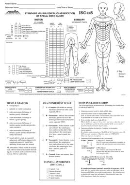 American Spinal Injury Association (ASIA) method for classifying spinal cord injury (SCI) by neurologic level.
See also Hypercalcemia and Spinal Cord Injury, Spinal Cord Injury and Aging, Rehabilitation of Persons With Spinal Cord Injuries, Central Cord Syndrome, Brown-Sequard Syndrome, and Cauda Equina and Conus Medullaris Syndromes.
Historical information in SCI classification
In 1982, ASIA first published standards for neurologic classification of patients with spinal injury, followed by further refinements to definitions of neurologic levels, identification of key muscles and sensory points corresponding to specific neurologic levels, and validation of the Frankel scale. In 1992, the International Medical Society of Paraplegia (IMSOP) adopted these guidelines to create true international standards, followed by further refinements. A standardized ASIA method for classifying spinal cord injury (SCI) by neurologic level was developed (see the image above).
Anatomy
The spinal cord is divided into 31 segments, each with a pair of anterior (motor) and dorsal (sensory) spinal nerve roots. On each side, the anterior and dorsal nerve roots combine to form the spinal nerve as it exits from the vertebral column through the neuroforamina. The spinal cord extends from the base of the skull and terminates near the lower margin of the L1 vertebral body. Thereafter, the spinal canal contains the lumbar, sacral, and coccygeal spinal nerves that comprise the cauda equina. As a result, injuries below L1 are not considered spinal cord injuries (SCIs) because they involve the segmental spinal nerves and/or cauda equina. Spinal injuries proximal to L1, above the termination of the spinal cord, often involve a combination of spinal cord lesions and segmental root or spinal nerve injuries.
Neuropathways
The spinal cord itself is organized into a series of tracts or neuropathways that carry motor (descending) and sensory (ascending) information. These tracts are organized somatotopically within the spinal cord. The corticospinal tracts are descending motor pathways located anteriorly within the spinal cord. Axons extend from the cerebral cortex in the brain as far as the corresponding segment, where they form synapses with motor neurons in the anterior (ventral) horn. They decussate (cross over) in the medulla before entering the spinal cord.
The dorsal columns are ascending sensory tracts that transmit light touch, proprioception, and vibration information to the sensory cortex. They do not decussate until they reach the medulla. The lateral spinothalamic tracts transmit pain and temperature sensation. These tracts usually decussate within 3 segments of their origin as they ascend. The anterior spinothalamic tract transmits light touch. Autonomic function traverses within the anterior interomedial tract. Sympathetic nervous system fibers exit the spinal cord between C7 and L1, whereas parasympathetic system pathways exit between S2 and S4.
Injury to the corticospinal tract or dorsal columns, respectively, results in ipsilateral paralysis or loss of sensation of light touch, proprioception, and vibration. Unlike injuries of the other tracts, injury to the lateral spinothalamic tract causes contralateral loss of pain and temperature sensation. Because the anterior spinothalamic tract also transmits light touch information, injury to the dorsal columns may result in complete loss of vibration sensation and proprioception but only partial loss of light touch sensation. Anterior cord injury causes paralysis and incomplete loss of light touch sensation.
Autonomic function is transmitted in the anterior interomedial tract. The sympathetic nervous system fibers exit from the spinal cord between C7 and L1. The parasympathetic system nerves exit between S2 and S4. Therefore, progressively higher spinal cord lesions or injury causes increasing degrees of autonomic dysfunction.
Vascular supply
The blood supply of the spinal cord consists of 1 anterior and 2 posterior spinal arteries. The anterior spinal artery supplies the anterior two thirds of the cord. Ischemic injury to this vessel results in dysfunction of the corticospinal, lateral spinothalamic, and autonomic interomedial pathways. Anterior spinal artery syndrome involves paraplegia, loss of pain and temperature sensation, and autonomic dysfunction. The posterior spinal arteries primarily supply the dorsal columns. The anterior and posterior spinal arteries arise from the vertebral arteries in the neck and descend from the base of the skull. Various radicular arteries branch off the thoracic and abdominal aorta to provide collateral flow.
The primary watershed area of the spinal cord is the midthoracic region. Vascular injury may cause a cord lesion at a level several segments higher than the level of spinal injury. For example, a lower cervical spine fracture may result in disruption of the vertebral artery that ascends through the affected vertebra. The resulting vascular injury may cause an ischemic high cervical cord injury. At any given level of the spinal cord, the central part is a watershed area. Cervical hyperextension injuries may cause ischemic injury to the central part of the cord, causing a central cord syndrome.
See also Topographic and Functional Anatomy of the Spinal Cord.
Pathophysiology
Spinal cord injury (SCI), as with acute stroke, is a dynamic process. In all acute cord syndromes, the full extent of injury may not be apparent initially. Incomplete cord lesions may evolve into more complete lesions. More commonly, the injury level rises 1 or 2 spinal levels during the hours to days after the initial event. A complex cascade of pathophysiologic events related to free radicals, vasogenic edema, and altered blood flow accounts for this clinical deterioration. Normal oxygenation, perfusion, and acid–base balance are required to prevent worsening of the SCI.
SCI can be sustained through different mechanisms, with the following 3 common abnormalities leading to tissue damage:
-
Destruction from direct trauma
-
Compression by bone fragments, hematoma, or disk material
-
Ischemia from damage or impingement on the spinal arteries
Edema could ensue subsequent to any of these types of damage.
Neurogenic shock
Neurogenic shock refers to the hemodynamic triad of hypotension, bradycardia, and peripheral vasodilation resulting from severe autonomic dysfunction and the interruption of sympathetic nervous system control in acute SCI. Hypothermia is also characteristic. This condition does not usually occur with SCI below the level of T6 and is more common in injuries above T6. Neurogenic shock is thought to occur secondary to the disruption of sympathetic outflow from T1-L2 and unopposed vagal tone. This results in a decrease in vascular resistance with the associated vascular dilatation. Neurogenic shock needs to be differentiated from spinal and hypovolemic shock. Hypovolemic shock tends to be associated with tachycardia. SCI patients may have multiple forms of shock occurring at the same time and the differential needs to remain broad during the early resuscitation period.
Spinal shock
Spinal shock is the complete loss of neurologic function, including reflexes, sensation, and rectal tone below the level of injury. It is a state of transient physiologic (rather than anatomic) reflex depression of cord function below the level of injury, with associated loss of all sensorimotor functions. Flaccid paralysis, including of the bowel and bladder, is observed, and sometimes sustained priapism develops. These symptoms tend to last several hours to days until the reflex arcs below the level of the injury begin to function again (eg, bulbocavernosus reflex, muscle stretch reflex [MSR]).
Primary vs secondary SCIs
SCIs may be primary or secondary. Primary SCI arises from mechanical disruption, transection, or distraction of neural elements. Secondary SCIs are potentially modifiable injuries that occur hours to days after the initial trauma. These include such pathologies as hypotension, infection, and thromboembolism. Anoxic or hypoxic effects can compound the extent of SCI.
Primary SCI usually occurs with fracture and/or dislocation of the spine. However, primary SCI may occur in the absence of spinal fracture or dislocation. Penetrating injuries due to bullets or weapons may also cause primary SCI through direct injury or propagation of a percussive wave. More commonly, displaced bony fragments cause penetrating spinal cord and/or segmental spinal nerve injuries.
Extradural pathology may also cause a primary SCI. Spinal epidural hematomas or abscesses cause acute cord compression and injury. Spinal cord compression from metastatic disease is a common oncologic emergency.
Longitudinal distraction with or without flexion and/or extension of the vertebral column may result in primary SCI without spinal fracture or dislocation. The spinal cord is tethered more securely than the vertebral column. Longitudinal distraction of the spinal cord with or without flexion and/or extension of the vertebral column may result in SCI without radiologic abnormality (SCIWORA).
SCIWORA was first coined in 1982 by Pang and Wilberger. Originally, it referred to SCI without radiographic or computed tomography (CT) scanning evidence of fracture or dislocation. However, with the advent of magnetic resonance imaging (MRI), the term has become ambiguous. Findings on MRI such as intervertebral disk rupture, spinal epidural hematoma, cord contusion, and hematomyelia have all been recognized as causing primary or secondary SCI. SCIWORA should now be more correctly renamed as "spinal cord injury without neuroimaging abnormality" and its prognosis recognized as actually better than that of patients with SCI and radiologic evidence of traumatic injury. [6, 7, 8]
Complete vs incomplete spinal cord syndrome
One of the goals of the physician is to classify the pattern of the neurologic deficit into one of the cord syndromes. Spinal cord syndromes may be complete or incomplete. In most clinical scenarios, physicians should use a best-fit model to classify the SCI syndrome.
A complete cord syndrome is characterized clinically as complete loss of motor and sensory function below the level of the traumatic lesion. Incomplete cord syndromes have variable neurologic findings with partial loss of sensory and/or motor function below the level of injury; these include the anterior cord syndrome, the Brown-Séquard syndrome, and the central cord syndrome.
Anterior cord syndrome involves a lesion causing variable loss of motor function and pain and/or temperature sensation, with preservation of proprioception.
Brown-Séquard syndrome, which is often associated with a hemisection lesion of the cord, involves a relatively greater ipsilateral loss of proprioception and motor function, with contralateral loss of pain and temperature sensation.
Central cord syndrome usually involves a cervical lesion, with greater motor weakness in the upper extremities than in the lower extremities, with sacral sensory sparing. The pattern of motor weakness shows greater distal involvement in the affected extremity than proximal muscle weakness. Sensory loss is variable, and the patient is more likely to lose pain and/or temperature sensation than proprioception and/or vibration. Dysesthesias, especially those in the upper extremities (eg, sensation of burning in the hands or arms), are common.
Other cord syndromes
Conus medullaris syndrome, cauda equina syndrome, and spinal cord concussion are briefly discussed below.
Conus medullaris syndrome is a sacral cord injury, with or without involvement of the lumbar nerve roots. This syndrome is characterized by areflexia in the bladder, bowel, and to a lesser degree, lower limbs, whereas the sacral segments occasionally may show preserved reflexes (eg, bulbocavernosus and micturition reflexes). Motor and sensory loss in the lower limbs is variable.
Cauda equina syndrome involves injury to the lumbosacral nerve roots in the spinal canal and is characterized by an areflexic bowel and/or bladder, with variable motor and sensory loss in the lower limbs. Because this syndrome is a nerve root injury rather than a true spinal cord injury, the affected limbs are areflexic. Cauda equina syndrome is usually caused by a central lumbar disk herniation.
A spinal cord concussion is characterized by a transient neurologic deficit localized to the spinal cord that fully recovers without any apparent structural damage.
Etiology
Since 2005, the most common causes of spinal cord injury (SCI) remain: (1) motor vehicle accidents (40.4%); (2) falls (27.9%), most common in those aged 45 years or older. Older females with osteoporosis have a propensity for vertebral fractures from falls with associated SCI; (3) interpersonal violence (primarily gunshot wounds) (15.0%), which is the most common cause in some US urban settings. Among patients who had suffered an assault, SCI from a penetrating injury tended to be worse than that from a blunt injury; [9] (4) and sports (8.0%), in which diving is the most common cause. [10] SCI due to trauma has major functional, medical, and financial effects on the injured person, as well as an important effect on the individual's psychosocial wellbeing. [11, 12, 13]
Other causes of SCI include the following:
-
Vascular disorders
-
Tumors [14]
-
Infectious conditions
-
Spondylosis
-
Iatrogenic injuries, especially after spinal injections and epidural catheter placement
-
Vertebral fractures secondary to osteoporosis
-
Developmental disorders
Injuries often associated with traumatic SCI also include bone fractures (29.3%), loss of consciousness (17.8%), and traumatic brain injury (TBI) affecting emotional/cognitive functioning (11.5%).
The rate of alcohol intoxication among individuals who sustain spinal cord injuries is 17%–49%.
Epidemiology
The incidence of spinal cord injury (SCI) in the United States is approximately 40 cases per million population, or about 12,000 patients per year based on data in the National Spinal Cord Injury database. [10] However, this estimate is based on older data from the 1990s as there has not been any new overall incidence studies completed. [10] Estimates from various studies suggest that the number of people in the United States alive in 2010 with SCI was about 265,000 persons (range, 232,000–316,000). [10]
SCIS occur most frequently in July and least commonly in February. The most common day on which these injuries occur is Saturday. SCIs also occur more frequently during daylight hours, which may be due to the increased frequency of motor vehicle accidents and of diving and other recreational sporting accidents during the day.
Racial, sexual, age-related differences in incidence
A significant trend over time has been observed in the racial distribution of persons with SCI. Since 2005, 66.5% are White, 26.8% are Black, 8.3% are Hispanic, and 2.0% are Asian. [10]
Males are approximately 4 times more likely than females to have SCIs. Overall, males account for 80.7% of reported injuries in the national database. [10]
Since 2005, the average age at injury is 40.7 years, reflecting the rise in the median age of the general population in the United States. [10] About 50% of SCIs occur between the ages of 16 and 30 years, 3.5% occur in children aged 15 years or younger, and about 11.5% occur in those older than 60 years (11.5%). Greater mortality is reported in older patients with SCI.
Pediatric SCI data
The pediatric data parallels that of the adult data on SCIs. Using information from the Kids' Inpatient Database (KID) and the National Trauma Database (NTDB), Vitale et al found that, with regard to the annual pediatric incidence rate, a significantly greater incidence of SCIs was found in Black children (1.53 cases per 100,000 children) than in Native American children (1.0 case per 100,000 children) and Hispanic children (0.87 case per 100,000 children), and the frequency in Asian children was significantly lower than that in all other races (0.36 per 100,000 children). [15] In addition, the likelihood that boys would suffer SCIs (2.79 cases per 100,000) was found to be more than twice that of girls (1.15 cases per 100,000). [15]
The overall incidence of pediatric SCI is 1.99 cases per 100,000 US children. As estimated from the above data, 1455 children are admitted to US hospitals annually for treatment of SCIs.
Vitale et al also looked at the major causative factors of pediatric cases, reporting the following incidences, [15] again paralleling adult data:
-
Motor vehicle accidents - 56%
-
Accidental falls - 14%
-
Firearm injuries - 9%
-
Sports injuries - 7%
Among children in the study, 67.7% of those injured in a motor vehicle accident were not wearing a seatbelt. [15] Alcohol and drugs were found to have played a role in 30% of all pediatric cases of SCIs.
Other epidemiologic data
Marital, educational, and employment status of patients with spinal cord injuries are discussed below.
Marital status
Single persons sustain SCIs more commonly than do married persons. Research has indicated that among persons with SCIs whose injury is approximately 15 years old, one third will remain single 20 years post injury. The marriage rate after SCI is annually about 59% below that of persons in the general population of comparable gender, age, and marital status.
Marriage is more likely if the patient is a college graduate, previously divorced, paraplegic (not tetraplegic), ambulatory, living in a private residence, and independent in the performance of activities of daily living (ADLs).
The divorce rate annually among individuals with SCI within the first 3 years following injury is approximately 2.5 times that of the general population, whereas the rate of marriages contracted after the injury is about 1.7 times that of the general population.
The divorce rate in those who were married at the time of their injury is higher if the patient is younger, female, Black, without children, nonambulatory, and previously divorced. The divorce rate among those who were married after the SCI is higher if the individual is male, has less than a college education, has a thoracic-level injury, and was previously divorced.
Educational status
The rate of injury differs according to educational status, as follows:
-
Less than a high school degree: 39.8%
-
High school degree: 49.9%
-
Associate degree: 1.6%
-
Bachelor's degree: 5.9%
-
Master's or doctorate degree: 2.1%
-
Other degree: 0.7%.
Employment status
Patients with SCI classified as American Spinal Injury Association (ASIA) level D are more likely to be employed than individuals with ASIA levels A, B, and C (see Neurologic level and extent of injury under Clinical). Persons employed tend to work full-time. Individuals who return to work within 1 year of injury tend to return to the same job. Those individuals who return to work after 1 year of injury tend to work for a different employer at a different job requiring retraining. [16]
The likelihood of employment after injury is greater in patients who are younger, male, and White and who have more formal education, higher reported intelligence quotient (IQ), greater functional capacity, and less severe injury. Patients with greater functional capacity, less severe injury, history of employment at the time of injury, greater motivation to return to work, nonviolent injury, and ability to drive are more likely to return to work, especially after more elapsed time following injury.
Prognosis
Patients with a complete spinal cord injury (SCI) have a less than 5% chance of recovery. If complete paralysis persists at 72 hours after injury, recovery is essentially zero. In the early 1900s, the mortality rate 1 year after injury in patients with complete lesions approached 100%. Much of the improvement since then can be attributed to the introduction of antibiotics to treat pneumonia and urinary tract infection (UTI).
The prognosis is much better for the incomplete cord syndromes.
If some sensory function is preserved, the chance that the patient will eventually be able walk is greater than 50%.
Ultimately, 90% of patients with SCI return to their homes and regain independence.
Providing an accurate prognosis for the patient with an acute SCI usually is not possible in the emergency department (ED) and is best avoided.
Life expectancy and mortality
Approximately 10–20% of patients who have sustained an SCI do not survive to reach acute hospitalization, whereas about 3% of patients die during acute hospitalization.
Originally, the leading cause of death in patients with SCI who survived their initial injury was renal failure; however, the current leading causes of death are pneumonia, pulmonary embolism, or septicemia. Heart disease, [17, 18] subsequent trauma, suicide, and alcohol-related deaths are also major causes of death in these patients. [19, 20] In persons with SCI, the suicide rate is higher among individuals who are younger than 25 years.
Among patients with incomplete paraplegia, the leading causes of death are cancer and suicide (1:1 ratio), whereas among persons with complete paraplegia, the leading cause of death is suicide, followed by heart disease.
Life expectancies for patients with SCI continues to increase but are still below the general population. Patients aged 20 years at the time they sustain these injuries have a life expectancy of approximately 35.7 years (patients with high tetraplegia [C1-C4]), 40 years (patients with low tetraplegia [C5-C8]), or 45.2 years (patients with paraplegia). [10] Individuals aged 60 years at the time of injury have a life expectancy of approximately 7.7 years (patients with high tetraplegia), 9.9 years (patients with low tetraplegia), and 12.8 years (patients with paraplegia).
A 2006 study by Strauss et al reported that among patients with SCI, during the critical first 2 years following injury, a 40% decline in mortality occurred between 1973 and 2004. [21] During that same 31-year period, there had been only a small, statistically insignificant reduction in mortality in the post 2-year period for these patients.
Life satisfaction
Studies have found that patients with SCI who suffer from pain have less life satisfaction than do patients in whom pain is well controlled; this may also affect patients' general outlook on life. [22, 23]
Rehabilitation
Patients younger than 65 years with muscle grade of 3 or greater in the myotome L3 and S1, and light touch sensation in the dermatome L3 and S1 within 15 days of injury (all within American Spinal Injury Association [ASIA] impairment scale D), are more likely to be independent indoor walkers within a year of injury. [24] Rehabilitation goals in this group should therefore be geared toward functional capacity and within expected independent walking.
Brain–computer interface for SCI
SCI can leave patients with severe or complete permanent paralysis. Brain–computer interface (BCI) can potentially restore or substitute for motor behaviors in patients with a high-cervical SCI. [25] Studies have shown that patients with SCI are able to control virtual keyboards, [26] a computer cursor, [25] and a limb prosthetic device [27] using BCI technologies. The BCI outputs are accomplished by acquiring neurophysiological signals associated with a motor process in the cerebral cortex, analyzing these signals in real time, and subsequently translating them into commands for a limb prosthesis. These are promising findings; in the future, BCI may provide a permanent solution for restoration of motor functions in SCI patients.
Walking assistance systems
In 2014, the FDA approved a wearable, motorized device to help individuals with paraplegia due to an SCI sit, stand, and walk with assistance from a companion. [28, 29] The device, which is intended for patients with SCIs at levels T7-L5 and for those with level T4-T6 injuries when used only in rehabilitation institutions, consists of the following:
-
Fitted metal brace that supports the legs and part of the upper body
-
Motors that provide movement to the hips, knees, and ankles
-
Tilt sensor
-
Computer and power supply worn on the back
Before using the device, caregivers and patients are required to undergo extensive training.
Patient Education
As part of inpatient therapy, patients with spinal cord injury (SCI) should receive a comprehensive program of physical and occupational therapy.
Prevention
Many SCIs result from incidents involving drunk driving, assaults, and alcohol or drug abuse. SCIs from industrial hazards, such as equipment failures or inadequate safety precautions, are potentially preventable causes. Unfenced, shallow, or empty swimming pools are known hazards.
-
American Spinal Injury Association (ASIA) method for classifying spinal cord injury (SCI) by neurologic level.


