Practice Essentials
The vertebrobasilar arterial system perfuses the medulla, cerebellum, pons, midbrain, thalamus, and occipital cortex. Occlusion of large vessels in this system usually leads to major disability or death. Vertebrobasilar stroke carries a mortality rate of more than 85%. Because of involvement of the brainstem and cerebellum, most survivors have multisystem dysfunction (eg, quadriplegia or hemiplegia, ataxia, dysphagia, dysarthria, gaze abnormalities, cranial neuropathies). [1]
However, many vertebrobasilar lesions arise from small vessel disease and are correspondingly small and discrete. The clinical correlates of these smaller lesions consist of a variety of focal neurologic deficits, depending on their location within the brainstem. Patients with small lesions usually have a benign prognosis with reasonable functional recovery.
See the images below regarding vertebrobasilar stroke.
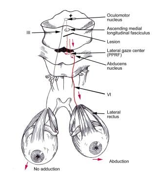 Lesion of the medial longitudinal fasciculus (MLF) resulting in internuclear ophthalmoplegia (INO). (Courtesy of BC Decker Inc.)
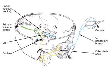 Illustration of afferent (CN V) and efferent (CN VII) limbs of the blink reflex. (Courtesy of BC Decker Inc.)
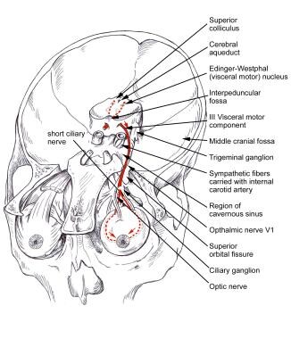 Visceral motor component of CN III and pathways involved in pupillary constriction. (Courtesy of BC Decker Inc.)
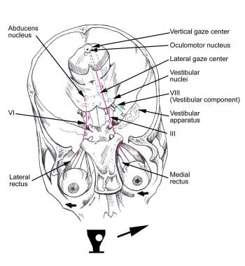
 Note the horizontal eye movement. Also note a topographic relationship of the center for vertical gaze. (Courtesy of BC Decker Inc.)
Distinction of vertebrobasilar and hemispheric stroke
Lesions in the vertebrobasilar system have some characteristic clinical features that distinguish them from lesions in the hemispheres, including the following [2] :
-
When cranial nerves or their nuclei are involved, the corresponding clinical signs are ipsilateral to the lesion and the corticospinal signs are crossed, involving the opposite arm and leg
-
Cerebellar signs (eg, dysmetria, ataxia) are frequent
-
Involvement of the ascending sensory pathways may affect the spinothalamic pathway or the medial lemniscus (dorsal columns), resulting in dissociated sensory loss (ie, loss of 1 sensory modality on one side and preservation of other sensory modalities in the opposite limbs)
-
Dysarthria and dysphagia typically are present
-
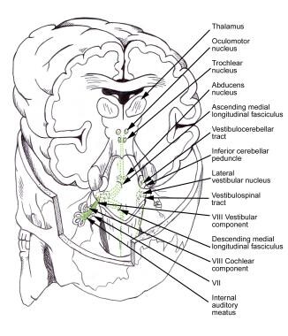
Vertigo, nausea, and vomiting, along with nystagmus, represent involvement of the vestibular system
-
Unilateral Horner syndrome occurs with brainstem lesions
-
Occipital lobe lesions result in visual field loss or visuospatial deficits
-
Cortical deficits, such as aphasia and cognitive impairments, are absent
Signs and symptoms of vertebrobasilar stroke
Commonly reported signs and symptoms associated with vertebrobasilar strokes include the following [2] :
-
Vertigo
-
Nausea and vomiting
-
Headache
-
Abnormalities in the level of consciousness
-
Abnormal oculomotor signs (eg, nystagmus, lateral gaze abnormalities, diplopia, pupillary changes)
-
Ipsilateral cranial nerve weakness (eg, dysarthria, dysphagia, dysphonia, weakness of facial muscles and tongue)
-
Sensory loss (in the face and scalp)
-
Ataxia
-
Contralateral motor weakness (eg, hemiparesis, quadriparesis)
-
Pain and temperature loss
-
Incontinence
-
Visual-field defects
-
Presence of central pain
-
Abnormal swelling
-
Sweating in the face or extremities
Workup in vertebrobasilar stroke
Laboratory studies
The laboratory workup should include the following:
-
Complete blood count (CBC)
-
Electrolytes
-
Blood urea nitrogen (BUN) and creatinine
-
Prothrombin time and activated partial thromboplastin time (aPTT)
-
Cholesterol level
-
Lipid profile
Imaging studies
Computed tomography (CT) scanning usually is the first imaging study performed, because it has a sensitivity of more than 95% when used in the identification intra-axial or extra-axial hemorrhage within the first 24 hours of onset. [3, 4] Other helpful findings include evidence of infarcts in the thalamus or occipital lobes (implicating involvement of the rostral basilar artery) and evidence that a hyperdense basilar artery is present (suggesting a probable occlusion). [5, 6]
Spiral CT angiography is used further to identify occluded and dolichoectatic vessels. [7, 8]
Magnetic resonance imaging (MRI) is more sensitive than CT scanning in the identification of ischemia (since bone does not degrade the image). Various techniques, including flow suppression and the production of diffusion-weighted and perfusion-weighted images, make MRI a very powerful tool for the exclusion of intraparenchymal hemorrhage or edema and for the identification of early and potentially reversible ischemia. [3, 4, 9, 10, 11]
MRI and magnetic resonance angiography (MRA) are very helpful in finding occult lesions, such as demyelinating plaques, tumors, vertebrobasilar dolichoectasia, or dissection. [12, 13, 14]
While the role of cerebral (catheter) angiography has changed due to the availability of noninvasive imaging modalities (eg, MRI, MRA, transcranial Doppler [TCD]), it still is considered the criterion standard for imaging.
TCD is helpful for purposes of follow-up once an initial evaluation has demonstrated the lesion. TCD has a sensitivity of 72% and a specificity of 94%, in patients with basilar artery disease. [15]
Echocardiography [16] should be considered in the following patients:
-
Those younger than 45 years
-
Those with explained basilar artery occlusion
Electrocardiography
Electrocardiography should be performed in all patients on initial evaluation. All patients should be monitored continuously for the first few days. Ischemic changes in the ECG should be investigated further with assays of serum creatine kinase, cardiac isoenzymes, and troponin, for reasons that include the following:
-
Up to 20% of patients with acute stroke have an arrhythmia
-
Myocardial infarction occurs in 2-3% of patients
-
The presence of arrhythmias (eg, atrial fibrillation) has an impact on long-term patient management related to stroke prevention
Management of vertebrobasilar stroke
Hemodynamic management
Hemodynamic management should be aimed at minimizing the ischemic injury.
Respiratory management
Endotracheal intubation may be considered in patients with a decreased level of consciousness and a Glasgow coma score of less than 8. Of the mechanical ventilation modes, pressure support ventilation (PSV) and synchronized intermittent mandatory ventilation are used most often.
For patients with poor respiratory drive, synchronized intermittent mandatory ventilation may be a better mode. This form of ventilation delivers a set number of breaths with a set tidal volume, which is synchronized with the patient's inspiratory effort while allowing the patient to take extra breaths. Adding PSV during the extra breaths can minimize the patient's respiratory effort when taking them.
Thrombolysis
Thrombolysis can be accomplished using tissue plasminogen activator (tPA).
Rehabilitation
Physical therapy (PT) and occupational therapy (OT) should be initiated soon after admission, depending on the condition of the patient. Once the symptoms have stabilized, patients should be mobilized out of bed, which will allow them to participate in full PT and OT activities.
Other rehabilitation strategies include the following:
-
Speech and swallowing therapy
-
Recreational therapy
Anatomy of the Vertebral and Basilar Arteries
The vertebral arteries arise from the subclavian arteries, and as they course cephalad in the neck, they pass through the costotransverse foramina of C6 to C2. They enter the skull through the foramen magnum and merge at the pontomedullary junction to form the basilar artery. Each vertebral artery usually gives off the posterior inferior cerebellar artery (PICA). At the top of the pons, the basilar artery divides into 2 posterior cerebral arteries (PCAs).
Proximal to its bifurcation into the terminal branches (PCAs), the basilar artery gives off the superior cerebellar arteries that supply the lateral aspect of the pons and midbrain, as well as the superior surface of the cerebellum. The cerebellum is supplied by long circumferential arteries, the PICA, and the anterior inferior and superior cerebellar arteries from the basilar artery.
The medulla is perfused by the PICA and by direct, smaller branches from the vertebral arteries. The pons is perfused by small, penetrating branches from the basilar artery and its major branches. Penetrating arteries from the PCAs perfuse the midbrain and thalamus, and the occipital cortex is perfused by the PCAs.
At the base of the brain, the carotid and basilar systems join to form a circle of large, communicating arteries known as the circle of Willis. Because of this arrangement of collateral vessels, even when one of the main arteries is occluded, adequate perfusion of the brain still may be possible. [17]
Pathophysiology of Vertebrobasilar Stroke
The most common vascular condition affecting the vertebrobasilar system is atherosclerosis, in which plaques cause narrowing and occlusion of the large vessels.
The pathology of small vessel disease (affecting arteries 50-200 µm in diameter) is different from that of atherosclerosis, because the small vessels become occluded by a process called lipohyalinosis, which frequently occurs in association with hypertension. Occlusions of these small vessels lead to small, round infarctions called lacunes, which may appear as single lesions or may be distributed as multiple lesions scattered widely throughout the subcortex and brainstem.
Lipohyalinosis weakens the vessel wall, and in hypertensive individuals, rupture of the artery may occur, resulting in a focal hemorrhage. Almost all intracerebral hemorrhages originate from the rupture of these small, penetrating vessels.
Because of the close anatomical relationship between the vertebral arteries and the cervical spine, chiropractic manipulation or neck rotation may traumatize the vertebral arteries in the neck. The damaged arteries may occlude with thrombus or undergo dissection. However, a retrospective study by Whedon et al of patients aged 66-99 years suggested that there is very little risk of vertebrobasilar stroke in association with chiropractic manipulation of the cervical spine. [18]
Embolic occlusion of the vertebrobasilar system is not common and usually is artery-to-artery with occlusion of the basilar artery. Donor sites for the emboli typically are the aortic arch, the subclavian artery, and the origin of the vertebral arteries.
Etiology of Vertebrobasilar Stroke
Vertebrobasilar insufficiency or stroke may be caused by a number of mechanisms, including thrombus, embolism, and hemorrhage (secondary to aneurysm or trauma). [19] In general, strokes occur because of ischemic events (80-85% of patients) or hemorrhage (15-20% of patients). A number of risk factors are associated with stroke, such as the following:
-
Increasing age
-
Family history
-
Race
-
Prior history of stroke
-
Coronary artery disease
-
Physical inactivity
A prospective study by Amin-Hanjani et al indicated that in patients with symptomatic atherosclerotic vertebrobasilar occlusive disease, angiographic evidence of low distal flow status signals a higher likelihood of subsequent vertebrobasilar stroke. The study used large-vessel quantitative magnetic resonance angiography to distinguish low from normal flow in patients who had suffered a recent vertebrobasilar transient ischemic attack or stroke and in whom at least 50% atherosclerotic stenosis or occlusion was present in vertebral and/or basilar arteries. Among patients with low distal flow status, the 12- and 24-month event-free survival rates were 78% and 70%, respectively, while in patients with normal flow, the rates were 96% and 87%, respectively. [20]
Another study by Amin-Hanjani et al indicated that in patients with vertebrobasilar disease and low blood flow, strict blood pressure control may actually raise the stroke risk. Again, the study’s patients had recently suffered a vertebrobasilar transient ischemic attack or stroke and had 50% or more atherosclerotic stenosis or occlusion of vertebral or basilar arteries. The investigators found the greatest risk of subsequent stroke to be in those individuals with a combination of low blood flow and blood pressure below 140/90 mm Hg. [21]
Epidemiology of Vertebrobasilar Stroke
The frequency, incidence, and prevalence of the vertebrobasilar syndromes vary, depending on the specific area and syndrome involved. Approximately 80-85% of all strokes are ischemic, and 20% of the lesions producing ischemic strokes occur in the vertebrobasilar system.
Overall, hemorrhage causes 15-20% of strokes. Although most intracerebral hemorrhages occur in the region of the putamen and thalamus, about 7% of all hemorrhagic lesions involve the cerebellum in the area of the dentate nucleus, and approximately 6% of hemorrhagic lesions involve the pons.
In most of the reported series, mortality patients with basilar artery occlusion has been consistently greater than 75-80%. [22] Most survivors of basilar artery occlusion have severe, persisting disability.
Clinical Presentation in Vertebrobasilar Stroke
Patient History
The onset and duration of symptoms in vertebrobasilar stroke depends, in large part, upon the etiology. Patients with basilar artery thrombosis typically have a waxing and waning course of symptoms, with as many as 50% of patients experiencing transient ischemic attacks for several days to weeks prior to the occlusion.
In contrast, embolic events are sudden, without prodrome or warning, with acute and dramatic presentation. Commonly reported signs and symptoms associated with vertebrobasilar strokes include the following [2] :
-
Vertigo
-
Nausea and vomiting
-
Headache
-
Abnormalities in the level of consciousness
-
Abnormal oculomotor signs (eg, nystagmus, lateral gaze abnormalities, diplopia, pupillary changes)
-
Ipsilateral cranial nerve weakness (eg, dysarthria, dysphagia, dysphonia, weakness of facial muscles and tongue)
-
Sensory loss (in the face and scalp)
-
Ataxia
-
Contralateral motor weakness (eg, hemiparesis, quadriparesis)
-
Pain and temperature loss
-
Incontinence
-
Visual-field defects
-
Presence of central pain
-
Abnormal swelling
-
Sweating in the face or extremities
Physical Examination
Common clinical findings observed in more than 70% of patients with vertebrobasilar stroke include an abnormal level of consciousness, as well as hemiparesis or quadriparesis, which usually is asymmetric. Pupillary abnormalities and oculomotor signs are common, and bulbar manifestations, such as facial weakness, dysphonia, dysarthria, and dysphagia, occur in more than 40% of patients.
Oculomotor signs usually reflect the involvement of the abducens nucleus; the horizontal gaze center located in the pontine paramedian reticular formation (PPRF), contiguous to the abducens nucleus; and/or the medial longitudinal fasciculus (MLF). Lesions to these structures result in ipsilateral lateral gaze or conjugate gaze palsy.
See the images below.
Lesion of the medial longitudinal fasciculus (MLF) resulting in internuclear ophthalmoplegia (INO). (Courtesy of BC Decker Inc.)
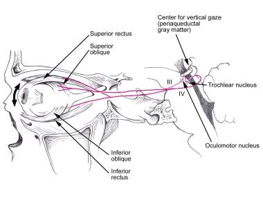 Center for vertical gaze and pathways involved in vertical eye movement (Courtesy of Cranial Nerves--Anatomy and Clinical Comments. BC Decker Inc; Toronto. 1988)
Illustration of afferent (CN V) and efferent (CN VII) limbs of the blink reflex. (Courtesy of BC Decker Inc.)
Center for vertical gaze and pathways involved in vertical eye movement (Courtesy of Cranial Nerves--Anatomy and Clinical Comments. BC Decker Inc; Toronto. 1988)
Illustration of afferent (CN V) and efferent (CN VII) limbs of the blink reflex. (Courtesy of BC Decker Inc.)
 Vestibular reflex illustrating horizontal eye movements only. (Courtesy of BC Decker Inc.)
Visceral motor component of CN III and pathways involved in pupillary constriction. (Courtesy of BC Decker Inc.)
Vestibular reflex illustrating horizontal eye movements only. (Courtesy of BC Decker Inc.)
Visceral motor component of CN III and pathways involved in pupillary constriction. (Courtesy of BC Decker Inc.)
Ocular bobbing is described as a brisk, downward movement of the eyeball with a subsequent return to the primary position. This deficit localizes the lesion to the pons. Other reported signs of pontine ischemia include ataxia and tremor associated with mild hemiparesis. The signs described can occur in different combinations, presenting a diagnostic challenge in lesion localization.
Certain constellations of findings may serve as clues to the location of the lesion, including the following examples:
-
Midbrain syndromes - cranial nerve (CN) III lesion and vertical gaze palsy
-
Pontine syndromes - CN VI lesion, horizontal gaze palsy, and VII nerve palsy
-
Medullary syndromes - ipsilateral facial pain and temperature loss, Horner syndrome, ipsilateral ataxia, contralateral loss of pain and temperature sensation, and ipsilateral paralysis of the tongue, soft palate, vocal cord, or sternocleidomastoid [SCM] muscle
-
Posterior cerebral artery - contralateral hemianopia with macular sparing
Complications
Potential complications of vertebrobasilar stroke include the following:
-
Aspiration pneumonia
-
Deep venous thrombosis
-
Pulmonary embolism
-
Myocardial infarction
A literature review by Yuan et al indicated that the top risk factor for lung infection in stroke patients is multiple vertebrobasilar strokes, followed by, among the other top 5 risk factors, a National Institutes of Health Stroke Scale score of over 15, the use of mechanical ventilation, the use of nasogastric tubes, and dysphagia. [23]
Vertebrobasilar Artery Stroke Syndromes
A variety of specific neurologic syndromes [24, 25] have been described in vertebrobasilar artery stroke, based on constellations of findings. Some examples are as follows:
-
Lateral medullary (Wallenberg) syndrome
-
Medial medullary (Dejerine) syndrome
-
Cerebellar infarction
-
Locked-in syndrome
-
Top-of-the-basilar syndrome
-
Internuclear ophthalmoplegia
-
One-and-a-half syndrome
-
Ventral pontine (Millard-Gubler) syndrome
-
Upper dorsal pontine (Raymond-Cestan) syndrome
-
Upper dorsal pontine (Raymond-Cestan) syndrome
-
Lower dorsal pontine (Foville) syndrome
-
Ventral midbrain (Weber) syndrome
-
Dorsal midbrain (Benedikt) syndrome
-
Posterior Cerebral Artery occlusion
Lateral medullary (Wallenberg) syndrome
This syndrome is most often due to vertebral artery occlusion or, less commonly, to posterior inferior cerebellar artery (PICA) occlusion. Patients present with nausea, vomiting, and vertigo from involvement of the vestibular system. [26]
Ipsilateral clinical features include the following:
-
Ataxia and dysmetria, due to damage to the inferior cerebellar peduncle and cerebellum
-
Horner syndrome (eg, ptosis, miosis, hypohidrosis or anhidrosis, enophthalmos), due to damage to descending sympathetic fibers
-
Facial pain and temperature loss
-
Reduced corneal reflex, from damage to the descending spinal tract and nucleus of CN V
-
Nystagmus
-
Hypoacusis (cochlear nucleus)
-
Dysarthria
-
Dysphagia
-
Paralysis of the pharynx, palate, and vocal cord
-
Loss of taste from the posterior third of the tongue (nuclei or fibers of CN IX and X)
Contralateral findings include the loss of pain and temperature sense in the body and extremities, indicating involvement of the lateral spinothalamic tract. Other findings include tachycardia and dyspnea (dorsal nucleus of CN X) and palatal myoclonus, a rhythmic involuntary jerking movement of the soft palate, pharyngeal muscles, and diaphragm. Palatal myoclonus sometimes follows infarction of the dentate nucleus of the cerebellum and inferior oliva.
A prospective study by Pavšič et al indicated that in the acute phase of unilateral lateral medullary infarction, sleep-disordered breathing is a common sign, with central events becoming reduced in frequency during the subacute phase. The apnea-hypopnea index during the acute phase was five or more per hour, 15 or more per hour, and 30 or more per hour in 79%, 68%, and 36% of patients, respectively. [27]
The prognosis of patients with the lateral medullary syndrome usually is quite good for functional outcome; however, patients may die in the acute phase from aspiration pneumonia, and death has been reported from sleep apnea in a number of cases.
Medial medullary (Dejerine) syndrome
This syndrome is an uncommon lesion resulting from occlusion of a vertebral artery or its branch to the anterior spinal artery; it involves the pyramid, the medial lemniscus, and, sometimes, the hypoglossal nerve. [28]
The clinical features include ipsilateral paresis of the tongue with deviation toward the lesion (lower motor neuron lesion of CN XII), contralateral hemiplegia with sparing of the face (corticospinal tract), and loss of ipsilateral vibration and proprioception (medial lemniscus). (See the image below.)
 LMN Lesion of the hypoglossal nerve producing tongue deviation to the side of the lesion. (Courtesy of BC Decker Inc.)
Cerebellar infarction
A stroke involving the cerebellum may result in a lack of coordination, clumsiness, intention tremor, ataxia, dysarthria, scanning speech, and even difficulties with memory and motor planning. Early diagnosis of cerebellar infarctions is important, because swelling may cause brainstem compression or hydrocephalus.
A study by Baek et al of 116 patients with basilar artery occlusion found the incidence of superior cerebellar artery occlusion to be 8.6% (10 patients). Eight of the 10 patients with superior cerebellar artery occlusion had had occlusion in the distal segment of the basilar artery. [29]
Locked-in syndrome
This dramatic clinical syndrome occurs when there is an infarction of the upper ventral pons. Locked-in syndrome can result from occlusion of the proximal and middle segments of the basilar artery or from hemorrhage involving that region. It can also be caused by trauma, central pontine myelinolysis, encephalitis, or a tumor. [30]
Bilateral ventral pontine lesions involving corticospinal and corticobulbar tracts lead to quadriplegia. The patient is unable to speak, to produce facial movement (damage to the corticobulbar tracts), or to look to either side (horizontal eye movement is impaired due to a lesion of bilateral CN VI nuclei). Because the tegmentum of the pons is spared, the patient's consciousness is preserved, with the patient fully awake, sensate, and aware. The only movements preserved are vertical eye movements and blinking. The patient is paralyzed completely and communicates only by blinking. Some recovery of facial muscle movement and horizontal gaze may occur with time or in an incomplete form of this syndrome.
Coma may occur with bilateral involvement of the pontine tegmentum or with lesions of the midbrain reticular formation. Coma generally is associated with oculomotor abnormalities, and motor abnormalities may be present. A comatose patient is unresponsive, and the coma may be prolonged when it is due to basilar artery occlusion. Sleep-wake cycles are absent in patients with coma.
Top-of-the-basilar syndrome
This syndrome is the manifestation of upper brainstem and diencephalic ischemia caused by occlusion of the rostral basilar artery; the occlusion usually results from an embolism. [31] Varying degrees of involvement of the midbrain, thalamus, and portions of the temporal and occipital lobes may occur and can produce severe disability.
Patients present with sudden changes in the level of consciousness, confusion, amnesia, and visual symptoms (eg, hemianopia, cortical blindness, abnormal color vision/color dysnomia). These patients can also demonstrate oculomotor abnormalities, most commonly of the vertical gaze, such as gaze palsy, skew deviation, convergence spasm resulting in pseudoabducens palsy, or convergence-retraction nystagmus.
CN III palsy and pupillary abnormalities, including small pupils with decreased light reactivity (diencephalic), large/mid-position and fixed pupils (midbrain), and ectopic or oval pupils, also are frequent.
Other abnormalities include varying degrees of weakness, sensory deficits, or posturing.
Internuclear ophthalmoplegia
Clinically, internuclear ophthalmoplegia (INO) is a horizontal gaze palsy; it results from a brainstem lesion affecting the MLF between the nuclei of CN VI and III, most commonly in the pons. (See the image below.)
Lesion of the medial longitudinal fasciculus (MLF) resulting in internuclear ophthalmoplegia (INO). (Courtesy of BC Decker Inc.)
When a patient with a lesion in the right MLF attempts to look to his/her left (ie, away from the involved side), he/she shows no adduction of the right eye and full abduction of the left eye with the end-point abduction nystagmus.
By the same logic, in the case of bilateral INO, there is no adduction to either side with nystagmus of the abducting eye in both directions. Convergence is preserved, because the nuclei of CN III and peripheral innervation of the medial recti muscles are intact.
Because horizontal gaze requires coordinated activity of the ipsilateral CN III and contralateral CN VI (relative to the lesion), disruption of the communication pathway (ie, the MLF) between the nuclei of CN III (in the midbrain) and CN VI (in the pons) results in the inability of the eye ipsilateral to the lesion to adduct and the contralateral eye to exhibit abduction nystagmus when looking away from the involved side.
In elderly patients, INO is caused most often by occlusion of the basilar artery or its paramedian branches. In younger adults, it may occur due to multiple sclerosis (MS), commonly with bilateral involvement.
One-and-a-half syndrome
This syndrome is caused by a lesion affecting the PPRF and MLF simultaneously, resulting in ipsilateral conjugate gaze palsy and INO [32] A patient with this syndrome is completely unable to move the ipsilateral eye, and is able only to abduct the contralateral eye, with resulting nystagmus; the ‘one’ in the syndrome name refers to the former, and the ‘half’ to the latter.
The patient with a lesion in the ipsilateral PPRF or abducens nucleus and MLF connecting to the contralateral CN VI exhibits horizontal gaze palsy when looking toward the side of the lesion and exhibits INO when looking away from the side of the lesion. Associated features may include vertical nystagmus, exotropia of the contralateral eye, and skew deviation. Vertical gaze and convergence generally are preserved.
Ventral pontine (Millard-Gubler) syndrome
This syndrome occurs after paramedian infarction in the pons and results in ipsilateral lateral rectus palsy (CN VI) with diplopia, complete facial paresis (unilateral CN VII palsy), and contralateral hemiparesis/hemiplegia (corticospinal tract involvement) with sparing of the face.
Upper dorsal pontine (Raymond-Cestan) syndrome
This syndrome is due to obstruction of flow in the long circumferential branches of the basilar artery. This occlusion results in ipsilateral ataxia and coarse intention tremor (indicating involvement of the superior and middle cerebellar peduncles), weakness of mastication and sensory loss in the face (suggesting sensory and motor trigeminal nuclei and tracts), and contralateral loss of all sensory modalities (due to damage to medial lemniscus and spinothalamic tract) with or without facial weakness and hemiparesis (corticospinal tract).
Horizontal gaze palsy also may occur.
Lower dorsal pontine (Foville) syndrome
This syndrome may result from lesions to the dorsal tegmentum of the lower pons. The patient exhibits ipsilateral paresis of the whole face (nucleus and fibers of CN VII), horizontal gaze palsy on the ipsilateral side (ie, PPRF with or without CN VI nucleus), and contralateral hemiplegia (corticospinal tract) with sparing of the face.
Ventral midbrain (Weber) syndrome
Weber syndrome occurs with an occlusion of the median and/or paramedian perforating branches of the basilar artery. Typical clinical findings include ipsilateral CN III palsy, ptosis, and mydriasis (ie, damage to parasympathetic fibers of CN III) with contralateral hemiplegia. Weakness of the lower face (corticospinal and corticobulbar tracts) may be noted.
Dorsal midbrain (Benedikt) syndrome
This syndrome is due to a lesion in the midbrain tegmentum resulting from occlusion of paramedian branches of the basilar artery, the PCA, or both.
The patient demonstrates ipsilateral oculomotor palsy, ptosis, and mydriasis (as in Weber syndrome), along with the contralateral involuntary movements, such as those of intention tremor, ataxia, or chorea (due to the involvement of the red nucleus).
Posterior cerebral artery occlusion
The most common finding is occipital lobe infarction leading to contralateral hemianopia with macular sparing. Clinical symptoms associated with occlusion of the PCA vary depending on the location of the occlusion and may include the thalamic syndrome, thalamic perforate syndrome, Weber syndrome, cortical blindness, color blindness, failure to see to-and-fro movements, verbal dyslexia, and hallucinations.
Diagnostic Considerations
The differential diagnosis of vertebrobasilar stroke includes the following:
-
Central pontine myelinolysis
-
Metastatic disease of the brain
-
Subarachnoid hemorrhage
-
Basilar meningitis
-
Basilar migraine
-
Cerebellopontine angle tumors
-
Supratentorial hemispheric mass lesions with mass effect, herniation, and brainstem compression
Laboratory Studies for Vertebrobasilar Artery Stroke
The laboratory workup should include the following:
-
Complete blood count (CBC)
-
Electrolytes
-
Blood urea nitrogen (BUN) and creatinine
-
Prothrombin time and activated partial thromboplastin time (aPTT)
-
Cholesterol level
-
Lipid profile
Patients who are younger than 45 years or who have no evidence of atherosclerosis should be investigated for the presence of hypercoagulable states, such as the following:
-
Lupus anticoagulant and anticardiolipin antibodies
-
Protein C, protein S, and antithrombin III deficiencies
-
Factor V Leiden mutation
Creatine kinase, cardiac isoenzymes, and troponin level should be tested in the following persons:
-
All patients with suggestive symptoms (eg, chest pain)
-
Patients with evidence of ischemic changes in the electrocardiogram (ECG; because of the high incidence of concomitant coronary artery disease) [33]
Computed Tomography
CT scanning usually is the first imaging study performed, because it has a sensitivity of more than 95% when used in the identification intra-axial or extra-axial hemorrhage within the first 24 hours of onset. [3, 4] Other helpful findings include evidence of infarcts in the thalamus or occipital lobes (implicating involvement of the rostral basilar artery) and evidence that a hyperdense basilar artery is present (suggesting a probable occlusion). [5, 6]
Spiral CT angiography is used further to identify occluded and dolichoectatic vessels. [7, 8]
The disadvantages of CT scanning include a low sensitivity for early ischemia and the presence of significant artifacts caused by the bony structures surrounding the brainstem and cerebellum.
Magnetic Resonance Imaging
MRI is more sensitive than CT scanning in the identification of ischemia (since bone does not degrade the image). Various techniques, including flow suppression and the production of diffusion-weighted and perfusion-weighted images, make MRI a very powerful tool for the exclusion of intraparenchymal hemorrhage or edema and for the identification of early and potentially reversible ischemia. [3, 4, 9, 10, 11]
MRI and magnetic resonance angiography (MRA) are very helpful in finding occult lesions, such as demyelinating plaques, tumors, vertebrobasilar dolichoectasia, or dissection. [12, 13, 14] MRA has a sensitivity of up to 97% and a specificity of up to 98% when used to identify vertebrobasilar occlusion.
A limitation of MRA is its tendency to overestimate the degree of stenosis. This overestimation occurs because the production of a vessel's image in MRA is a based on a flow-related phenomenon; hence, the presence of severe stenosis with significant flow compromise may result in poor visualization of a vessel and cause the MRA image to resemble vascular occlusion.
Angiography
While the role of cerebral (catheter) angiography has changed due to the availability of noninvasive imaging modalities (eg, MRI, MRA, TCD), it still is considered the criterion standard for imaging. Catheter cerebral angiography is performed in the following circumstances:
-
The patient has an absolute contraindication to MRA (eg, a pacemaker, metallic implant)
-
The quality of noninvasive studies is not satisfactory
-
The results of noninvasive studies do not explain the clinical findings
Angiography should be considered a first-line diagnostic test after a CT scan, once it has been decided that recanalization with thrombolysis should be completed. The most important goal of the workup is to establish the type of vascular lesion and the mechanism of the stroke.
Ultrasonography
Transcranial Doppler (TCD) is used in the evaluation of cerebrovascular disease, but it often is inaccurate. Absence of signal in an initial examination does not necessarily mean occlusion.
TCD is helpful for purposes of follow-up once an initial evaluation has demonstrated the lesion. TCD has a sensitivity of 72% and a specificity of 94%, in patients with basilar artery disease. [15]
Electrocardiography
Electrocardiography should be performed in all patients on initial evaluation. All patients should be monitored continuously for the first few days. Ischemic changes in the ECG should be investigated further with assays of serum creatine kinase, cardiac isoenzymes, and troponin, for reasons that include the following:
-
Up to 20% of patients with acute stroke have an arrhythmia
-
Myocardial infarction occurs in 2-3% of patients
-
The presence of arrhythmias (eg, atrial fibrillation) has an impact on long-term patient management related to stroke prevention
Echocardiography
Echocardiography [16] should be considered in the following patients:
-
Those younger than 45 years
-
Those with explained basilar artery occlusion
Findings that may affect management include valvular disorders, vegetations, intramural or extramural thrombi, ventricular aneurisms, cardiac tumors (myxoma), right-to-left shunts, and poor ejection fraction.
Treatment of Acute Vertebrobasilar Artery Stroke
Ideally, all patients who have suffered a vertebrobasilar stroke should be admitted to a unit specializing in the care of stroke patients. Admission to a neurologic intensive care unit (ICU) is indicated for patients who are candidates for interventional therapies (eg, thrombolysis) or who have any of the following [34] :
-
Unstable or fluctuating neurologic symptoms
-
Decreased level of consciousness
-
Hemodynamic instability
-
Active cardiac or respiratory problems
Hemodynamic management
Hemodynamic management should be aimed at minimizing the ischemic injury. Cerebral ischemia impairs the brain’s ability to autoregulate its circulation through vasoconstriction and vasodilatation. Therefore, under ischemic conditions, the cerebral blood flow becomes blood pressure–dependent. [35] An increase in the mean arterial pressure (MAP) results in vasoconstriction. This response limits the perfusion pressure and the blood volume. A decrease in the MAP results in vasodilatation.
In normotensive patients, the limits of autoregulation are within the range of 50-150 mm Hg of the MAP. In chronic hypertensive patients, the curve of autoregulation is shifted upward. In the patients with severe cerebral vascular occlusive disease, the MAP and the cerebral perfusion pressure (CPP) become critical in maintaining the cerebral blood flow. CPP is equal to MAP less intracranial pressure (ICP) (ie, CPP = MAP-ICP). Therefore, overzealous treatment of hypertension should be avoided, because it can decrease the cerebral perfusion pressure and exacerbate the ongoing ischemia.
No existing information from randomized trials indicates whether treating hypertension is better than not treating it. Based on evidence from experimental models and on data from clinical experience, hypertension should not be treated unless there is evidence of end-organ damage, such as hypertensive encephalopathy, unstable angina, acute myocardial infarction, heart failure, or acute renal failure.
Hypertension should be treated when the diastolic blood pressure is greater than 120 mm Hg or when the systolic blood pressure is over 200 mm Hg. Otherwise, when thrombolysis is a strong consideration, the treatment parameters become 110 mm Hg or more for diastolic blood pressure or greater than 180 mm Hg for systolic blood pressure.
Commonly used antihypertensives are labetalol and nitroprusside. When diastolic blood pressure is greater than 140 mm Hg, nitroprusside is the preferred drug, provided that no contraindications exist.
Patients with hypotension need to be treated to optimize the MAP and, consequently, the blood pressure–dependent cerebral blood flow. Maximal effort should be made to maintain a normal intravascular volume using isotonic solutions. If the MAP continues to be low despite fluid management, vasopressors, such as dopamine, dobutamine, and phenylephrine, should be used.
In patients with unknown intravascular volume status or those with complications, such as congestive heart failure and pulmonary edema, a pulmonary artery catheter should be placed to monitor the central venous pressure and the pulmonary capillary wedge pressure. This approach would improve monitoring of the intravascular volume to avoid overload.
Respiratory management
Early assessment and management of the airway are critical due to the frequent involvement of lower cranial nerves and the impairment of consciousness in patients with brainstem ischemia. Assessment of the respiratory drive, gag reflex, and ability to handle secretions with a forceful cough also is of great importance.
Endotracheal intubation may be considered in patients with a decreased level of consciousness and a Glasgow coma score of less than 8. Of the mechanical ventilation modes, pressure support ventilation (PSV) and synchronized intermittent mandatory ventilation are used most often. For patients with good respiratory drive, the most comfortable mode is PSV. In this mode, the ventilator does not deliver a set of breaths but provides enough pressure support to maintain the desired tidal volume, usually in the range of 5-8 mL/kg. Most patients with no pulmonary comorbidities reach this goal with a PSV of 5-10.
For patients with poor respiratory drive, synchronized intermittent mandatory ventilation may be a better mode. This form of ventilation delivers a set number of breaths with a set tidal volume, which is synchronized with the patient's inspiratory effort while allowing the patient to take extra breaths. Adding PSV during the extra breaths can minimize the patient's respiratory effort when taking them.
Sedation and paralysis should be avoided, because they may obscure the neurologic assessment. Circumstances may exist that require the use of sedation and paralysis (eg, neurogenic hyperventilation) to avoid hypocarbia and worsening of the ischemic process.
Thrombolysis
In 1996, The US Food and Drug Administration (FDA) approved tissue plasminogen activator (tPA) for the treatment of acute ischemic stroke within the first 3 hours of onset. [3] Approval was based on data from the National Institute of Neurological Disorders and Stroke trial, which showed that a higher number of treated versus untreated patients had minimal deficit and minimal or no disability.
However, this trial did not include patients in stupor or coma, and that criterion probably excluded patients who suffered a basilar artery occlusion. Moreover, the trial did not study the vascular anatomy systematically in all patients. From experimental evidence and thrombolytic trials, it is apparent that recanalization improves outcome. [22, 36, 37]
In 2009, the American Heart Association/American Stroke Association (AHA/ASA) published a science advisory recommending that the time window for tPA administration be increased to 4.5 hours after a stroke, although this change has not been approved by the FDA. [38] Research indicates that tPA is effective in patients even when administered within the 3- to 4.5-hour window, [39, 40, 41] but the AHA/ASA stated that, despite its recommendation, the effectiveness of tPA administration in comparison with other treatments for thrombosis, within that time period, is not yet known.
The eligibility criteria for treatment between 3 and 4.5 hours are similar to those employed for treatment prior to 3 hours, as established in the AHA/ASA's 2007 guidelines, [42] but with the exclusion criteria expanded to include any of the following patient characteristics:
-
Age greater than 80 years
-
Use of oral anticoagulants
-
Baseline National Institutes of Health (NIH) Stroke Scale score >25
-
A history of both stroke and diabetes
In the early 1980s, Nenci and colleagues reported the first 4 cases of local thrombolysis for vertebrobasilar occlusion, establishing a trend to treat patients with intra-arterial thrombolysis. [3, 43] To date, several case series have been published. The average time to treatment has ranged from 8-48 hours. Overall mortality has decreased from 46-75% to 26-60%. The patient's condition at presentation appears to be the major prognostic factor; patients with quadriplegia and/or coma have demonstrated the least favorable outcomes. Despite the above efforts, intra-arterial thrombolysis for vertebrobasilar occlusion has not been studied systematically in randomized, controlled trials.
Of the different agents currently used for thrombolysis (urokinase, prourokinase, streptokinase, tPA), prourokinase and tPA seem to have more selectivity for thrombi. Streptokinase has not been used for stroke since the multicenter European and Australian trials documented a greater mortality in the treated patients. Because of concerns with its production, urokinase is not currently available in the United States.
Prourokinase was tested in a prospective, randomized fashion, including only patients with middle cerebral artery stem occlusion. Results showed a better outcome in treated patients, but prourokinase has not been approved for use in acute stroke.
At this time, the only viable option for thrombolysis in the United States continues to be tPA. This drug has been studied prospectively in trials involving combined intravenous and intra-arterial therapy, in doses of 0.3 mg/kg, with a maximum of 10-20 mg. Limited experience with the use of GPIIb/IIIa inhibitors, such as abciximab, to block the platelet function and rethrombosis has shown an overall reocclusion rate of approximately 30%.
Anticoagulation
Anticoagulation therapy with heparin has been used, but there is no evidence that it has an impact on outcome. Results from a trial using low–molecular weight heparin intravenously in patients with acute stroke, although negative overall, did show a better outcome at 7 days for patients with large vessel disease.
Angioplasty
Angioplasty has been performed to treat patients with atherosclerotic basilar artery stenosis. The use of angioplasty is based on the tendency of thrombosis to occur in stenosed arterial segments. Reports describe angioplasty performed in patients with acute vertebrobasilar occlusion, as well as electively. The published case series report a morbidity rate of 0-16% and a mortality rate of up to 33%; however, the role of angioplasty in the treatment of vertebrobasilar occlusion is not well defined.
A study by Hatano and Tsukahara indicated that endovascular treatment with balloon angioplasty can be safe and effective in selected patients with intracranial vertebrobasilar artery stenosis. In the report, 44 patients with the condition who fit certain criteria—ie, those who were medically refractory and symptomatic, with lesions below 15 mm in length and, as revealed on angiography, greater than 60% stenosis—underwent balloon angioplasty under local anesthesia. Stent placement was also used, if dilation was insufficient or if dissection or restenosis occurred. By 30 days after treatment, the stroke and death rate was just 2.3%. [44]
Within six months following treatment, restenosis had occurred in nine patients, with four of them being symptomatic, but subsequent balloon angioplasty or stenting successfully relieved the problem in the symptomatic patients. Hatano and Tsukahara cautioned that although their study indicates that endovascular therapy can effectively treat intracranial vertebrobasilar artery stenosis, clinical and radiologic follow-up is necessary owing to the possibility of restenosis. [44]
Other Treatment
Other aspects of treatment for vertebrobasilar stroke should include the following:
-
Aggressive pulmonary toilet to prevent mucous congestion and pneumonia
-
Prevention of aspiration pneumonitis
-
Early establishment of bowel and bladder programs
-
Monitoring of skin and all indwelling catheters for signs of infection
-
Control of body temperature (fever may worsen the outcome in stroke patients)
-
Tight blood glucose control
-
Heel protectors or L'Nard Multi Podus boots with regular skin inspection for breakdown/decubitus
-
Deep vein thrombosis prophylaxis with sequential compression devices or arteriovenous pumps and/or anticoagulants (eg, low–molecular weight heparin; adjusted-dose, subcutaneous heparin; warfarin), provided that there are no contraindications
A study by Alexander et al indicated that symptomatic vertebrobasilar atherosclerosis can be safely and effectively treated with endovascular therapy. The study involved interventions for 136 lesions (122 patients), including 13 treatments for acute stroke. The investigators found that treatment was technically successful for 123 of the lesions (90.4%), with better technical results achieved in cases of extracranial disease. Patients with nonprogressive symptoms in the subacute period and individuals who underwent statin treatment had better clinical results. [45]
Similarly, a retrospective study by Piechowiak et al indicated that endovascular treatment is a safe and effective therapy for tandem vertebrobasilar occlusion (ie, a basilar artery occlusion combined with proximal stenosis). The investigators found that of 52 patients with acute vertebrobasilar occlusive stroke who underwent mechanical thrombectomy, 8 of the 15 individuals (53.3%) with a tandem occlusion had a favorable outcome, while favorable results were obtained in 4 out of 14 patients (28.6%) with a single basilar artery occlusion with underlying stenosis and 5 out of 23 patients (21.7%) with an isolated embolic basilar artery occlusion. [46]
A study by Laible et al indicated that in patients with vertebrobasilar stroke who undergo thrombectomy, the presence of renal dysfunction increases the chance for posttreatment intracerebral hemorrhage (odds ratio = 3.54). However, the investigators did not find that either renal dysfunction or intracerebral hemorrhage was associated with an increased 3-month mortality risk in these patients. [47]
The aforementioned study by Baek and colleagues suggested that following thrombectomy for basilar artery occlusion, it may be unnecessary to try to recanalize remaining superior cerebellar artery occlusions. According to the report, out of 12 such occlusions (in 10 patients), nine recanalized spontaneously. The investigators found that none of the 10 patients “experienced symptomatic cerebellar hemorrhage or malignant cerebellar infarction.” [29]
Further inpatient care
Most patients with vertebrobasilar stroke have a significant degree of disability, due to involvement of the brainstem and cerebellum, with resultant multisystem dysfunction (eg, quadriplegia or hemiplegia, ataxia, dysphagia, dysarthria, gaze abnormalities, cranial neuropathies). They often require ongoing, acute rehabilitation, with attention paid to specific patient issues and the formulation of short-term and long-term care plans. The rehabilitation and planning are performed best in a multidisciplinary and interdisciplinary setting.
Rehabilitation After Vertebrobasilar Artery Stroke
Rehabilitation services have been shown to play a critical role in recovery from acute stroke. Physicians and nurses play crucial roles on the rehabilitation team; nurses often are the first to suggest initiation of therapy services, because they have the most extensive involvement with the patient. Prior to a discussion of the specific therapy disciplines, address nursing issues in the care of patients with vertebrobasilar stroke.
Nursing issues
A wide variation in symptoms may be seen with stroke, depending on the severity of the brain damage. Initial nursing intervention involves maintaining skin integrity, establishing a bowel and bladder program, maintaining nutrition, and ensuring the person's safety from injury.
Other important nursing issues include communication with the treating clinician in order to initiate therapy services for the assessment of ambulation, transfers, swallowing function, and the performance of activities of daily living (ADL). In some patients, the severity of the deficits makes ambulation impossible; however, patients should be mobilized out of bed and should be actively involved in physical and occupational therapy.
Positioning in bed and in a chair assures the patient's comfort and prevents complications from skin breakdown. If the upper extremity is flaccid or paretic, positioning is critical to the prevention of shoulder subluxation and pain from shoulder-hand syndrome.
Nursing staff always should involve family members in the care of a person who has sustained a stroke. The patient and family members may be unfamiliar with stroke and its effects. Education must be provided to make the patient and his/her family members aware of the importance of continuing with activities, of appropriate precautions, and of continuing therapy upon discharge to home.
Some patients have fluctuating symptoms and signs, which often are related to position. Because of this possibility, precautions are necessary with activities that can be undertaken until the symptoms have stabilized.
Physical therapy (PT) and occupational therapy (OT) should be initiated soon after admission, depending on the condition of the patient. Once the symptoms have stabilized, patients should be mobilized out of bed, which will allow them to participate in full PT and OT activities.
Physical therapy
The physical therapist is responsible for retraining of gross motor skills, such as gait, balance, transfers, bed mobility, and wheelchair mobility. The physical or occupational therapist may be involved with assessing the patient for the proper wheelchair and seating system.
The physical therapist also develops a PT program and instructs the patient in general strengthening and range of motion. Training of the patient and family members in the use of lower extremity orthotics may be necessary to provide for functional mobility.
Vestibular evaluation and training are very important, due to a high prevalence of vestibular and cerebellar involvement in vertebrobasilar strokes. Patients often need extensive balance and gait training. Evaluation always should begin with a detailed and focused history. A premorbid vestibular status determination is of great importance, because dizziness is the third most frequent complaint during physician visits from patients aged 65 years and the most frequent complaint from patients aged 75 years and older.
Further clinical testing may include the following:
-
Oculomotor examination - Visual tracking, convergence/divergence, saccades and smooth pursuit movement, spontaneous and gaze-evoked nystagmus, static/dynamic visual acuity, and vestibulo-ocular reflex (VOR)
-
Positional testing - Hallpike-Dix maneuver
-
Static balance - Romberg, sharpened Romberg, and single leg stance (each test is performed on even and uneven surfaces, with eyes open and closed)
-
Dynamic balance - Thorough gait assessment, including head turning, tandem gait, retro walking, negotiating obstacles, and turning
An exercise-based approach has been successful in the treatment of vestibular disorders, due to several possible mechanisms. These include adaptation, substitution, habituation, and repositioning.
With adaptation by the central vestibular system, the brain modulates the gain of the vestibular response, attempting to correct for a retinal slip (error signal) caused by the decreased gain of the VOR. The VOR training strategy includes focusing on a stationary or moving target while rotating the head, resulting in a retinal slip that facilitates adaptation.
Substitution for the loss of function by the remaining intact visual and somatosensory systems is used in treating patients with bilateral vestibular lesions (complete or partial loss of both labyrinths).
Habituation for postural vertigo results in decreased response to repeated provoking stimuli. Patients move into the provoking position 2-3 times during each session, and repeat these sessions 3-5 times per day.
Repositioning maneuvers (eg, Epley maneuver) are used for positional vertigo, based on the mechanical displacement of the debris from the affected canal(s) by a series of head movements. Alternating eye patches or prisms can help diplopia.
General conditioning also is incorporated into the overall rehabilitation plan, encouraging an increase in the performance of ADL as tolerated.
For patients with locked-in syndrome, improved function can be sought by boosting stability of the head, neck, and trunk through targeted rehabilitation, while mobility can be aided via proper fitting of an appropriate wheelchair. Distal motor control interventions and upright tolerance training, with subsequent balance and mobility exercises, are also important to rehabilitation. [48]
Occupational therapy
OT is used for retraining fine motor skills that are needed to perform ADL (eg, dressing, bathing, grooming), as well as for improving hand and arm function. OT also is involved in general strengthening, wheelchair mobility, upper extremity orthotics, and the evaluation of needs for adaptive equipment, as well as in family training and cognitive retraining for safety and ADL.
Speech and swallowing therapy
Speech therapy (ST) is used for cognitive retraining, speech and language skills, safety skills, swallowing assessment, and family training. In patients with dysphagia from brainstem lesions, the cricopharyngeus muscle may fail to open sufficiently, resulting in an impaired passage of the bolus from the pharynx to the esophagus. Increased pooling of a bolus in the vallecula and/or pyriform sinuses, which spills into the airway, poses a significant risk for aspiration and pneumonia.
Evaluation of these patients should be thorough and should include a videofluoroscopy with a modified barium swallow to assess for silent aspiration. The speech and language therapist often performs the initial swallowing evaluation and determines the risk for aspiration and the consistency of the patient's diet.
The patient's vocalization and possible reading, writing, and processing deficits also are addressed. Interventions for the prevention of aspiration include compensatory strategies, such as oromotor exercises and postural changes while swallowing, as well as facilitative strategies (eg, modification of bolus consistency, volume, delivery).
Surface electromyography biofeedback for dysphagia has shown promising results. Surface electromyography is used in training a patient to perform maneuvers that compensate for the weak swallow.
The Mendelsohn maneuver, for example, requires voluntary maintenance of the thyroid cartilage in an elevated position for a few seconds, resulting in further widening of the opening of the cricopharyngeus muscle and easier passage of the food bolus through to the esophagus. The patient observes the plateau (as opposed to the peak) of the generated waveform on the screen, reinforcing the concept of muscle activation in the desired position (thyroid cartilage elevation).
The patient should be on a nothing-by-mouth restriction until the swallowing mechanism has been assessed and cleared and the airway has been protected. If there is a high risk of aspiration, a nasogastric or nasoduodenal tube should be placed, although neither completely eliminates the aspiration risk. If the swallowing abnormalities are so severe that recovery is expected to take weeks or months, then a gastrostomy tube should be placed either surgically or percutaneously.
If there is persistent cricopharyngeal dysfunction on videofluoroscopic modified barium swallow, then cricopharyngeal myotomy can be considered. [49]
Recreational therapy
The recreational therapist should concentrate on finding alternative recreational activities for patients who are unable to perform at their premorbid level. Engaging in these activities provides a creative outlet and a positive emotional gain that potentially enhance the patient's psychological recovery.
Other consultations
In addition to consultations with physical, occupational, and speech therapists, consultation with a neuropsychologist and a social services worker may also be required in the management of patients with vertebrobasilar stroke.
Evaluation by a neuropsychologist is recommended to screen for depression, family dysfunction, coping skills, and subtle cognitive, memory, or processing deficits. All of these may affect future participation in and compliance with rehabilitation.
The social services department is responsible for coordinating intake and planning discharge. Depending on the setting, the social services representative may be a licensed social worker or may instead be someone with a more limited background. Home health agencies typically employ licensed social workers, but in nursing homes, the social worker usually is not licensed or certified.
Follow-up After Vertebrobasilar Stroke
Patients should follow up with the primary care provider, neurologist, and other specialists, including the physiatrist, and continue with the outpatient rehabilitation program. The patient requires continued reassessment of various factors (eg, functional gains, psychological status, mood, the need for further equipment, home and other modifications, skin care, bowel and bladder function, spasticity management, pain, vocational needs, and social issues).
Prevention of recurrent stroke
Strict risk factor control is important to decrease the risk of stroke recurrence. [50] Prevention strategies depend on the primary cause of the stroke. Patients with a definite cardioembolic source, such as atrial fibrillation, should be treated with warfarin to maintain an international normalized ratio of 2-3.
Treatment of patients with basilar artery stenosis and, for that matter, vertebral artery stenosis is less clear. Retrospective evidence suggests that warfarin is better than aspirin for the prevention of stroke recurrence in patients with greater than 50% basilar artery stenosis. The ongoing warfarin-aspirin trial for symptomatic intracranial disease will provide valuable information in that regard.
Several oral anticoagulant medications are in various stages of clinical trials for the prophylaxis of ischemic thromboembolic stroke. [51] If approved for use, the potential of such drugs in the arena of stroke treatment is significant.
Prognosis
Patients with acute basilar artery occlusion have a mortality rate of more than 85%. Survivors usually are left with significant neurologic deficit. For symptomatic patients who survive, the risk of recurrent stroke is 10-15%.
A study by Kim et al indicated that in patients who receive endovascular treatment for acute vertebrobasilar stroke, outcomes are worse in those whose occlusion is caused by intracranial atherosclerotic disease (IAD) rather than by embolism. The investigators found that among patients in the IAD group, the National Institutes of Health Stroke Scale score was higher than that for the embolic group at 7 days postprocedure (21 vs 8, respectively). [52]
Looking at 3-month outcomes, another study indicated that in patients with minor posterior circulation infarction (PCI) and those with minor anterior circulation infarction, with or without large vessel disease, the risk of disability was considerably greater in patients with minor PCI who had vertebrobasilar large vessel disease. [53]
In a study by Bouslama et al, multivariate logistic regression analysis indicated that in patients with acute posterior circulation stroke, good outcome after endovascular treatment is associated with smoking status, a low baseline National Institutes of Health Stroke Scale score, and successful reperfusion status. [54]
Patient Education
For patient education information, see the Stroke Center, as well as Stroke.
Questions & Answers
Overview
What is vertebrobasilar stroke?
How is vertebrobasilar stroke differentiated from hemispheric stroke?
What is the anatomy of vertebral and basilar arteries relative to vertebrobasilar stroke?
What is the pathophysiology of vertebrobasilar stroke?
What are the risk factors for vertebrobasilar stroke?
What is the prevalence of vertebrobasilar stroke?
Which clinical history findings are characteristic of vertebrobasilar stroke?
What are the signs and symptoms of vertebrobasilar stroke?
Which physical findings are characteristic of vertebrobasilar stroke?
Which physical findings help locate the lesions of vertebrobasilar stroke?
What are the possible complications of vertebrobasilar stroke?
Which neurologic syndromes are associated with vertebrobasilar artery stroke?
What is the prognosis of lateral medullary (Wallenberg) syndrome?
What are the signs and symptoms of medial medullary syndrome in vertebrobasilar stroke?
What are the signs and symptoms of cerebellar infarction in vertebrobasilar stroke?
What are the signs and symptoms of locked-in syndrome in vertebrobasilar stroke?
What are the signs and symptoms of top-of-the-basilar syndrome in vertebrobasilar stroke?
What are the signs and symptoms of internuclear ophthalmoplegia (INO in vertebrobasilar stroke?
What are the signs and symptoms of one-and-a-half syndrome in vertebrobasilar stroke?
What are the signs and symptoms of ventral midbrain (Weber) syndrome in vertebrobasilar stroke?
What are the signs and symptoms of dorsal midbrain syndrome in vertebrobasilar stroke?
What are the signs and symptoms of posterior cerebral artery occlusion in vertebrobasilar stroke?
Which conditions should be included in the differential diagnoses of vertebrobasilar stroke?
Which lab studies are performed in the workup of vertebrobasilar stroke?
What is the role of CT scanning in the diagnosis of vertebrobasilar stroke?
What is the role of MRI in the diagnosis of vertebrobasilar stroke?
What is the role of angiography in the diagnosis of vertebrobasilar stroke?
What is the role of ultrasonography in the diagnosis of vertebrobasilar stroke?
What is the role of electrocardiography in the diagnosis of vertebrobasilar stroke?
What is the role of echocardiography in the workup of vertebrobasilar stroke?
When is admission to the ICU indicated for the treatment of vertebrobasilar stroke?
What is included in hemodynamic management in patients with vertebrobasilar stroke?
What is included in respiratory management for the treatment of vertebrobasilar stroke?
What is the role of thrombolytic therapy in the treatment of vertebrobasilar stroke?
What are the eligibility criteria for thrombolytic therapy in vertebrobasilar stroke?
What is the efficacy of thrombolytic therapy for vertebrobasilar stroke?
What is the role of anticoagulation therapy in the treatment of vertebrobasilar stroke?
What is the role of angioplasty in the treatment of vertebrobasilar stroke?
How is vertebrobasilar stroke treated?
What is included in the long-term inpatient care for vertebrobasilar stroke?
What is the role of rehabilitation after a vertebrobasilar artery stroke?
What is included in nursing care of patients with vertebrobasilar stroke?
What is the role of physical therapy in the treatment of vertebrobasilar stroke?
Which clinical tests should be performed during rehabilitation of a vertebrobasilar stroke?
What is included in an exercise-based approach to rehabilitation of vertebrobasilar stroke?
What is the role of occupational therapy in the treatment of vertebrobasilar stroke?
What is the role of speech and swallowing therapy in the treatment of vertebrobasilar stroke?
What is the role of recreational therapy in the treatment of vertebrobasilar stroke?
Which consultations are beneficial to patients with vertebrobasilar stroke?
What is included in the follow-up care of patients with vertebrobasilar stroke?
How are recurrent vertebrobasilar strokes prevented?
What is the prognosis of vertebrobasilar stroke?
-
Lesion of the medial longitudinal fasciculus (MLF) resulting in internuclear ophthalmoplegia (INO). (Courtesy of BC Decker Inc.)
-
Center for vertical gaze and pathways involved in vertical eye movement (Courtesy of Cranial Nerves--Anatomy and Clinical Comments. BC Decker Inc; Toronto. 1988)
-
Illustration of afferent (CN V) and efferent (CN VII) limbs of the blink reflex. (Courtesy of BC Decker Inc.)
-
Vestibular reflex illustrating horizontal eye movements only. (Courtesy of BC Decker Inc.)
-
Visceral motor component of CN III and pathways involved in pupillary constriction. (Courtesy of BC Decker Inc.)
-
Connections of the primary visual cortex. (Courtesy of BC Decker Inc.)
-
LMN Lesion of the hypoglossal nerve producing tongue deviation to the side of the lesion. (Courtesy of BC Decker Inc.)
-
Note the horizontal eye movement. Also note a topographic relationship of the center for vertical gaze. (Courtesy of BC Decker Inc.)
-
Vestibular nuclei and their connections. (Courtesy of BC Decker Inc.)
Tables

What would you like to print?
- Practice Essentials
- Anatomy of the Vertebral and Basilar Arteries
- Pathophysiology of Vertebrobasilar Stroke
- Etiology of Vertebrobasilar Stroke
- Epidemiology of Vertebrobasilar Stroke
- Clinical Presentation in Vertebrobasilar Stroke
- Vertebrobasilar Artery Stroke Syndromes
- Diagnostic Considerations
- Laboratory Studies for Vertebrobasilar Artery Stroke
- Computed Tomography
- Magnetic Resonance Imaging
- Angiography
- Ultrasonography
- Electrocardiography
- Echocardiography
- Treatment of Acute Vertebrobasilar Artery Stroke
- Rehabilitation After Vertebrobasilar Artery Stroke
- Follow-up After Vertebrobasilar Stroke
- Prognosis
- Patient Education
- Questions & Answers
- Show All
- Media Gallery
- References


