Practice Essentials
First described by Bastian in 1867, posttraumatic syringomyelia (PTS) refers to the development and progression of a cyst filled with cerebrospinal fluid (CSF) within the spinal cord. PTS is a relatively infrequent, but potentially devastating, complication following traumatic spinal cord injury (SCI). PTS is characterized clinically by the often insidious progression of pain and loss of sensorimotor function that may manifest many years after traumatic SCI. If left untreated, PTS can result in loss of function, chronic pain, respiratory failure, or death. (See images below.)
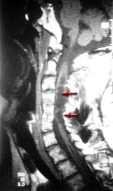 This illustration shows a T1-weighted, cervical magnetic resonance imaging (MRI) scan of multiple syrinx cavities (arrows). Note the lowest thin cavity extending into the thoracic spinal cord.
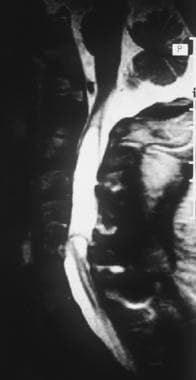 This T2-weighted magnetic resonance imaging (MRI) scan (same patient as above) delineates the syrinx cavity. Note the spinal cord edema extending rostrally from the upper limit of the cavity.
Signs and symptoms of posttraumatic syringomyelia
Pain, the most commonly reported symptom of PTS, may be localized or diffuse and frequently is characterized as a dull ache or a burning or stabbing sensation.
Other symptoms include increased weakness, numbness, increased spasticity, and hyperhidrosis (increased sweating).
Ascending sensory level and sensory dissociation (selective loss of pain and temperature sensation) are very sensitive indicators for detecting progressive PTS.
Other signs may include a complete or partial Horner syndrome or other evidence of dysautonomia (eg, labile blood pressure, hyperhidrosis).
Workup of posttraumatic syringomyelia
Pulmonary function tests, especially vital capacity, should be ordered on any patient with symptoms or suggested respiratory impairment.
Magnetic resonance imaging (MRI), myelography-enhanced computed tomography (CT-myelography), and plain radiography of the spine are useful in the diagnosis and management of PTS. MRI is the preferred initial imaging study for the diagnosis of the condition. Most PTS develops around the site of the original spinal cord lesion. T1 and T2 sequences provide differentiation between CSF and normal spinal cord tissue and areas of spinal cord edema, myelomalacia, or gliosis. Serial examinations are necessary to evaluate for changes in cavity size over time; there is a marked lack of correlation between cavity size and severity of clinical symptoms.
Management
The focus of physical therapy in patients with syringomyelia should be preservation of range of motion and maintenance of function, including transfers, wheelchair mobility, and gait if applicable. Selection of appropriate assistive devices also is important. The occupational therapist (OT) is helpful in assessing and treating the function of the person in performance of activities of daily living.
Surgery frequently is performed to prevent further syringomyelia expansion and collapse syrinx cavities. Neurologic deterioration, pain, or autonomic dysreflexia may be indications for surgery. [1] No surgical procedure has been uniformly successful in relief of symptoms or resolution of radiographic abnormalities.
Surgical treatment has included simple drainage, a variety of shunting procedures, [2] and decompressive laminectomy with expansion duraplasty. [3, 4, 5, 6] Cordectomy has also been performed. [7]
Pathophysiology
The pathophysiology is not understood fully (see Causes). Formation of a cavity within the spinal cord is common after traumatic SCI. Factors related to initial cavity formation include liquefaction of intraparenchymal hematoma, ischemia due to tethering, arterial or venous obstruction, release of intracellular lysosomal enzymes and excitatory amino acids, and mechanical damage from cord compression. Cavity formation alone is not considered PTS.
In PTS, cavity formation is followed by enlargement and extension of the cystic cavity. Rostral or caudal cyst extension may occur due to turbulent CSF flow or a "one-way valve" phenomenon that allows CSF into, but not out of, the cyst cavity. Tethering of the spinal cord, which results in impaired CSF circulation around the traumatized segment of spinal cord, occurs as a sequela of bleeding-induced arachnoiditis, scarring, spinal canal stenosis, or kyphotic deformity.
Syringomyelia can be categorized as “communicating” (dilation of the central canal) and “noncommunicating” (eccentrically located within the substance of the spinal cord). Congenital conditions, such as Chiari malformations, are associated with either communicating or noncommunicating syrinxes. However, PTS is generally considered noncommunicating. [8]
The "slosh-and-suck" theory proposes that increased epidural venous flow occurring during activities (eg, coughing, sneezing) that produce effects like the Valsalva maneuver results in increased pressure around the spinal cord, which cannot be dissipated because of disruptions in CSF flow. This pressure may force CSF into the cyst, resulting in expansion and extension.
A model developed by Carpenter et al suggests that a cough or sneeze can produce a pressure wave that would, in turn, give rise to a shocklike elastic jump. [9, 10] According to the model, the elastic jump could create a transient high-pressure region in the spinal cord, resulting in fluid accumulation. However, in an analysis of the model, Elliott et al maintained that the effect of an elastic jump would probably be too weak for fluid accumulation to result and that "the polarity of the pressure differential set up by cough-type impulses opposes the tenets of the elastic-jump hypothesis." [11] The authors conclude that, based on their analysis, cough-based pressure impulses cannot cause syringomyelia.
A study by Krebs et al of 138 patients indicated that complete SCI and patient age over 30 years are risk factors for developing syrinx early (within 5 years of injury). The study also found that in almost 60% of patients with PTS, the cervical spine was involved. [12]
Epidemiology
Frequency
United States
Approximately 3-4% of persons with traumatic SCI develop clinically symptomatic PTS (although that rate has also been listed as about 1-7% [12, 13] ). A larger percentage of persons have clinically silent syrinx cavities diagnosed by imaging techniques.
Mortality/Morbidity
Morbidity is associated with weakness, loss of function, and chronic pain. Mortality can occur from involvement of brainstem respiratory centers or surgical complications.
Race
No racial differences are known for development of PTS.
Sex
The incidence of PTS is higher in men due to the increased frequency of SCI in males; however, there is no association of manifestations of the condition with the patient's sex.
Age
Development of PTS can occur at any age, and may begin at any time after traumatic SCI. Cases are reported as early as 1 month or as late as 45 years following injury. The risk of syrinx development within 5 years following traumatic SCI is greater in persons over age 30 years. [12]
Prognosis
Some patients remain clinically stable for long periods of time, despite large syrinx cavities. In other persons, sensorimotor impairment progresses.
Patient Education
Patients should be educated regarding signs and symptoms to look for. These may include subtle changes in pain and with regard to the bowel, the bladder, sexual function, sensory or motor function, and functional abilities.
-
This illustration shows a T1-weighted, cervical magnetic resonance imaging (MRI) scan of multiple syrinx cavities (arrows). Note the lowest thin cavity extending into the thoracic spinal cord.
-
This T2-weighted magnetic resonance imaging (MRI) scan (same patient as above) delineates the syrinx cavity. Note the spinal cord edema extending rostrally from the upper limit of the cavity.
-
T2-weighted magnetic resonance imaging (MRI) scan (same patient as above) after patient underwent expansile duraplasty. Note dramatic reduction in size of the main syrinx cavity (white).
-
T2-weighted sagittal image of large, multiloculated cervical syrinx extending into brainstem. Patient had preserved functional status.
-
T1-weighted magnetic resonance imaging (MRI) scan of a slender syrinx (arrow) extending from the C5 vertebral level. This syrinx extends beyond the image to an area of spinal cord disruption at the T3 vertebral level.
-
Same patient as in image above, with the magnetic resonance imaging (MRI) scan slightly farther down the cervicothoracic region of the spine
-
T2 proton density magnetic resonance imaging (MRI) scan demonstrating syrinx cavity (arrow) extending from approximately C6-C7 to T2. The syrinx cavity is 9 mm at its widest dimension. The spinal cord is reduced to a thin membrane at this level and is atrophic below.
-
T1-weighted image demonstrating a large, multiloculated cervical syrinx cavity. This is a recurrent syrinx, having come back despite an attempt at drainage utilizing expansile duraplasty.


