Practice Essentials
There are two common stress injuries to bone: (1) insufficiency fractures, which occur typically when osteoporotic bone is subjected to normal stress, and (2) stress fractures, which can occur when normal bone is subjected to abnormal activity. [1] Stress fractures are overuse injuries of bone. These fractures, which may be nascent or complete, result from repetitive subthreshold loading that, over time, exceeds the bone's intrinsic ability to repair itself. Computed tomography (CT) scanning is a useful diagnostic imaging tool in stress fracture, as is magnetic resonance imaging (MRI). [2] The foundation of treatment for symptomatic stress injury is activity modification.
Briefhaupt originally described stress fractures in military recruits in 1855. Our present understanding of the pathophysiology of stress fractures and of bone's response to loading has been advanced by numerous studies investigating the epidemiology of stress fractures in military recruits and in athletes. [3, 4, 5, 6]
Stress fractures most commonly occur in the lower limbs as a result of the ground-reaction forces (GRFs) that must be dissipated during running, [7] walking, marching, or jumping. Stress fractures of the vertebral arch, upper limbs, ribs, and even the scapula have also been described and are not uncommon in some sports. [8]
Milner et al investigated whether hip, knee, and rearfoot kinematics in female distance runners who had previously suffered tibial stress fractures (n = 30 women) differed from those of female distance runners who had not sustained such injuries (n = 30 women). A motion capture system and force platform were used to gather kinematic and kinetic data from the study subjects. The authors found the rearfoot eversion angles and peak hip adduction to be greater in the stress-fracture group than in the other runners. Milner et al suggested that as a result of these differences, lower-extremity load distribution in members of the first group differed from that in the controls, increasing the former's likelihood of fractures. [9]
Signs and symptoms of stress fractures
The most salient historical feature in the diagnosis of stress fracture is the insidious onset of activity-related pain. Early on, the pain typically is mild and occurs toward the end of the inciting activity.
Subsequently, the pain may worsen and occur earlier, limiting participation in sports activities. While rest may provide transient relief of symptoms in the early stages, as the stress injury progresses, the athlete's pain may persist even after cessation of activity. Night pain is a frequent complaint. Pain resulting from long-bone fractures is thought to be localized, while pain associated with stress injury of trabecular bone is characteristically described as more diffuse.
Inspection of the injury site may reveal localized swelling and, possibly, erythema.
Workup of stress fractures
With regard to CT scanning and MRI, these cross-sectional imaging modes may be helpful in defining the extent of a suspected fracture.
A 3-phase bone scan (scintigraphy) may be indicated if conventional radiographic findings are negative or nondiagnostic and the clinical suspicion of stress fracture remains high. The bone scan is diagnostic of stress fracture if focal isotope uptake occurs in the area of clinical interest on the third phase of the scan. Drawbacks of scintigraphy include a relative lack of specificity and anatomic resolution.
High-resolution ultrasonography may detect bone stress injury or occult fractures before they are evident on radiographs. [10]
Management of stress fractures
To create an environment conducive to healing the stress injury, interrupting the cycle of repetitive overload is essential. For athletes, this typically results in time lost from competition and intensive training. For most stress fractures, the period of relative rest may be expected to last from 4-12 weeks.
Rehabilitation of the individual with a stress fracture should include a program of muscle strengthening and generalized conditioning. Strong, well-conditioned muscles help to dissipate ground-reaction forces (GRFs) that otherwise would be transmitted to bones and joints along the kinetic chain.
If pain persists or becomes further limiting, analgesics may be helpful. Nonsteroidal anti-inflammatory drugs (NSAIDs) are prescribed frequently, but some clinicians believe that these agents should be relatively contraindicated in this setting. NSAIDs inhibit the production of prostaglandins, which are demonstrated to be involved in normal bone remodeling and fracture healing. In addition to pharmacotherapy, physical therapy modalities, such as ice or interferential current, may be used to help treat symptoms.
Pathophysiology
Bone, like muscle, is an adaptable tissue capable of repair, regeneration, and remodeling in response to environmental (particularly mechanical) signals. Bones are exposed to both stress (ie, load) and strain (ie, deformation) with weight-bearing exercise. One measure of load is GRF, which can approach 12 times body weight during jumping and landing. Factors influencing the local skeletal response to loading include bone geometry and bone density. For example, cortical (ie, long) bones are generally more resistant to compressive forces than trabecular bones, but long bones also experience more strain in response to torsion or bending forces. In addition, a bone's strength is roughly proportional to the square of its mineral density; thus, osteopenic bone is weaker than bone of normal density.
Wolff's law states that bone develops the structure most suited to resist the forces acting upon it. The ability of bone to remodel has tremendous clinical consequences. For example, an individual on prolonged bed rest quickly begins to lose bone mineral density (BMD). Conversely, an athlete who engages in a sporting discipline that requires repetitive jumping and landing is likely to have a higher BMD than a sedentary person. Such adaptation is the result of a continuous process of bone resorption and subsequent repair mediated at the cellular level by osteoclasts and osteoblasts, respectively.
Advanced cross sectional imaging has demonstrated that bone responds to repetitive loading via a continuum of stress responses that precede the onset of clinical symptoms. In their study involving a cohort of military recruits, Kiuru et al reported that only 40% of the magnetic resonance imaging (MRI) findings suggestive of a low-grade bone stress injury correlated with clinical symptoms. [11] The vast majority of the radiographically detected areas of bone stress reaction remained clinically silent despite uninterrupted training, and disappeared upon follow-up imaging at the conclusion of the 5-month training program.
Therefore, under normal circumstances, bone appears able to keep up with necessary repairs without manifesting clinically significant injury as it remodels in accordance with Wolff's law. However, when a bone's reparative and adaptive capacity is overwhelmed by chronic overload, damage can begin to accumulate. If allowed to progress, this multifactorial process may eventually result in a stress fracture.
Animal studies have demonstrated that bone subjected to repetitive cyclical loading develops what has been termed microdamage. Furthermore, a physiological threshold appears to exist, below which such microdamage is not detectable. Increased osteoclastic activity at sites of bone stress or strain may cause transient weakening of the bone locally, predisposing the area to microdamage. Unless given appropriate time for healing and osteoblastic-mediated bone deposition, adjacent sites of microdamage are thought to coalesce, giving rise to an area of stress reaction or injury. At this stage, the individual may be minimally symptomatic and conventional radiographs are likely to appear normal. With progressive overload, the bone becomes increasingly vulnerable and the individual proceeds to develop symptoms that are thought to reflect the extent of underlying bone injury.
If uninterrupted, the process may culminate in a stress fracture. Some clinicians prefer to distinguish between stress fractures of normal bone that becomes fatigued through abnormal loading (ie, fatigue fractures) and stress fractures of pathologic bone that may fail even under comparatively normal loads (ie, insufficiency fractures). However, both processes are characterized by disrupted bone homeostasis and inadequate repair in the face of repetitive overload. An example of an insufficiency fracture is shown in the images below.
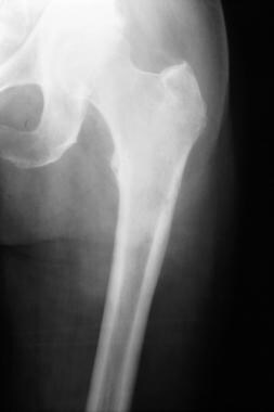 A 63-year-old man with metastatic thyroid carcinoma went for a walk and awoke the following morning with left hip girdle pain. Plain film imaging revealed a subtle area of linear cortical lucency at the proximal left femoral metadiaphysis, consistent with an insufficiency fracture through pathologic bone. The patient subsequently underwent internal fixation. Courtesy of Michael Spieth, MD, and Nandita Bhattacharjee, MD, MHA; Marshfield Clinic Department of Radiology.
 Enlarged view of the fracture shown in the above image. Plain film imaging revealed a subtle area of linear cortical lucency at the proximal left femoral metadiaphysis, consistent with an insufficiency fracture through pathologic bone. The patient subsequently underwent internal fixation. Courtesy of Michael Spieth, MD, and Nandita Bhattacharjee, MD, MHA; Marshfield Clinic Department of Radiology.
Epidemiology
Frequency
United States
Estimates of the annual incidence of stress fractures among athletes and military recruits range from 5-30%. [12] Stress fractures are among the 5 most common injuries suffered by runners and have been reported to account for up to half of the injuries sustained by military recruits. The interaction between genetic susceptibility and other internal and external risk factors determines an individual's likelihood of sustaining a bony stress injury.
Race
Stress fractures probably occur less frequently among African Americans than among whites by virtue of the generally higher BMD found in African Americans.
Sex
Most studies suggest that females are at increased risk of developing stress fractures compared with males. For example, using a national administrative claims database, a study by Kale et al indicated that lower-extremity stress fractures are found in a greater proportion of women than men, with 74.1% of 41,257 identified stress fractures occurring in females. [13]
The incidence of stress fractures among female military recruits and athletes has been reported to be twice that of their male counterparts. Disordered eating places females at higher risk of developing stress fractures.
The clinician should be mindful that a stress fracture may herald the existence of underlying amenorrhea, disordered eating, and osteoporosis (the "female athlete's triad"). Therefore, diagnosis of a stress fracture in a female should prompt the clinician to obtain a dietary history to ensure adequate intake of both energy (calories) and calcium. Finally, in the proper clinical context, a stress fracture should alert the clinician to the possibility of osteoporosis or other underlying skeletal pathology.
Age
Stress fractures typically affect individuals who are more active, and the incidence probably increases with age due to age-related reduction in BMD. By no means, however, should the diagnosis be dismissed in children, whose bones have not reached peak density and strength. [14]
Interestingly, some evidence suggests that the risk of stress fracture may be lower among adult runners who have a broad athletic background that includes childhood participation in "ball sports." This finding provides additional incentive for coaches and parents to avoid promoting early sport-specialization among young athletes.
Among US high school athletes, an estimated 1.54 stress fractures occur per 100,000 athlete-exposures, with the highest rates being seen in girls' and boys' cross country and in girls' gymnastics. [15] Female sex-specific risk factors include late menarche, low body mass index, and participation in gymnastics and dance. [16]
-
This image is of a 17-year-old male wrestler with a 2-month history of left-sided low back pain, worse with extension. Total body scintigraphy findings were unremarkable. Courtesy of Michael Spieth, MD, and Nandita Bhattacharjee, MD, MHA; Marshfield Clinic Department of Radiology.
-
Same patient as in the above image. Single-photon emission computed tomography (SPECT) images demonstrate abnormal delayed uptake in the posterior elements of L5. Courtesy of Michael Spieth, MD, and Nandita Bhattacharjee, MD, MHA; Marshfield Clinic Department of Radiology.
-
Same patient as in the above 2 images. Subsequent MRI revealed an area of bright signal in the left pars interarticularis of L5 on T2-weighted images, confirming the diagnosis of acute unilateral spondylolysis. The patient was treated successfully with activity restriction and bracing with a lumbar corset for 3 months, at which point he was asymptomatic. Plain film imaging at follow-up (not shown) was unremarkable, with no evidence of spondylolysis on oblique views. Courtesy of Michael Spieth, MD, and Nandita Bhattacharjee, MD, MHA; Marshfield Clinic Department of Radiology.
-
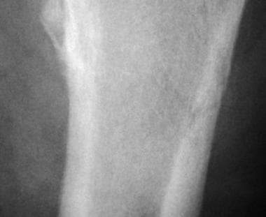
A 17-year-old female dancer with a 2-week history of left shin pain. Plain film imaging was unremarkable. Three-phase bone scanning demonstrated an area of linear uptake in the posterior medial aspect of the left tibia on blood pool images, but delayed images were considered normal. This scintigraphic pattern is consistent with medial tibial stress syndrome (shin splints), but not with stress fracture. Courtesy of Michael Spieth, MD, and Nandita Bhattacharjee, MD, MHA; Marshfield Clinic Department of Radiology.
-
This is a 55-year-old female industrial worker with a 1-week history of right foot pain. Plain film imaging was unremarkable. Bone scanning revealed a stress fracture of the second metatarsal. Courtesy of Michael Spieth, MD, and Nandita Bhattacharjee, MD, MHA; Marshfield Clinic Department of Radiology.
-
This image is of an 18-year-old female soccer player with a 3-week history of left leg pain, which was worse at night and with activity. Upon examination, she reported tenderness in response to palpation over the midtibia. Bilateral pes planus was noted. Plain film radiography failed to demonstrate a fracture. Bone scanning revealed a focal area of delayed uptake on the posterior medial aspect of the proximal third of the left tibia, confirming the diagnosis of stress fracture. Courtesy of Michael Spieth, MD, and Nandita Bhattacharjee, MD, MHA; Marshfield Clinic Department of Radiology.
-
A 63-year-old man with metastatic thyroid carcinoma went for a walk and awoke the following morning with left hip girdle pain. Plain film imaging revealed a subtle area of linear cortical lucency at the proximal left femoral metadiaphysis, consistent with an insufficiency fracture through pathologic bone. The patient subsequently underwent internal fixation. Courtesy of Michael Spieth, MD, and Nandita Bhattacharjee, MD, MHA; Marshfield Clinic Department of Radiology.
-
Enlarged view of the fracture shown in the above image. Plain film imaging revealed a subtle area of linear cortical lucency at the proximal left femoral metadiaphysis, consistent with an insufficiency fracture through pathologic bone. The patient subsequently underwent internal fixation. Courtesy of Michael Spieth, MD, and Nandita Bhattacharjee, MD, MHA; Marshfield Clinic Department of Radiology.
-
This case involves a 16-year-old female basketball player with a 2-year history of left foot pain refractory to casting and reduced weight bearing. Bone scanning revealed a focal area of delayed uptake lateral to the left first metatarsal phalangeal joint, which corresponded to a bipartite sesamoid on plain films. Courtesy of Michael Spieth, MD, and Nandita Bhattacharjee, MD, MHA; Marshfield Clinic Department of Radiology.
-
Sesamoid stress fractures are prone to nonunion, and sesamoidectomy is indicated for patients who do not respond to conservative management. Some clinicians recommend bone grafting as an alternative to complete or partial sesamoidectomy. Courtesy of Michael Spieth, MD, and Nandita Bhattacharjee, MD, MHA; Marshfield Clinic Department of Radiology.