Background
Primary and revision rhinoplasty are challenging procedures. In order to obtain aesthetically pleasing results, ensure patient satisfaction, and minimize complications, the rhinoplasty surgeon must possess a thorough knowledge of nasal anatomy and ideal facial aesthetic proportions. The rhinoplasty surgeon must also be familiar with all types of graft material and the current methods to correct nasal deformities. This article addresses augmentation of the deficient nose, as seen in the images below.
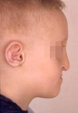 Young boy with bilateral cleft lip and palate who also has midface hypoplasia and requires nasal augmentation.
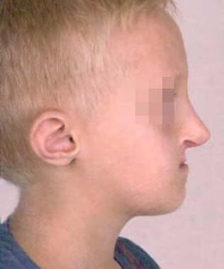 Young boy (same patient as in previous image) with bilateral cleft lip and palate who also had midface hypoplasia and required nasal augmentation, shown after dorsal nasal augmentation was accomplished using autologous rib grafts. It was fashioned slightly larger than needed to account for the patient's future facial growth.
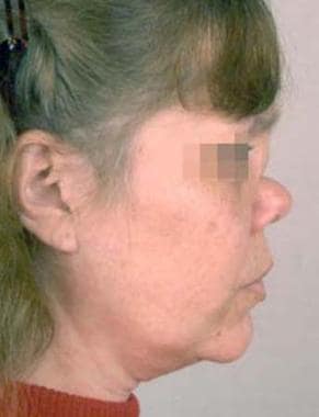 Woman with a subtype of midline granulomatous disease that caused nasal collapse. Augmentation was required.
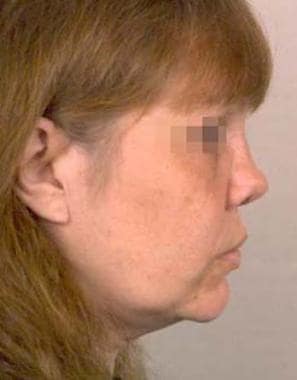 Woman (same patient as in previous image) with a subtype of midline granulomatous disease that caused nasal collapse, shown after augmentation with both autologous rib and cartilage grafts. AlloDerm was also used for additional augmentation.
History of the Procedure
Rhinoplasty originated as a reconstructive procedure, essentially in the form of nasal augmentation. In 500 BCE, Sushruta pioneered nasal reconstruction with the use of the nasolabial flap. In the 1800s, two German surgeons, Carl von Grafe and Johann Dieffenbach, made significant advances in rhinoplasty techniques. In subsequent years, Dr John Roe described the endonasal approach in 1887, and Drs. Joseph Safian and Gustave Aufricht described advanced techniques. Several publications continue to propose innovative ways to correct nasal deformities and improve outcomes. In addition to autologous materials, many new synthetic and allogenic materials have become available for nasal augmentation surgery.
Presentation
Rhinoplasty requires a careful preoperative analysis of the patient's problem during the office visit and meticulous surgical execution in the operating room. The surgeon must elicit the expectations, concerns, and motivations of the rhinoplasty patient. Establishing a good rapport with the patient can help increase the potential for postoperative satisfaction. An accurate nasal history should alert the surgeon to a previous history of nasal trauma, nasal obstruction, sinus disease, and allergic disorders.
A detailed history of any comorbid conditions, current prescribed medications, over-the-counter medications, illicit drugs, and herbal supplements must also be elicited. Any agents such as aspirin, anticoagulants, and herbal supplements (eg, St. John’s wart, ginkgo, ginger, vitamin E) should be discontinued at least 2 weeks prior to surgery to minimize the risk of intraoperative and postoperative bruising and bleeding.
The anatomic examination should include an analysis of the external nasal deformity and internal nasal anatomy. The endonasal examination should be performed after adequate decongestion with a vasoconstrictor. Specific attention should be given to any external nasal valve collapse upon inspiration, the adequacy of the internal nasal valve, tip ptosis, septal deviation, the size of the inferior turbinates, septal perforation, and the existence of any purulence in the middle meatus. Preoperative photographs must be obtained. They should include a frontal view, lateral view in the Frankfort horizontal plane, basal view, and oblique view. Informed consent must be obtained and documented. Any questions or concerns the patient has should be addressed prior to the surgical procedure.
Indications
Functional concerns, aesthetic concerns, or both may prompt a patient to seek rhinoplasty surgery. The nose functions as an airway conduit. It filters and humidifies air as it passes through the nasal cavity. Any obstruction to the airflow may be bothersome to patients. To effectively treat the problem, anatomical and/or medical causes of nasal obstruction must be sought. Several publications describe algorithms that can help the surgeon with the differential diagnosis. Canady proposed an algorithm that can help determine whether a patient has an allergic problem, a mucosal problem, or other anatomical problem that is causing nasal obstruction. [1]
Nasal obstruction secondary to anatomic deformities or variations from the ideal nasal aesthetics can be improved with surgical intervention. Examples include internal nasal valve and/or external nasal valve collapse upon inspiration. The internal nasal valve is formed by the septum medially and the upper lateral cartilages superiorly. The ideal angle for adequate airflow has been reported to be 10-15°. Inadequate airflow can occur when this angle is more acute. In addition, encroachment of the inferior turbinate laterally at the internal nasal valve can further worsen the obstruction.
Internal nasal valve collapse is observed in some patients who have previously undergone a nasal dorsal reduction during a rhinoplasty. For more information, see Rhinoplasty, Postrhinoplasty Nasal Obstruction. Overresection of the nasal dorsum and the upper lateral cartilages can lead to loss of structural support at the internal nasal valve, which can result in obstruction. Correction of this condition usually requires reestablishing the normal angle with the use of spreader grafts between the septum and the upper lateral cartilages. Patients also may require external nasal valve augmentation with batten grafts, rim grafts, or other structural support for a patent airway. The inferior turbinates may need to be reduced or partially excised.
Tip ptosis can also lead to nasal obstruction. Tip ptosis can result from aging, trauma, or previous surgical intervention that caused a loss of the crucial tip-supporting mechanisms. Augmentation of the nasal tip with grafts and restoration of the tip-supporting mechanisms may be required to alleviate functional nasal obstruction.
Another indication for augmentation of the nose is a deficient nasal dorsum. Nasal dorsal deficiency can result from overresection of the nasal dorsum during a prior rhinoplasty, in patients with Wegener granulomatosis or Binder syndrome (maxillonasal dysplasia), or secondary to septal necrosis from a septal hematoma or cocaine abuse. Such factors can cause a saddle-nose deformity due to the loss of underlying septal support. These patients require nasal dorsal augmentation.
Augmentation materials
Several different materials can be used to augment the nose. Available augmentation materials can be classified as autologous grafts and nonautologous graft materials. Autologous materials consist of bone, cartilage, or both harvested from the nasal septum, ear, rib, inferior turbinate, and calvaria. [2, 3] The use of tensor fascia lata has also been described. [4] The main nonautologous materials include Medpor, silicone, silastic, GORE-TEX, and the human acellular dermis product marketed as AlloDerm or porcine acellular dermal product marketed as Permacol or Stratus. In addition injectables such as hyaluronic acid have been utilized for temporary and minor corrections [5] . Opinions regarding the indications for and outcomes with using these materials vary.
Autologous tissue is readily available, and the morbidity associated with harvesting the autologous grafts is low. Many prominent surgeons believe the aesthetic and long-term functional outcomes are superior and the complication rates are low. Therefore, they advocate the use of autologous grafts over nonautologous materials. Bone is usually needed only in patients with very deficient dorsi with saddle-nose deformities.
Autologous tissues may also be required in patients with severe nasal defects in whom near-total reconstruction is planned. Bottini et al presented a 79% satisfaction rate in their series of 132 patients who underwent reconstruction of nasal defects using various autologous graft techniques. [6] Gentile and Cervelli reviewed 123 cases in which 84% of treated patients showed cosmetic improvements, with results satisfactory to both surgeon and patient, and 94% of the operated cases resulted in functional improvement. [7] Stuzin and Kawamoto reported their use of cranial bone grafts to correct saddle-nose deformities. [8] These grafts require careful placement and rigid fixation. The cartilage grafts can be used as struts, [9] anatomic replicas, or morselized/crushed grafts wrapped in fascia, Surgicel, or other components. In some cases, a combination of autologous and nonautologous materials may be necessary.
One concern with bone grafts is the potential for resorption. Powell and Riley reviewed 850 calvarial bone grafts to the nose in 170 patients and found resorption rates of as high as 30%. [10] Given this concern and the increased morbidity of harvesting bone grafts, most surgeons prefer using cartilaginous grafts.
Septal cartilage should be harvested with caution. At least a 10-mm dorsal and caudal strut is very important for nasal structural support. In revision rhinoplasty cases, septal cartilage may not be available. [11] In such cases, ear cartilage, rib cartilage or both can be harvested. [12] Some inherent disadvantages with the use of auricular cartilage are the inherent curves in ear cartilage and the fragility of the cartilage compared with septal or rib cartilage. However, ear cartilage is ideally suited for alar rim and nasal tip reconstruction. The amount of ear cartilage available is usually inadequate for dorsal augmentation. Ear cartilage can be harvested via an anterior or posterior approach. The resultant morbidity is minimal in either approach.
When more structural support or rigidity is needed for augmentation, rib cartilage is a great alternative. [13] The ninth floating rib is an ideal location to harvest the graft. This location provides the surgeon with a long, straight piece of structurally strong cartilage. Adherence to the Gibson principles of balanced cross-sectional harvesting leads to an ideally shaped graft that holds its final shape without distortion or warping. This involves amputation of an equal amount of cartilage from both sides to minimize warping of the graft. Meticulous surgical technique is required to prevent inadvertent entry into the thoracic cavity. With adherence to sound surgical principles, the rate of donor site complications is usually very low. Some advocate making an anatomic replica using an osteocartilaginous graft.
Tosun et al presented a decade of experience using allogenous cartilage grafts. [14] They compared allogenous grafts to autogenous grafts and found no differences in resorption and complications. Kridel et al reviewed 357 cases in which allogenous cartilage was used over 24-y period; they found it to be a stable graft in rhinoplasty and with a very low complication rate (3.25%). [15] They believe that the allograft should be considered when septal or auricular cartilage is not available or even as primary grafting material, given their findings.
Godin et al reviewed 137 patients who underwent nasal augmentation with GORE-TEX patches. [16] The infection rate was low (2.2%), and none of the patients required further augmentation. The authors were pleased with the results and listed technical points they felt were important for success. Taylor and Owsley also related their satisfaction with GORE-TEX for nasal augmentation. In their study of 106 patients, they found no complications related to the graft. [17]
More recently, Conrad et al reviewed 521 patients over a 17-y period who underwent GORE-TEX augmentation in rhinoplasty and found it to be a safe, inexpensive, and predictable alternative to autografts. [18] They found that complications requiring implant removal occurred in only 1.9% of patients and included infection, soft tissue swelling, migration, and extrusion. Studies on other implant materials have revealed mixed results. However, Rohrich and Muzaffar, in addition to many other rhinoplasty surgeons, prefer autologous materials over nonautologous materials for nasal augmentation. [19]
El-Shazly and El-Shafley recommended soft implants for both aesthetic and reconstructive surgeries because the result in a dorsum with a smoother contour and pad. In addition, soft implants have fewer complications and higher satisfaction rates. [20]
Hopkins et al reviewed usage of the porcine acellular dermal matrix marketed as Permacol in 58 patients. Complications included one postoperative infection that cleared with oral antibiotics but resulted in a suboptimal cosmetic result and one cyst that developed one year postoperatively, which was drained and resulted in a good cosmetic result. [21]
A retrospective, single-surgeon study by Park et al indicated that a cross-linked human acellular dermal matrix is an effective tool in either primary or revision dorsal augmentation rhinoplasty. In both groups, significant postoperative changes were achieved in the ratio of the dorsal height and radix height to the nasal length (ie, the degree of augmentation). Moreover, these ratios did not significantly differ between the primary and revision patients. No serious complications occurred in either group, and microscopic examination found the removed matrix to have “abundant collagen tissue with newly formed vessels,” as well as no evidence of a foreign body reaction. [22]
Relevant Anatomy
The nose is composed of skin, subcutaneous tissue, nasal mucosa, cartilage, and bone. The upper third of the nose is composed of the nasal bones, the middle third is composed of the nasal septum and the upper lateral cartilages, and the lower third encompasses the lower lateral cartilages and the caudal aspect of the cartilaginous septum. The midline septal cartilage provides underlying support to the lower two thirds of the nose. While this is the basic anatomic framework, variations and asymmetry are present in many individuals.
The nasal bones vary in thickness and width from the nasofrontal suture line cephalically to the end of the nasal bones caudally. They are thick and widest at the nasofrontal suture, narrow at the nasofrontal angle before they widen, and become thinner approximately 9-12 mm below the nasofrontal angle. These anatomic variations in thickness and width are important to remember when osteotomies are planned. The thickness of the soft tissue and skin of the nose also varies at different anatomic points. The skin and soft tissue covering the nasal skeleton is thickest in the supratip region, while it is thinnest at the bony cartilaginous junction of the nasal dorsum. These variations in skin and soft tissue thickness affect the final nasal contour and profile after nasal augmentation.
For a detailed and illustrated description of nasal anatomy, see the Medscape article Nasal Anatomy.
Contraindications
Augmentation rhinoplasty has relatively few contraindications. Any medical condition that inhibits patient safety during the procedure is a contraindication. Patients should not be taking any anticoagulants (eg, enoxaparin [Lovenox], warfarin [Coumadin], heparin), aspirin, or nonsteroidal anti-inflammatory drugs before the surgical procedure. Any herbal supplements such as ginger, ginkgo, or vitamin E should also be avoided. Allergies to any type of nonautologous material must be identified prior to the procedure. Patients with inappropriate motivations for seeking rhinoplasty must be carefully screened.
-
Young boy with bilateral cleft lip and palate who also has midface hypoplasia and requires nasal augmentation.
-
Young boy (same patient as in previous image) with bilateral cleft lip and palate who also had midface hypoplasia and required nasal augmentation, shown after dorsal nasal augmentation was accomplished using autologous rib grafts. It was fashioned slightly larger than needed to account for the patient's future facial growth.
-
Woman with a subtype of midline granulomatous disease that caused nasal collapse. Augmentation was required.
-
Woman (same patient as in previous image) with a subtype of midline granulomatous disease that caused nasal collapse, shown after augmentation with both autologous rib and cartilage grafts. AlloDerm was also used for additional augmentation.


