Continually Updated Clinical Reference
 
 
  All Sources     eMedicine     Medscape     Drug Reference     MEDLINE
 
eMedicine - Nasal Implants : Article by

Quick Find
Authors & Editors
Introduction
Indications
Relevant Anatomy
Contraindications
Workup
Treatment
Complications
Outcome and Prognosis
Multimedia
References


Patient Education
Click here for patient education.


Author: Garrett A Wirth, MD, Assistant Clinical Professor, Department of Plastic Surgery, Aesthetic and Plastic Surgery Institute, Orange, California

Garrett Wirth is a member of the following medical societies: Alpha Omega Alpha, American Society of Plastic Surgeons, Association for Academic Surgery, and Plastic Surgery Research Council

Coauthor(s): James G Hoehn, MD, Program Director, Professor, Department of Surgery, Division of Plastic Surgery, Albany Medical Center Hospital

Editors: Gregory Caputy, MD, PhD, Chief, Department of Plastic Surgery, Aesthetica Plastic and Laser Surgery Center of Honolulu; Francisco Talavera, PharmD, PhD, Senior Pharmacy Editor, eMedicine; Edward Owen Terino, MD, Director, Department of Plastic Surgery, Los Robles Medical Center; Nicolas (Nick) G Slenkovich, MD, Practice Director, Colorado Plastic Surgery Center at Swedish Medical Center; Al Aly, MD, FACS, Consulting Surgeon, Iowa City Plastic Surgery

Author and Editor Disclosure

Synonyms and related keywords: nose implants, nose deformities, nasal deformities, nasal reconstruction

The nose is critically involved in appearance, both to oneself and to others, and it is significantly involved in the perception of beauty both publicly and privately. Because of its central location on the face, plane of projection, and relatively weak chondrocutaneous support structure, the nose is susceptible to injury, and deformities are readily apparent.

Whatever the circumstances that led to the nasal deformity, the complex tasks of assessing the patient's nasal anatomy, pathologic defect, aesthetic qualities, baseline perception, planning the reconstruction, and preparing the patient for the possible positive and negative outcomes can be daunting. In planning for nasal reconstruction requiring an implant, careful decisions need to be made regarding the types of materials to substitute for support. Final soft-tissue coverage of the planned reconstruction is of paramount importance in the preparation.

History of the Procedure

In northern India, during the sixth century BC, Susruta focused on soft-tissue augmentation for amputation injuries and nasal reconstruction (see Image 1). In the 16th and 17th centuries, Tagliocozzi used a tubed pedicle flap (see Image 2) and Carpue, an Englishman, reinstituted the forehead flap (see Image 3). A variety of nonautogenous materials have been employed over the centuries to improve reconstruction of the nose by providing bridge support. Early attempts at finding the ideal or most suitable nasal implant for bridge construction included trials of materials such as paraffin, gold, silver, aluminum, ivory, cork, stones from the Black Sea, polyethylene, rubber, silicone, lead, and a toothbrush handle.

Currently, general categories of available materials include autografts, homografts, and allografts. This article covers the variety of materials commonly employed. The pros and cons of various materials are detailed and illustrated.

Problem

The choice of nasal implants to be placed in reconstructive surgery of the nose is a difficult but important component in the patient's care. The degree of loss noted in tip support, bridge contour, or nasal valve collapse may mandate implant use in nasal reconstruction. Each of the various materials has benefits and pitfalls, which must be balanced against the surgeon's familiarity with each material. Because of the nose's central location, minor defects of reconstruction may be particularly noticeable to any casual observer but may play a much larger role psychologically in the patient's perception of self. Based on the authors' review of the literature and clinical experience, autogenous cartilage and/or bone are recommended as the primary choices for nasal implants.


Although the major underlying cause of nasal reconstruction is trauma, a number of pathologic entities traditionally have required surgical nasal reconstruction (eg, congenital malformations, malignant destruction, septal perforations, granulomatous disease, congenital syphilis, leishmaniasis, leprosy). Whatever the cause of the defect, nasal reconstruction with or without various implant materials allows restoration of function along with restoration of an aesthetically critical component of the face.


A prerequisite to nasal reconstruction is familiarity with nasal anatomy and proportions. The nose is generally divided by anatomic units into thirds. The upper third of the nose, or bony vault, is represented by the paired nasal bones that overlie the nasal spine of the frontal bone. The cartilaginous vault represents the lower two thirds of the nose, with the middle third being the region of the upper lateral cartilages, and the lower third involving the nasal tip, septum, and lower lateral (alar) cartilages. The general function of the nose is to warm and humidify incoming air. To achieve both form and function, the nasal vestibule should comprise approximately one half to two thirds of the nasal lobule as viewed from the basal projection. Attention should be dedicated to reconstructing the columella, tip, and ala to form an adequate nostril internally while incorporating aesthetics externally.


Ongoing infection or conditions requiring further therapy (eg, serial débridements) are examples of contraindications to reconstruction. Address patient stabilization and optimization, including nutritional status when possible, prior to surgical intervention. Therapies such as radiation or chemotherapy should be completed prior to reconstruction.


Lab Studies

  • Standard preoperative testing still is required for patient safety during anesthesia for nasal reconstruction.
  • Should any continuing process be suspected, institute preoperative workup and therapy prior to placing implants or performing reconstruction, whether this involves serial débridements, treatment of an infectious process, ongoing medical therapies, or other therapies.


Surgical Therapy

Materials Currently in Use

Cartilage

Autogenous cartilage grafts are the most frequently used material for nasal augmentation and are considered the preferred material by many authors. The various reasons for its success and preferred status include ease of use, availability, and biocompatibility. Maas et al stated that cartilage is firm yet flexible and can be carved easily to fit the desired nasal contour. It also provides good structural support while maintaining the consistency of the normal nasal cartilaginous skeleton.

McCarthy and Ortiz-Monasterio et al emphasize that autogenous cartilage grafts have decreased resorption rates and low infection rates. Resorption rates of septal cartilage grafts are estimated at 12-50%. Absorption of autogenous cartilage follows a typical pattern. Absorption begins early, is of short duration, and appears to subside after the initial postoperative period.

Generally, accepted donor sites available for implantation are the nasal septum, auricular conchal cartilage, and rib. Cartilage from these donor sites varies in texture, rigidity, and contour. Use of cartilage from each site is chosen based on need (see Image 4). Clearly, the availability of septal cartilage may be influenced by the underlying cause of the deformity (eg, trauma). Septal cartilage generally does not warp easily and has the advantage of tolerating careful manipulations such as gentle crushing to alter thickness and/or contours.

Unfortunately, cartilage grafts that have been crushed to facilitate contouring tend to undergo significantly higher rates of resorption. However, the compliant nature of septal cartilage that lends itself to providing structural support without the rigid appearance of bone makes it a nearly ideal nasal augmentation material.

If only a small amount of cartilage is required, or when a gentle curvature facilitates reconstruction, the conchal auricular cartilage can be harvested from either the anterior or posterior approach (see Image 15). The posterior approach generally offers easier access to larger amounts of cartilage without significant scars. The contour of auricular cartilage makes it ideal for internal nasal valve grafts and replacement of the lower lateral cartilages. Its softness makes it less noticeable when used as an onlay graft. Costal cartilage is available in the largest quantity, but conversely offers much greater donor site morbidity. Of the autogenous cartilages available, costal cartilage is the most likely to deform when carved to thin dimensions.

Recently, an increase has occurred in the use of diced autologous cartilage grafts wrapped either in Surgicel, which is manufactured by Johnson & Johnson, (Erol, 2000; Elahi, 2003) or in deep temporal fascia (Daniel, 2004). Controversy exists as to which technique allows better maintenance of the graft, contour correction, and cartilage viability (Erol, 2005; Daniel, 2005). Survivability of diced cartilage grafts was addressed by Brenner et al (2006). Diced cartilage and bone chips, as well as cartilage allografts, have also been used and are addressed below.

Collagen gel matrix

Britt and Park stated that tissue-engineered cartilage can be produced reliably and that predetermination of graft shape is possible. They further concluded that histologically, allografted rib cartilage represents composites of mature cartilage infiltrated by vasculature and fibrous tissue with delayed osteoid formation. Of note, the tensile strength of the allografted rib cartilage is less than that of native cartilage. However, this histologic finding suggests that tissue-engineered cartilage is a feasible option for a future graft material.

Further evidence that tissue-engineered cartilage is a future material for nasal reconstruction was presented by Lindsey et al using a rat model and an osteoconductive collagen gel matrix. They demonstrated histologically that native, isotonic, neutral pH, space-filling collagen gel implants revealed restoration of the anatomy with a thin plate of immature bone spanning the defect in continuity with the cartilage of the nasal septum and with apparent preservation of maxillonasal suture lines.

The feasibility of using collagen gel for nasal augmentation primarily is due to its ready availability and ability to be molded. The material can be (1) brought to the operative field as a liquid and poured into defects, forming a gelatin at warmer temperatures within the defect; (2) molded at the operating table and then placed into the defect; or (3) arrive preshaped.

Finally, rodent models also are used to study fibrin sealants augmented with osteoprogenitor cells for repair of osseous facial critical size defects. Clearly, these materials merit further investigation and evaluation as their final role in reconstructive surgery of the nose has not been elucidated fully.

Bone

Bone, like cartilage, can be used for augmentation and as a supportive structural framework for nasal tip and internal nasal valve support. Generally considered a second choice after autogenous cartilage, autogenous bone grafts offer advantages of stability, availability, accessibility, and low infection rates in nasal reconstructions. Bone grafts have gained attention in the past few years as alternative grafting material, particularly for reconstruction of total nasal defects and the collapsed nose when structural support is needed. Sites available for bone graft donation include split calvarial grafts (cortical bone), iliac crest (cancellous, cortical), and rib (see Image 4), (see Images 12-14), (see Image 20).

Romo and Jablonski stated that the determination to use calvarial grafts was made on the basis of the lack of available auricular and nasal cartilage or the lack of patient consent to use autogenous auricular cartilage. Split calvarial bone grafts can provide a large source of material for grafting, having further advantages of excellent structural support and a high level of tolerance (see Images 11-17). Another advantage is that calvarial bone graft is harvested from a donor site in the same operative field as in the nasal operation. Conversely, the stiffness of bone can make it difficult to shape and more susceptible to fracture, increased morbidity is associated with the donor site (eg, significantly more pain), and a less natural appearance may result.

During the past few decades, calvarial bone grafts have become increasingly popular because of their widespread use in cranial vault reconstruction. This is in contrast to using either rib or iliac crest bone for nasal reconstructions. Carter first introduced the use of rib as a donor site in 1911. The donor site usually can be hidden within the inframammary fold at approximately the fifth rib or in a more inferior and lateral position using the ninth rib (see Images 4-10, Images 18-23). As a substitute, Macomber first reported in 1949 the use of iliac bone for the correction of depressed deformities of the nose. Both the iliac and rib sites carry considerable donor site morbidities, not the least of which is the patient's discomfort level.

The risk of morbidity associated with bone grafts applies to the split calvarial graft as well as iliac crest or rib graft. Split calvarial grafts also carry a slight risk of cerebral injury. This is a rare but significant complication and should be weighed in the decision to harvest from this site. Zins and Whitaker noted that animal model studies suggest that membranous bone, such as a cranial bone graft, is resorbed less compared to endochondral bone. Powell et al noted that the resorption rates of calvarial bone grafts are estimated at 20-30%. Rates of resorption vary for reasons that remain a mystery although it appears that arguments about the merits of fixation of grafts versus onlay alone merit continued investigation.

Several authors reiterate that bone is more difficult to shape to the recipient site. Romo and Jablonski (1992) stated, "We believe that autogenous cartilage is still the ideal replacement material for structural defects of the nose; however, in cases in which cartilage is not available, split calvarial bone grafts are an excellent substitute." Celik et al (2004) published results using bone chips and diced cartilage for treatment of the nasal dorsum. Results showed satisfactory and durable results correcting deficiencies on the bony part, cartilage part, or both parts.

Temporalis fascia graft

Nasal reconstruction may include the use of temporalis fascial grafts. The fascia generally has been used to augment or complete the contouring of the nose, as it may be used over bone or for filling the soft tissue lower and lateral aspects of the nose (see Images 24-27). Temporalis fascial grafts also may be used in the reconstruction of the nasal septum during a large reconstruction or for an individual with a septal perforation. Guerrerosantos noted that temporalis fascial grafts are used to cover the osteocartilaginous framework (including cartilage or bone grafts), aiming to prevent possible irregularities or sharp edges.

Miller noted that these grafts can be used as the sole source of contour augmentation of facial depressions in primary as well as secondary rhinoplasty. Such grafts undergo an initial uniform shrinkage (approximately 20%) during the first 4-6 weeks postoperatively because of compaction and condensation of the fibrous tissue of the fascia; after this shrinkage, the grafts stabilize and become firm. Guerrerosantos stated that the most common complications relating to augmentation rhinoplasty with fascia and cartilage grafts are erythema and resorption of the fascia.

Homografts/Allografts

Homografts, or allografts, refer to those materials that are procured from a similar species (but different patient) for donation and use in reconstruction for the recipient patient. The benefits of homografts include the availability of significant quantities of tissues and the ability to obtain tissue that still contains the property of matrix invasion and adjacent connective tissue replacement without donor site morbidity. The risk of infection associated with these products can present greater problems than for autogenous material. In an effort to minimize the risk of infection, irradiated tissue has been used. Kridel and Konior, as well as Welling et al, have suggested that irradiated cartilage in the nasal dorsum, being relatively immobile, does not demonstrate as much evidence of resorption as in other facial regions.

Irradiated cartilage is shaped and carved easily and can be used in areas where tissue strength is important for structural support, such as in improving nasal valve collapse or improvement of nasal tip projection. In principle, the homograft's pathogens and viruses are destroyed using gamma wave radiation. The graft is stored in a sterile saline solution and then reconstituted in an antibiotic solution prior to implantation. Any questionable material can be further debrided at the time of surgery.

Welling et al concluded that irradiated homologous cartilage grafts in humans are resorbed progressively, complete resorption may be expected, the amount of time necessary for complete resorption varies with each graft but eventual resorption appears inevitable, and fibrous scar tissue provides bulk that may result in a satisfactory aesthetic result. However, irradiated homologous cartilage grafts cannot be recommended for long-term structural support. A contrary opinion has been expressed by Demirkan et al (2003); they proposed that irradiated homologous costal cartilage is a versatile grafting material for rhinoplasty. These authors utilized irradiated costal cartilage for various nasal defects. Results demonstrated no significant resorption, and they concluded that irradiated costal cartilage is a safe and reliable source of cartilage graft for both primary and secondary septorhinoplasties when autogenous septal cartilage is either insufficient or unsuitable (Demirkan, 2003).

Burke et al reported that the longevity of irradiated homologous rib cartilage grafts has been favorable for functional, structural, and cosmetic nasal reconstruction, with low levels of resorption identified clinically (Burke, 2004). Strauch and Wallach (2003) stated that "use of irradiated homograft costal cartilage for functional, traumatic, and aesthetic reconstruction of the nose is recommended." They summarized that this cartilage is recommended for correction of contour defects and structural support, has yielded a low rate of complications, and has the added advantages of absence of donor-site morbidity and shorter surgical time required in the absence of surgical harvesting (Strauch, 2003).

AlloDerm (manufactured by LifeCell Corporation, Palo Alto, Calif) is an acellular allograft dermal matrix. In recent years, some interest has focused on the development of a nonimmunogenic, extracellular tissue matrix for permanent transplantation.

Originally popularized for use in full-thickness burns, the material may be used successfully for lip repairs, nasal reconstructions, and repair of septal perforations. This material is prepared from fresh human cadaveric skin. The processing technique of this material results in an acellular dermal matrix with normal collagen bundling and organization with intact basement membrane. The full spectrum of uses and long-term studies has yet to be evaluated, but this material has been available to the plastic and reconstructive surgeon. The use of AlloDerm has been reported in nasal procedures. For example, Ayshford et al (2003) reported its successful use with endoscopic repair of nasal septal perforations while also utilizing an inferior turbinate flap. Multiple authors have also used this material for the correction of nasal contour deformities and in secondary rhinoplasty (Jackson, 2001; Gryskiewicz, 2001; Gryskiewicz, 2005).

Originally, the use of AlloDerm was reported as "encouraging," with long-term "good results, though partial graft resorption occurred in some patients" (Gryskiewicz, 2001). The use of AlloDerm in secondary rhinoplasty showed that absorption did not seem to relate to the number of layers used and that the material did not appear to shift but still showed the persistent disadvantage of partial, but not complete, resorption (Gryskiewicz, 2005). Porcine dermal collagen (Permacol, Tissue Science Laboratories, Aldershot, UK) has also been reported for coverage of osseocartilaginous irregularities in secondary nasal surgery (Saray, 2003). Its limited use still warrants further investigation.

Prosthetic implants

Porous implants have a greater risk of immediate infection, as increased surface area is present for adherence of bacterial polysaccharide cell surface components. However, porous implants have fewer late-stage infections, as the incorporation of host tissue into implant pores allows access to the site for immune response mediators. Thus, surgeons must account for the chemical and biologic nature of implant materials and the tissue characteristics of the implantation site while paying meticulous attention to sterile implantation technique.

  • Mersilene: Meshed polymers have the advantage of inciting prolific fibrous ingrowth, but this stability comes at the price of extreme difficulty in removing these implants should they become infected. Polyethylene terephthalate (Mersilene) did not gain widespread acceptance among plastic and reconstructive surgeons as a nasal implant material. Colton and others have continued to support the use of Mersilene based on properties such as relative ease of use, ability to tolerate folding, ability to be sutured in place, and relative early stability. Infection rates as high as 4% have been noted, necessitating removal in 2% of patients. As a polyester mesh, Mersilene does not undergo hydrolytic degradation when implanted.
  • Proplast: A porous polymer known as Proplast formerly was used in temporomandibular joint reconstructions but was noted to shred under vigorous mechanical forces. The resultant particulate matter caused a response that resulted in intense inflammation and scarring, precipitating removal of its marketing clearance by the Food and Drug Administration. However, in nasal reconstruction, its ability to be "carved" into the desired shape made it popular (see Images 4-10) (see Images 28-33).
  • Medpor: Medpor is another porous polymer. The material can be molded while hot, but it is not sculpted easily once it begins to harden. Once this product sets, it is noted to become very hard, and, as a nasal implant, it may appear unnatural. Medpor is available as a strut or as a sheet. Sclafani et al concluded that because of differences in pore size, Medpor promotes faster fibrovascular ingrowth, and the presence of vascularized host tissue in and around the implant lends stability and resistance to experimentally induced infection. They suggested that conservative treatment of clinical implant infections should be considered if bacterial seeding occurs after substantial fibrovascular ingrowth is present.

    Niechajev supported the use of Medpor for secondary rhinoplasties and confirmed histologically the rapid vascularization and both soft tissue ingrowth and collagen deposition with the use of the porous polyethylene implant. Romo et al have reported multiple uses of Medpor as a nasal implant (Romo, Arch Facial Plast Surg, 2003; Romo, Facial Plast Surg, 2003; Romo and Litner, 2003). In cleft lip rhinoplasty (nasal reconstruction), the authors reported that the implant was well tolerated and achieved excellent long-term aesthetic results through fibrovascular ingrowth to the surrounding tissue (Romo, Arch Facial Plast Surg, 2003; Romo, Facial Plast Surg, 2003). Romo also reported safe utilization of polyethylene alloimplants in multiple locations as a nasal implant.

  • Gore-Tex: Gore-Tex (expanded polytetrafluoroethylene [ePTFE]) can be included within the polymer category. The porous nature of the material permits cellular attachment and tissue infiltration with minimal capsule formation. Its ability to be used within the body is well accepted, primarily based on its long-standing history of use in vascular surgery. That said, it has a relatively short history of use for nasal surgery.

    Long-term studies have not established its true infection rate, extrusion rate, or morbidity at this time, but early information suggests approximately a 1% extrusion rate. Ham et al (2003) reviewed the literature on ePTFE implants in rhinoplasty and noted that 20 out of 769 procedures produced inflammation or infection that required removal at a rate of 2.6%. Maas et al concluded the ePTFE soft-tissue patch appears to be safe and reliable material for augmentation, demonstrating high biocompatibility, low tissue reactivity, and increasing stability over time.

    Godin et al concluded that Gore-Tex is a safe and effective implant material to use in primary and revision rhinoplasty when augmentation is needed and autogenous material is not available or desirable. Ham (2003) concurs with this by stating that "although the infection rates of Gore-Tex are much higher than the rate of 0.1% for autogenous grafts, Gore-Tex may have a role in carefully selected patients. Only with time will the long-term results be revealed."

Silicone

Silicone in various states of viscosity was a popular alloplast. The body is unable to infiltrate the material and tends to establish a fibrous tissue capsule around the implant. Without tissue infiltration, the implant tended to remain slightly mobile and was noted to have a significant extrusion rate. On a positive note, since no tissue ingrowth occurs, silicone implants may be removed easily if necessary (see Images 18-23). Silicone also has been used as a columellar strut implant, generally using medium grade silicone rubber. The placement of a silicone columellar strut serves to correct columellar retrusion, improve the nasolabial angle, and increase tip projection by elevating the medial crura of the lower lateral cartilages.

The advantages of the silicone columellar strut are as follows:

  • Low extrusion rate when properly implanted
  • Ability to sculpt and implant the prosthesis to assess the immediate effect of size and shape intraoperatively
  • Use of a harder grade of silicone rubber to permit the insertion of relatively small implants
  • Lack of resorption and elimination of a second procedure to obtain a graft
  • Ability to increase tip projection without disruption of the tip cartilages
  • Easy removal, causing minimal disruption of surrounding tissues

Potential disadvantages include extrusion, infection, migration, and asymmetry. Improved outcomes have been reported with a subperiosteal placement of the silicone, but this operation is more difficult to perform and requires special instruments (Zeng, 2003). Solid silicone implants have not survived as popular implants but remain a viable option within the reconstructive surgeon's armamentarium.

Metals and alloys

Metals and alloys such as titanium and vitallium can play an interesting role as nasal implants. These materials have a unique ability to facilitate osteointegration or direct bone adhesion and ingrowth in perforated implants. They are rigid and have limited value in standard nasal reconstruction but may be an excellent alternative to calvarial bone grafts in total nasal reconstruction. A combination of alloplastic materials such as vitallium or titanium and autogenous tissues may be considered in selected patients. Bikhazi et al describe using vitallium or titanium mesh for the dorsal framework formation, a tissue-expanded paramedian forehead flap for soft tissue coverage, and composite chondrocutaneous auricular grafts for tip reconstruction as an example of a combination reconstruction.

Hydroxyapatite, ceramic, and nonceramic implants, and calcium phosphate cement

Other options for nasal implants include surface-active ceramics such as solid hydroxyapatite, bioglass, and nonceramic hydroxyapatite. Nonceramic hydroxyapatite has the advantage of resorption and subsequent replacement by bone. A paste form of hydroxyapatite can be mixed in the operating theater as a combination of tetracalcium phosphate and dicalcium phosphate anhydrous in a water solute. As the material sets it can be molded; it sets rapidly and within hours converts to hydroxyapatite. Okada et al (2004) have reported on the use of calcium phosphate cement for nasal augmentation. This material is injected as a paste after elevating the periosteum to create a cavity. The authors reported that this material seems to be suitable for minor augmentation rhinoplasty in the Asian patient, but that its long-term safety and reliability require proof with longer follow-up periods (Okada, 2004).

Preoperative Details

Several factors must be considered to reach an acceptable outcome in nasal reconstruction. Consider a careful analysis of each patient's individual nasal condition as well as combinations of materials, their relative roles, and required techniques. The fundamental physician-patient relationship must be based on understanding of the overall goal and specific objectives to achieve physician and, more importantly, patient satisfaction. In addition, the limitations of the procedures must be understood clearly.


Complications are discussed individually within each material section. Depending on the material used, complications range from infection to extravasation. This may necessitate antibiotic therapy, surgical revision, removal of the implant, or a combination of these therapeutic modalities.


Optimal results include a combination of form and function. The final result may be achieved with a single surgery or may require staged procedures, combinations of implants, surgical revision, or a combination thereof. The choice of nasal implants to be placed in reconstructive surgery of the nose is a difficult but important component in the patient's care. The degree of loss of tip support, bridge contour, or nasal valve collapse may mandate nasal implant reconstruction.

Various materials each have their benefits and pitfalls, which must be balanced against the surgeon's familiarity with each material. Implantation of alloplasts in the nose is controversial; some authors believe the risk of complication, albeit low, does not justify their use.

Because of the nose's central location, minor defects of reconstruction may be particularly noticeable to any casual observer but may play a much larger psychological role in the patient's perception of self. Based on the authors' review of the literature and clinical experience, autogenous cartilage and/or bone are recommended as the primary choices for nasal implants.


Media file 1:  Nasal implant. 
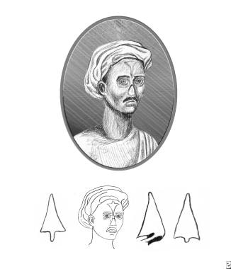
Reproduction of plate from Susruta showing style of nasal reconstruction. Note that support for the nasal bridge is absent. The intrinsic rigidity of the dermis was relied upon for structural support. (Susruta, An English Translation of the Susrita Samhita, based on original Sanskrit text. Edited and published by Kaniraj Kunjabal Blushagratna. Calcutta: Bose, 1916).
Click to see larger pictureClick to see detailView Full Size Image
Media type:  Image

Media file 2:  Nasal implant. 
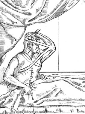
Reproduction of plate from Tagliocozzi as depicted by Gnudi and Webster. Skin of the inner upper arm has minimal intrinsic rigidity and always requires internal structural support.
Click to see larger pictureClick to see detailView Full Size Image
Media type:  Image

Media file 3:  Nasal implant. Reproduction of plate from Carpue manuscript. Note refinements in flap design.
Click to see larger pictureClick to see detailView Full Size Image
Media type:  Image

Media file 4:  Nasal implant. Rib cartilage graft and Proplast bridge graft. A 46-year-old woman with a congenital deficiency of the nasal bridge underwent a series of reconstruction attempts using septal and conchal cartilage grafts. Front view.
Click to see larger pictureClick to see detailView Full Size Image
Media type:  Photo

Media file 5:  Nasal implant. Rib cartilage graft and Proplast bridge graft. A 46-year-old woman with a congenital deficiency of the nasal bridge underwent a series of reconstruction attempts using septal and conchal cartilage grafts. Right side view.
Click to see larger pictureClick to see detailView Full Size Image
Media type:  Photo

Media file 6:  Nasal implant. Rib cartilage graft and Proplast bridge graft. A 46-year-old woman with a congenital deficiency of the nasal bridge underwent a series of reconstruction attempts using septal and conchal cartilage grafts. Harvesting of the rib graft.
Click to see larger pictureClick to see detailView Full Size Image
Media type:  Photo

Media file 7:  Nasal implant. Rib cartilage graft and Proplast bridge graft. A 46-year-old woman with a congenital deficiency of the nasal bridge underwent a series of reconstruction attempts using septal and conchal cartilage grafts. Postoperative right side view.
Click to see larger pictureClick to see detailView Full Size Image
Media type:  Photo

Media file 8:  Nasal implant. Rib cartilage graft and Proplast bridge graft. A 46-year-old woman with a congenital deficiency of the nasal bridge underwent a series of reconstruction attempts using septal and conchal cartilage grafts. Removal of deformed rib cartilage 6 years after reconstruction.
Click to see larger pictureClick to see detailView Full Size Image
Media type:  Photo

Media file 9:  Nasal implant. Rib cartilage graft and Proplast bridge graft. A 46-year-old woman with a congenital deficiency of the nasal bridge underwent a series of reconstruction attempts using septal and conchal cartilage grafts. Tailored carved Proplast bridge graft to replace deformed rib cartilage.
Click to see larger pictureClick to see detailView Full Size Image
Media type:  Photo

Media file 10:  Nasal implant. Rib cartilage graft and Proplast bridge graft. A 46-year-old woman with a congenital deficiency of the nasal bridge underwent a series of reconstruction attempts using septal and conchal cartilage grafts. Postoperative right side view 4 years after Proplast bridge graft.
Click to see larger pictureClick to see detailView Full Size Image
Media type:  Photo

Media file 11:  Nasal implant. Calvarial bone graft and auricular conchal cartilage. A 61-year-old man required a subtotal nasal reconstruction of a septal squamous cell carcinoma and left cervical radical lymphadenectomy. Reconstruction began with a forehead flap of tissue expanded skin. Right lateral view.
Click to see larger pictureClick to see detailView Full Size Image
Media type:  Photo

Media file 12:  Nasal implant. Calvarial bone graft and auricular conchal cartilage. A 61-year-old man required a subtotal nasal reconstruction of a septal squamous cell carcinoma and left cervical radical lymphadenectomy. Reconstruction began with a forehead flap of tissue expanded skin. Calvarial bone graft harvest site.
Click to see larger pictureClick to see detailView Full Size Image
Media type:  Photo

Media file 13:  Nasal implant. Calvarial bone graft and auricular conchal cartilage. A 61-year-old man required a subtotal nasal reconstruction of a septal squamous cell carcinoma and left cervical radical lymphadenectomy. Reconstruction began with a forehead flap of tissue expanded skin. Sculpted calvarial bone graft.
Click to see larger pictureClick to see detailView Full Size Image
Media type:  Photo

Media file 14:  Nasal implant. Calvarial bone graft and auricular conchal cartilage. A 61-year-old man required a subtotal nasal reconstruction of a septal squamous cell carcinoma and left cervical radical lymphadenectomy. Reconstruction began with a forehead flap of tissue expanded skin. Calvarial bone graft in place and anchored to glabellar area of frontal bone.
Click to see larger pictureClick to see detailView Full Size Image
Media type:  Photo

Media file 15:  Nasal implant. Calvarial bone graft and auricular conchal cartilage. Harvest of auricular conchal cartilage graft from anterior approach. A 61-year-old man required a subtotal nasal reconstruction of a septal squamous cell carcinoma and left cervical radical lymphadenectomy. Reconstruction began with a forehead flap of tissue expanded skin.
Click to see larger pictureClick to see detailView Full Size Image
Media type:  Photo

Media file 16:  Nasal implant. Calvarial bone graft and auricular conchal cartilage. A 61-year-old man required a subtotal nasal reconstruction of a septal squamous cell carcinoma and left cervical radical lymphadenectomy. Reconstruction began with a forehead flap of tissue expanded skin. Sculpted conchal cartilage graft on nasal tip.
Click to see larger pictureClick to see detailView Full Size Image
Media type:  Photo

Media file 17:  Nasal implant. Calvarial bone graft and auricular conchal cartilage. Postoperative frontal view at 1 year. A 61-year-old man required a subtotal nasal reconstruction of a septal squamous cell carcinoma and left cervical radical lymphadenectomy. Reconstruction began with a forehead flap of tissue expanded skin.
Click to see larger pictureClick to see detailView Full Size Image
Media type:  Photo

Media file 18:  Nasal implant. Silastic graft and rib bone graft. Preoperative frontal view. A 37-year-old woman presented 10 years following reconstruction of an unreduced naso-orbitoethmoid fracture with silastic H-shaped graft.
Click to see larger pictureClick to see detailView Full Size Image
Media type:  Photo

Media file 19:  Nasal implant. Silastic graft and rib bone graft. Preoperative oblique view demonstrating the distortion of the silastic graft secondary to scar tissue. A 37-year-old woman presented 10 years following reconstruction of an unreduced naso-orbitoethmoid fracture with silastic H-shaped graft.
Click to see larger pictureClick to see detailView Full Size Image
Media type:  Photo

Media file 20:  Nasal implant. Silastic graft and rib bone graft. Harvest of the rib bone graft. A 37-year-old woman presented 10 years following reconstruction of an unreduced naso-orbitoethmoid fracture with silastic H-shaped graft.
Click to see larger pictureClick to see detailView Full Size Image
Media type:  Photo

Media file 21:  Nasal implant. Silastic graft and rib bone graft. A 37-year-old woman presented 10 years following reconstruction of an unreduced naso-orbitoethmoid fracture with silastic H-shaped graft. Fabrication of the osseous H-shaped graft. Removed silastic graft serves as a pattern.
Click to see larger pictureClick to see detailView Full Size Image
Media type:  Photo

Media file 22:  Nasal implant. Silastic graft and rib bone graft. A 37-year-old woman presented 10 years following reconstruction of an unreduced naso-orbito-ethmoid fracture with silastic H-shaped graft. Postoperative frontal view at 10.5 years postreconstruction.
Click to see larger pictureClick to see detailView Full Size Image
Media type:  Photo

Media file 23:  Silastic graft and rib bone graft. A 37-year-old woman presented 10 years following reconstruction of an unreduced naso-orbitoethmoid fracture with silastic H-shaped graft. Postoperative side view at 10.5 years postreconstruction.
Click to see larger pictureClick to see detailView Full Size Image
Media type:  Photo

Media file 24:  Nasal implant. Fascial grafts. A 55-year-old woman sought improvement of radix and upper nasal bridge following an aesthetic rhinoplasty. Preoperative side view. Note the shallow glabella.
Click to see larger pictureClick to see detailView Full Size Image
Media type:  Photo

Media file 25:  Nasal implant. Fascial grafts. A 55-year-old woman sought improvement of radix and upper nasal bridge following an aesthetic rhinoplasty. Temporal fascial graft prior to placement.
Click to see larger pictureClick to see detailView Full Size Image
Media type:  Photo

Media file 26:  Nasal implant. Fascial grafts. A 55-year-old woman sought improvement of radix and upper nasal bridge following an aesthetic rhinoplasty. Right temporal donor site closure.
Click to see larger pictureClick to see detailView Full Size Image
Media type:  Photo

Media file 27:  Nasal implant. Fascial grafts. A 55-year-old woman sought improvement of radix and upper nasal bridge following an aesthetic rhinoplasty. Postoperative side view at 3 months.
Click to see larger pictureClick to see detailView Full Size Image
Media type:  Photo

Media file 28:  Nasal implant. Proplast nasal grafts. A 20-year-old man with a posttraumatic nasal deformity with septal collapse. Preoperative side view.
Click to see larger pictureClick to see detailView Full Size Image
Media type:  Photo

Media file 29:  Nasal implant. Proplast nasal grafts. A 20-year-old man with a posttraumatic nasal deformity with septal collapse. Carved Proplast graft prior to insertion.
Click to see larger pictureClick to see detailView Full Size Image
Media type:  Photo

Media file 30:  Nasal implant. Proplast nasal grafts. A 20-year-old man with a posttraumatic nasal deformity with septal collapse. Postoperative side view.
Click to see larger pictureClick to see detailView Full Size Image
Media type:  Photo

Media file 31:  Nasal implant. Proplast nasal grafts. A 20-year-old man with a posttraumatic nasal deformity with septal collapse. Carved Proplast graft to place on alveolar ridge to augment the columellar base.
Click to see larger pictureClick to see detailView Full Size Image
Media type:  Photo

Media file 32:  Nasal implant. Proplast nasal grafts. A 20-year-old man with a posttraumatic nasal deformity with septal collapse. Proplast alveolar graft in place.
Click to see larger pictureClick to see detailView Full Size Image
Media type:  Photo

Media file 33:  Nasal implant. Proplast nasal grafts. A 20-year-old man with a posttraumatic nasal deformity with septal collapse. Postoperative side view.
Click to see larger pictureClick to see detailView Full Size Image
Media type:  Photo


  • Ayshford CA, Shykhon M, Uppal HS, Wake, M. Endoscopic repair of nasal septal perforation with acellular human dermal allograft and an inferior turbinate flap. Clin Otolaryngol Allied Sci. Feb 2003;28(1):29-33. [Medline].
  • Beekhuis GJ. Saddle nose deformity: etiology, prevention, and treatment; augmentation rhinoplasty with polyamide. Laryngoscope. Jan 1974;84(1):2-42. [Medline].
  • Bikhazi NB, Chow AW, Maas CS. Nasal reconstruction using a combination of alloplastic materials and autogenous tissues: a surgical alternative. Laryngoscope. Aug 1997;107(8):1086-93. [Medline].
  • Brenner KA, McConnell MP, Evans GR. Survival of diced cartilage grafts: an experimental study. Plast Reconstr Surg. Jan 2006;117(1):105-15. [Medline].
  • Brent B. The versatile cartilage autograft: current trends in clinical transplantation. Clin Plast Surg. Apr 1979;6(2):163-80. [Medline].
  • Britt JC, Park SS. Autogenous tissue-engineered cartilage: evaluation as an implant material. Arch Otolaryngol Head Neck Surg. Jun 1998;124(6):671-7. [Medline].
  • Brown BL, Neel HB, Kern EB. Implants of Supramid, Proplast, Plasti-Pore, and Silastic. Arch Otolaryngol. Oct 1979;105(10):605-9. [Medline].
  • Bujia J. Determination of the viability of crushed cartilage grafts: clinical implications for wound healing in nasal surgery. Ann Plast Surg. Mar 1994;32(3):261-5. [Medline].
  • Burke AJC, Wang TD, Cook TA. Irradiated homograft rib cartilage in facial reconstruction. Arch Facial Plast Surg Sept/Oct. 6:334-341.
  • Burke AJC, Wang TD, Cook TA. Irradiated homograft rib cartilage in facial reconstruction. Arch Facial Plast Surg Sept/Oct. 6:334-341.
  • Carpue JC. An account of two successful operations for restoring a host nose. In: The Classics of Medicine Library. Birmingham:. 1981.
  • Carter WW. Transplantation of bone for the correction of depressed deformities of the nose, with report of cases. Laryngoscope. 1911;21:94-97.
  • Celik M, Haliloglu T, Baycin N. Bone chips and diced cartilage: an anatomically adopted graft for the nasal dorsum. Aesthetic Plast Surg. Jan-Feb 2004;28(1):8-12. [Medline].
  • Colton JJ, Beekhuis GJ. Use of Mersilene mesh in nasal augmentation. Facial Plast Surg. Jul 1992;8(3):149-56. [Medline].
  • Daniel RK. Rhinoplasty and rib grafts: evolving a flexible operative technique. Plast Reconstr Surg. Oct 1994;94(5):597-609; discussion 610-1. [Medline].
  • Daniel RK, Calvert JW. Diced cartilage grafts in rhinoplasty surgery. Plast Reconstr Surg. Jun 2004;113(7):2156-71. [Medline].
  • Daniel RK. Diced cartilage grafts in rhinoplasty surgery, Plast Reconstr Surg (Letters and Viewpoints). Sept 2005;116(4):1171-1173.
  • Demirkan F, Arslan E, Unal S. Irradiated homologous costal cartilage: versatile grafting material for rhinoplasty. Aesthetic Plast Surg. May-Jun 2003;27(3):213-20. [Medline].
  • Elahi MM, Jackson IT, Moreira-Gonzalez A, Yamini, D. Nasal augmentation with Surgicel-wrapped diced cartilage: a review of 67 consecutive cases. Plast Reconstr Surg. Mar 2003;111(3):1309-18; discussion 1319-21. [Medline].
  • Erol OO. The Turkish delight: a pliable graft for rhinoplasty. Plast Reconstr Surg. May 2000;105(6):2229-41; discussion 2242-3. [Medline].
  • Erol OO, Gundogan H. Diced cartilage grafts in rhinoplasty surgery. Plast Reconstr Surg. Sep 15 2005;116(4):1169-71; author reply 1171-3. [Medline].
  • Frodel JL, Marentette LJ, Quatela VC, Weinstein GS. Calvarial bone graft harvest. Techniques, considerations, and morbidity. Arch Otolaryngol Head Neck Surg. Jan 1993;119(1):17-23. [Medline].
  • Gnudi MT, Webster JP. The life and lives of Gaspar Tagliacozzi. Los Angeles:. Zeitlin & Ver Brugge;1976.
  • Godin MS, Waldman SR, Johnson CM. The use of expanded polytetrafluoroethylene (Gore-Tex) in rhinoplasty. A 6-year experience. Arch Otolaryngol Head Neck Surg. Oct 1995;121(10):1131-6. [Medline].
  • Gryskiewicz JM, Rohrich RJ, Reagan BJ. The use of alloderm for the correction of nasal contour deformities. Plast Reconstr Surg. Feb 2001;107(2):561-70; discussion 571. [Medline].
  • Gryskiewicz JM. Waste not, want not: the use of AlloDerm in secondary rhinoplasty. Plast Reconstr Surg. Dec 2005;116(7):1999-2004. [Medline].
  • Guerrerosantos J. Temporoparietal free fascia grafts in rhinoplasty. Plast Reconstr Surg. Oct 1984;74(4):465-75. [Medline].
  • Ham J, Miller PJ. Expanded polytetrafluoroethylene implants in rhinoplasty: literature review, operative techniques, and outcome. Facial Plast Surg. Nov 2003;19(4):331-9. [Medline].
  • Kline RM, Wolfe SA. Complications associated with the harvesting of cranial bone grafts. Plast Reconstr Surg. Jan 1995;95(1):5-13; discussion 14-20. [Medline].
  • Kridel RW, Kraus WM. Grafts and implants in revision rhinoplasty. Facial Plast Surg Clin North Am. 1995;3:473-486.
  • Kridel RW, Konior RJ. Irradiated cartilage grafts in the nose. A preliminary report. Arch Otolaryngol Head Neck Surg. Jan 1993;119(1):24-30; discussion 30-1. [Medline].
  • Lawson W, Reino AJ, Seidman D. The silicone columellar strut. Plast Reconstr Surg. Apr 1996;97(5):938-43. [Medline].
  • Lindsey WH, Ogle RC, Morgan RF, et al. Nasal reconstruction using an osteoconductive collagen gel matrix. Arch Otolaryngol Head Neck Surg. Jan 1996;122(1):37-40. [Medline].
  • Maas CS, Monhian N, Shah SB. Implants in rhinoplasty. Facial Plast Surg. Oct 1997;13(4):279-90. [Medline].
  • Maas CS, Gnepp DR, Bumpous J. Expanded polytetrafluoroethylene (Gore-Tex soft-tissue patch) in facial augmentation. Arch Otolaryngol Head Neck Surg. Sep 1993;119(9):1008-14. [Medline].
  • Maas CS, Gnepp DR, Bumpous J. Expanded polytetrafluoroethylene (Gore-Tex soft-tissue patch) in facial augmentation. Arch Otolaryngol Head Neck Surg. Sep 1993;119(9):1008-14. [Medline].
  • Macomber DW. Cancellous iliac bone in depression of forehead, nose and chin. Plast Reconstr Surg. 1949;4:157-162.
  • McDowell F, Valone JA, Brown JB. Bibliography and historical note on plastic surgery of the nose. Plast Reconstr Surg. 1952;10:149-186.
  • Miller TA. Temporalis fascia grafts for facial and nasal contour augmentation. Plast Reconstr Surg. Apr 1988;81(4):524-33. [Medline].
  • Niechajev I. Porous polyethylene implants for nasal reconstruction: clinical and histologic studies. Aesthetic Plast Surg. Nov-Dec 1999;23(6):395-402. [Medline].
  • Okada E, Maruyama Y, Hayashi A. Nasal augmentation using calcium phosphate cement. J Craniofac Surg. Jan 2004;15(1):102-5. [Medline].
  • Ortiz-Monasterio F, Olmedo A, Oscoy LO. The use of cartilage grafts in primary aesthetic rhinoplasty. Plast Reconstr Surg. May 1981;67(5):597-605. [Medline].
  • Powell NB, Riley RW. Facial contouring with outer-table calvarial bone. A 4-year experience. Arch Otolaryngol Head Neck Surg. Dec 1989;115(12):1454-8. [Medline].
  • Romo T, Jablonski RD. Nasal reconstruction using split calvarial grafts. Otolaryngol Head Neck Surg. Nov 1992;107(5):622-30. [Medline].
  • Romo T, Choe KS, Sclafani AP. Cleft lip nasal reconstruction using porous high-density polyethylene. Arch Facial Plast Surg. Mar-Apr 2003;5(2):175-9. [Medline].
  • Romo T, Choe KS, Sclafani AP. Secondary cleft-lip rhinoplasty utilizing porous high-density polyethylene. Facial Plast Surg. Nov 2003;19(4):369-77. [Medline].
  • Romo T, Litner JA, Sclafani AP. Management of the severe bulbous nasal tip using porous polyethylene alloimplants. Facial Plast Surg. Nov 2003;19(4):341-8. [Medline].
  • Saray A. Porcine dermal collagen (Permacol) for facial contour augmentation: preliminary report. Aesthetic Plast Surg. Sep-Oct 2003;27(5):368-75. [Medline].
  • Sclafani AP, Thomas JR, Cox AJ, Cooper MH. Clinical and histologic response of subcutaneous expanded polytetrafluoroethylene (Gore-Tex) and porous high-density polyethylene (Medpor) implants to acute and early infection. Arch Otolaryngol Head Neck Surg. Mar 1997;123(3):328-36. [Medline].
  • Sheen JH. Secondary rhinoplasty. In: McCarthy JG, ed. Plastic Surgery: The Face, Part II. Vol 3. Philadelphia:. WB Saunders Co;1990:1895-1923.
  • Silver WE, Goldberg J. Nasal grafts and implants. Facial Plast Surg Clin North Am. 1994;2:477-499.
  • Strauch B, Wallach SG. Reconstruction with irradiated homograft costal cartilage. Plast Reconstr Surg. Jun 2003;111(7):2405-11; discussion 2412-3. [Medline].
  • Stucker FJ. Auto-Alloplast: a contemporary concept in facial implantation. Facial Plast Surg. 1986;3:137-139.
  • Susruta AN. English translation of the Susrita Samhita, based on original Sanskrit text. Edited and published by Kaniraj Kunjabal Blushagratna. Calcutta:. 1916.
  • Tardy ME, Denneny J, Fritsch MH. The versatile cartilage autograft in reconstruction of the nose and face. Laryngoscope. May 1985;95(5):523-33. [Medline].
  • Taylor IT, Yavuzer R. Alloderm for dorsal nasal irregularities. Plast Reconstr Surg,. Feb 2001;107(2):553-558.
  • Tholpady SS, Schlosser R, Spotnitz W, et al. Repair of an osseous facial critical-size defect using augmented fibrin sealant. Laryngoscope. Oct 1999;109(10):1585-8. [Medline].
  • Welling DB, Maves MD, Schuller DE, Bardach J. Irradiated homologous cartilage grafts. Long-term results. Arch Otolaryngol Head Neck Surg. Mar 1988;114(3):291-5. [Medline].
  • Zeng YJ, Sun XP, Yang J, et al. Mechanical properties of nasal fascia and periosteum. Clin Biomech (Bristol, Avon). Oct 2003;18(8):760-4. [Medline].
  • Zins JE, Whitaker LA. Membranous versus endochondral bone: implications for craniofacial reconstruction. Plast Reconstr Surg. Dec 1983;72(6):778-85. [Medline].

Nasal Implants excerpt

Article Last Updated: Jun 29, 2006