Overview
Since the first replant more than 60 years ago, thousands of severed body parts have been reattached, preserving the quality of life for thousands of patients through improved function and appearance that the void remaining after amputation cannot provide. Ronald Malt performed the first replantation, on May 23, 1962, at Massachusetts General Hospital; the surgery was carried out on a boy aged 12 years who had his right arm amputated in a train accident. [1, 2] This amputation occurred at the level of the humeral neck.
This first replantation was performed with an intramedullary steel rod for internal fixation and involved repair of the brachial artery; both communicating brachial veins; and the median, ulnar, and radial nerves. Malt performed another replant and reported on these in The Journal of the American Medical Association in 1964. [2] In a later report, this original patient was described to achieve some functional recovery of the replanted right arm after tendon transfers and wrist arthrodesis. [3]
Since Malt's first replant, technological advances and the use of the microscope have made possible the replantation of other parts, including thumbs, fingers, ears, scalps, facial parts, and genitalia. [4, 5, 6, 7, 8, 9, 10, 11, 12, 13, 14, 15, 16]
A study by Reavey et al found that although the number of finger amputations occurring in the United States in 2000 was similar to that occurring in 2010 (26,668 vs 24,215, respectively), the number of replantations declined significantly over this period (930 in 2001 vs 445 in 2011). The investigators suggested that this reduction reflects changes in clinical decision making. [17]
For more information on amputation of specific body parts, please see the articles Elbow and Above-Elbow Amputations, Digital Amputations, Wrist and Forearm Amputations, and Amputations of the Lower Extremity.
See the images below.
Indications
Although replantation has improved the lives of many, not all persons who have experienced amputation benefit from or are candidates for replantation. The replantation procedure represents the culmination of the field of hand surgery as a successful replantation often requires repairing blood vessels, nerves, and tendons.
The decision to replant a severed part has evolved and is influenced by many factors, including the importance of the part, level of injury, expected return of function, and mechanism of injury. Because hand function is severely compromised if the thumb or multiple fingers are not present to oppose each other, thumb and multiple-finger replants can be considered for replantation. [18, 19, 20, 21]
Functional outcomes following replantation vary with the level of injury. Replants of the fingers distal to the flexor superficialis insertion, the hand at the wrist, and the upper extremity at the distal forearm can achieve good function. [22, 23, 24, 25, 26] Replantation of the upper extremity at the proximal arm should be attempted in order to preserve the elbow.
If nerve regeneration is inadequate after upper-arm replantation, elbow preservation allows for a forearm amputation and a below-elbow prosthesis. [22] A below-elbow prosthesis with a gravity-activated grip is more functional than an above-elbow prosthesis. Patients who undergo replantation of a zone II finger proximal to the flexor superficialis insertion and a zone VI forearm amputation at the muscle belly and elbow level can be expected to recover less function.
A single digit, amputated within the zone II level, is relatively contraindicated for replantation because the expected stiffness and rehabilitation significantly delays the patient's return to work and the procedure offers minimal or no functional benefit. [27] Interestingly, replantation of zone II finger amputations has been justified in Japan to avoid confusion with a yakuza member (Japanese gangster) who has amputated his finger as a symbol of devotion to the mob boss.
Amputations through the elbow or forearm musculature are relative contraindications to replantation because of the impaired functional recovery of these injuries. When the amputation is through the elbow, the muscles just proximal and distal to the injury are sometimes irreversibly denervated and devascularized and, therefore, may not be expected to recover much function. Also, the elbow joint may not be reconstructible. Amputations at the forearm musculature can also be irreversibly denervated and devascularized and may not recover function.
The mechanism of injury may be the most predictive variable for successful replantation; studies have demonstrated significantly higher success rates with replantations of guillotine versus avulsion amputations. [28] The replantation of severely crushed and mangled body parts is sometimes contraindicated if the part has the red line sign or ribbon sign of the vessels because grafts cannot be used to reestablish flow outside the zone of injury in these circumstances.
Fingers with the red line in the skin over both neurovascular bundles, resulting from hematomas from transections of the branches off the proper digital vessels, cannot successfully perfuse the skin of the replanted part, even if the digital vessels can be repaired. Another predictor of the inability to graft distal to the zone of injury is when the ribbon sign is found to involve the length of both digital arteries, as this often results in intimal injury up the length of the vessel, which is thrombogenic, even if microanastomosis can be performed. [29]
Other contraindications to replantation include amputations with vascular and nerve injuries at multiple levels. Patients who are mentally unstable are poor candidates for replantation because they are prone to reamputate the replanted part. A frustrating example of this is a patient who reamputated his replanted penis in the recovery room after hours of difficult surgery. [11]
However, the only absolute contraindication to replantation exists when associated injuries or preexisting illnesses preclude a prolonged and complex operation. In this circumstance, temporary ectopic implantation has been described for preservation of the amputated extremity before eventual elective replantation later. [30, 31, 32] However, the indication for such an ectopic implantation is rare.
Preoperative Considerations
Amputated parts should be retrieved and brought to the emergency department because they can provide a valuable source of tissue, even if the part is not replantable. Bleeding vessels in the stump should not be clamped. Hemostatic control of the stump can be achieved with a compressive dressing and elevation. The amputated part should be wrapped in a saline-moistened gauze sponge placed in a plastic bag. The plastic bag should be sealed and placed on ice. The amputated part should not be placed directly on ice because this could result in a frostbite injury to the vessels. [33] The part should not be immersed in water; Urbaniak has demonstrated that this makes digital vessel repair more difficult and less reliable. [34, 35]
Recommended ischemia times for reliable success with replantation are 12 hours of warm and 24 hours of cold ischemia for digits and 6 hours of warm and 12 hours of cold ischemia for major replants, although successful replantation has been reported after longer ischemia times. [36, 37, 38, 39, 40, 41] In 1986, May et al reported a successful digit replantation after 39 hours of cold ischemia, the seventh of a 7-finger replant. [38] Then, in 1988, Wei et al reported successful digital replantations after 84, 86, and 94 hours of cold ischemia. [39] Minimizing ischemia times is more critical when performing replantation of major body parts with muscle, and this minimization can be achieved with shunting, which is discussed in more detail in the Major replants section of Special Considerations and Specific Cases below. [30, 31, 32]
Preoperative preparation of the patient includes radiography of both amputated and stump parts to determine the level of injury and suitability for replantation. For medical-legal protection, one should obtain a photographic record of both parts. In obtaining the informed consent, the positive and negative aspects of the procedure should be discussed with the patient and family. These issues include failure rates; the duration of rehabilitation; and a realistic expectation of sensation, mobility, and function. Furthermore, preoperative preparation includes consideration of prophylactic antibiotics, updating the patient's tetanus status, initiating fluid resuscitation to prevent hypotension, warming of the patient to prevent hypothermia and proximal vessel vasoconstriction/spasm, inserting a Foley catheter for volume monitoring, and protecting pressure points in expectation of a long operation.
Operative Considerations
In order to minimize ischemia time, the amputated part can be prepared on a back table before the patient is brought to the operating room. If the amputated part is grossly contaminated, it can be prepared with gentle irrigation. The neurovascular structures of the fingers can be exposed by making either bilateral longitudinal incisions in the midaxial line or volar zigzag and dorsal veins with dorsal longitudinal incisions. [42, 43] However, volar zig-zag incisions can potentially complicate revision amputation closure techniques if the replant should fail.
The neurovascular structures are identified and tagged with sutures to expedite identification later. Furthermore, when preparing the amputated part, note any signs of arterial damage, including telescope, cobweb, and ribbon signs or terminal thrombosis, which require freshening of the vessel. [43] Resection of the vessel distal to the zone of injury may result in a defect that requires a vein graft; this vein graft should be harvested before osteosynthesis in order to minimize warm ischemia time.
Once the neurovascular structures of the amputated part have been identified and tagged, they can be carefully retracted for bone shortening. Approximately 5-10 mm of bone shortening is necessary for tension-free vessel repairs and to avoid neurovascular defects and subsequent grafting. [44] Reducing the tension across the nerve repairs can improve nerve regeneration and recovery of sensation. Bone shortening should be performed on the amputated part, not the stump, to retain the length of the stump should the replant fail. If the amputation level is near the joint on the amputated part, bone can be resected on the stump side for the fingers but not for the thumb, because length preservation of this digit is more critical. Hand function is compromised with thumb loss proximal to the interphalangeal joint. If the amputation level is through the joint of the digits (thumb or fingers), fusion in the functional position is required.
Primary implant arthroplasty has been described in replantation, but this is associated with an increased risk of infection. [45] The next step is to place retrograde Kirschner wires (K wires) or intraosseous wires through the bone on the amputated part. Intraosseous wires can allow for earlier motion and more reliable healing of the fracture (higher union rates). However, K wires are preferred for thumb replants because they are technically easier, they are potentially safer, and, since thumb motion is less important, early motion is not critical.
Usually, preparation of the amputated part can be performed before the patient arrives in the operating room, or a second team can be recruited to begin the preparation of the stump. Neurovascular structures are identified in the same manner as for the amputated part but under tourniquet control. Then, the tourniquet is deflated to assess inflow pressure by the proximal vessel spurt. [43] If the spurt is inadequate, additional vessel shortening is required. Then, the neurovascular structures are tagged with sutures as on the amputated part to facilitate identification of these structures when needed for repair later.
Sometimes, when longer operations are encountered, such as multiple-finger replantations, a pseudocoagulum makes re-identification of these vessels challenging. When preparing the stump, exposing the proximal flexor tendon for placement of a core suture is easier performed at this point in the procedure with shot gun barrel exposure than after bone fixation. Also, it is easier to be able to pull the tendon suture rather than to have to retrieve the retracted flexor later needed for repair. In general when performing multiple-finger replantation procedures, it is advantageous to replant each finger separately to minimize warm ischemia to the digits, which can be kept on ice until ready for that digit. The assembly-line technique should be avoided with multiple-digit replantation procedures.
The order of repair for the various structures is based on surgeon preference and the specific patient. The authors' preferred sequence of bone repair is extensors, veins, dorsal skin, arteries, nerves, and flexors because this sequence allows efficient repair of all the dorsal structures and then the volar structures. [28] If the warm ischemia time is unusually long, the arteries can be repaired earlier.
For bone fixation, many prefer K wires, which are quick and safe and can be placed in a cross or axial configuration. However, better union rates are reported with intraosseous wires, either in combination with a K wire as described by Lister [46] or as 90-90 wires, which are 2 intraosseous wires placed perpendicular to each other. [47] The 90-90 wire procedure has lower nonunion rates. [47]
The hand is pronated. Of the dorsal structures, the extensor is repaired first. If the amputation is at the proximal phalanx level, repairing the lateral slips is important to prevent loss of extension at interphalangeal joints. The veins are repaired next. At least 2 veins should be repaired in finger replants, especially for replants proximal to the proximal interphalangeal joint.
Dorsal veins are preferred because they are larger and do not interfere with later repair of volar structures. Occasionally, arterial repair is required first to locate veins by back bleeding, especially for digital replantations. Then, the dorsal skin is repaired.
The hand is supinated to repair the injured volar structures. At least one digital artery is repaired. Several anastomotic techniques have been described. This is a well-accepted technique that involves placing the first 2 sutures at the 10- and 2-o'clock positions and then the 12-o'clock position. The vessel is then turned 180 degrees, and additional simple interrupted sutures are placed in sequence. An alternative sequence involves placing the initial suture in the 6-o’clock position, followed by 7-o’clock and 8-o’clock, until the 11-, 12-, and 1-o’clock sutures, which are left untied until the end to allow for better visibility of the inside of the vessel to avoid backwalling.
After completing the digital artery repairs, deflating the tourniquet, and removing the clamp, patency of the arterial anastomosis is assessed with the milk test, capillary refill test, and the pinprick bleeding test. If arterial flow appears inadequate, confirm that the patient has adequate blood pressure and volume and that the tourniquet has been deflated. Bathing the vessel with papaverine, lidocaine, and warm irrigation solution has been described to counteract vasospasm. [48]
Finally, the hand can even be placed in the dependent position to increase inflow pressure via gravity. [49] Allow at least 10 minutes of observation time for resolution of vasospasm before manipulating the anastomosis. If the milk test result is abnormal or if the petechia-of-measles sign or the ballooning-of-the-sausage sign is encountered, consider thrombosis as the cause and redo the anastomosis. [50, 43] Frequently, further resection of the vessels is necessary. If the additional vessel resection places the vessel repair under tension, an interposition vein graft is required. Even if flow is reestablished through this anastomosis, a second artery should be repaired as a safeguard.
At this point, the digital nerves are coapted. This can be performed after the tourniquet has been deflated, since the nerves are superficial within the wound. Of the different digital nerve repairs, the epineurial technique is preferred and can be performed with as few as 3 sutures. If the nerve cannot be repaired primarily, a nerve graft may be required. If the nerve defect is less than 2 cm, a nerve conduit, such as unused vein graft or polyglycolic nerve tubes, can be used. If the defect is larger, nerve grafts can be used. Upper extremity donor nerve grafts include the medial antebrachial and posterior interosseous nerves. The posterior interosseous nerve is the same caliber as the digital nerve and leaves no donor sensation defect when harvested distally at the level of the wrist where it innervates the dorsal wrist capsule.
The last part of a digit replant involves repairing the flexor tendon; the previously placed proximal and distal core sutures are tied. Performing this later allows for finger extension and better exposure for microsurgical repair of the digital arteries and digital nerves for zone II replants.
If a vein graft is needed, potential harvest sites for distal digital replants include the palmar forearm/wrist, which is preferred by many because of their vessel size match. [51, 52, 29, 53] The leg or contralateral arm may be used to harvest vein grafts of similar size match for major replants of the hand, forearm, or multiple fingers; a second team can harvest these simultaneously. Vein grafts must be reversed for arterial interposition. Common indications for arterial interposition vein grafts include thumb replants and ring avulsions. [54, 55, 56, 20]
Special Considerations and Specific Cases
Thumb replants
In the thumb, the ulnar aspect digital artery is of larger caliber than the radial digital artery. [57, 58] Therefore, arterial revascularization in thumb replantation is more reliable if based on repair of the ulnar digital artery. However, this vessel is difficult to expose and requires extreme pronation for microanastomosis. Repairing the ulnar digital artery before performing the osteosynthesis usually allows for better exposure of the vessels and a flat field of focus for the microsurgery. Using an arterial interposition vein graft from the radial artery in the anatomic snuffbox to the distal end of the ulnar aspect artery in the amputated thumb or digital artery repair before osteosynthesis may help avoid difficult exposure of the ulnar digital artery of the thumb, especially for more proximal level arterial repairs when the volar skin obscures the ulnar digital artery. [57, 58, 49, 59]
When the thumb has been amputated at or near the metacarpophalangeal (MCP) joint and the proximal digital artery has retracted and is difficult to expose, an end-to-side vein graft can be used from the radial artery in the snuffbox to the distal ulnar digital artery in the amputated part. First, retrograde K wires and core sutures are placed in the flexor pollicis longus (FPL) tendon proximally and distally. The digital nerves are labeled with long sutures for easier identification later. A subcutaneous tunnel is created from the ulnar aspect of the thumb base to the snuffbox. The radial artery is exposed in the snuffbox, and double Pott ties are placed on the radial artery proximally and distally in preparation for an end-to-side vein graft to radial artery anastomosis.
The vein graft is then repaired end-to-end to the distal ulnar aspect digital artery of the amputated part on the back table, with the microscope. This provides much better exposure for the microanastomosis, a flat surface for resting the hand, and a more uniform field of focus for performing microsurgery. Then, the vein graft is carefully pulled distal to proximal through the subcutaneous tunnel to the radial artery in the anatomic snuffbox, making sure not to pull apart the ulnar artery vein graft microanastomosis. Also, in harvesting the vein graft, it is important to clip all branches to avoid blind bleeding into tunnel, later. The ulnar digital nerve is repaired. Then, the osteosynthesis is performed by carefully passing the previously placed K wires in a retrograde manner through the proximal bone. At this point, the radial digital nerve, extensor tendon, and dorsal veins are repaired. Then, the proximal end of the vein graft is repaired end-to-side to theradialartery in the snuffbox.
If the amputation is distal to the MCP joint and the proximal end of the ulnar aspect digital artery is well exposed, a primary arterial anastomosis can occasionally be performed (a graft is not needed). A technique that has been described to optimize exposure of the ulnar aspect digital artery during microanastomosis involves performing the microanastomosis before the osteosynthesis. [60, 61] This allows for better exposure, as the field is flat, so the field of focus for the microsurgery is more uniform.
First, the amputated part and stump are prepared. Longitudinal K wires are placed in the amputated part. The ulnar aspect digital artery and nerve are repaired with the hand in supination, with excellent flat exposure for the microsurgery. At this point, the bones are manually reduced, which can be confirmed by fluoroscopy, and the K wires are passed in retrograde fashion. The K wires previously placed in the distal amputated part are carefully pushed retrograde into the stump using a Penrose to protect the neurovascular repairs. The digital artery clamps are left in place until the radial digital nerve, extensor tendon, and dorsal veins are repaired. Then, the flexor core sutures are carefully tied and the skin is loosely approximated.
Multiple-finger replantation
When considering multiple-finger replantation, the finger with the best chance for successful replantation, best expected recovery, and most significant contribution to function should be repaired first. If all the fingers are injured equally and have the same chance for successful repair, the authors prefer to repair the middle, then index, then ring, and, lastly, the small finger, in order of contribution of function. If the index finger is stiff or insensate, the patient bypasses this to use the middle finger. When all the fingers are stiff, the index finger can actually impede the function and opposition of the other fingers to the thumb. Replanting as many fingers as possible is important in case any of the replants fail.
Each finger is replanted separately to minimize warm ischemia time, as the other fingers can be kept cold until needed. The assembly line technique of performing osteosynthesis for each finger, and then digital artery for each finger, and so on, increases warm ischemia time. Minimizing ischemia time is essential when performing multiple-digit replantations.
The amputated fingers should be brought to the operating room as soon as possible because the digital vessels, nerves, and tendons can be identified and tagged with sutures or clips, which saves time and minimizes ischemia. The order for repairs can be improvised with multiple replantations.
Initially, the osteosynthesis, extensor tendon, one dorsal vein, and one digital artery can be repaired for each finger to minimize overall ischemia time, then another dorsal vein, the digital nerves, and the flexor tendon core sutures can be repaired later, once blood flow has been reestablished to the fingers.
Major replants
The order of the replant procedure is modified for major replantations of the hand and upper extremity. Early use of shunting has been described to minimize muscle ischemia time. [30, 32, 49] Of critical importance to avoid muscle necrosis is minimizing warm ischemia time to less than 4 hours. Intravenous tubing or carotid shunts can be used to infuse and return blood to and from the amputated part.
Fasciotomies are required with major limb replantation and, to save time, can be performed during the shunting reperfusion. Bone shortening is critical to avoid the need for nerve and vein grafting and to allow for soft tissue closure over the repairs. Because muscle along the amputation line is often devascularized, reevaluation of muscle viability and debridement is required 48-72 hours postoperatively.
Upper extremity replants
The amputated part is brought to the operating room, irrigated, and debrided. Neurovascular structures are identified and tagged. Shunting is performed to minimize ischemia. To save time, fasciotomies can be performed during the shunting. Bone shortening is important to avoid grafts and to allow for coverage of repairs. With humeral-level replants, the brachial artery and brachial venae comitante are repaired. The ulnar, median, and radial nerves are repaired. The skin is lightly approximated over the skin.
These upper extremity replants require several operating room debridements at 48-hour intervals to remove devascularized, nonviable muscle at the suture line. Amputations at this level often denervate the biceps muscles and require latissimus or pectoralis muscle transfers later to provide for active elbow flexion.
Hand replants
Vessels, nerves, and tendons are identified and tagged, and K wires are placed in retrograde fashion into the amputated part in the operating room before the patient is transported from the emergency department. Ischemia time can be minimized by shunting, during which fasciotomies can be performed. The most time-consuming part of a hand replantation is tendon repair. The exposure of the proximal flexor tendon ends can be optimized by placing a longitudinal incision up the mid portion of the forearm. Also, tagging of the proximal and distal flexor tendon ends before osteosynthesis can facilitate the repair of the flexor tendons by shotgun barrel exposure.
Importantly, understand the stacked array of the flexor tendons, with the middle and ring flexor superficialis volar to the index and small finger superficialis flexors. Replantation of hand amputations at the wrist level may necessitate bone shortening with proximal-row carpectomy, for example, to avoid nerve and vein grafts. Overall, the ulnar and radial arteries; 4 veins; and median, ulnar, and superficial radial nerves are repaired in addition to as many tendons as possible.
Perform repair of at least the 4 flexor profundus tendons, flexor carpi radialis, flexor carpi ulnaris tendons, 4 extensor digiti communis tendons, extensor carpi ulnaris, extensor carpi radialis tendons, extensor pollicis longus, and FPL tendons. In general, replantations at this level can achieve very good results.
Cross-hand transfer
A cross-hand transfer may be considered under special circumstances, like bilateral hand upper extremity crush avulsion amputations with significant soft tissue or bone destruction that precludes bilateral ipsilateral replantation. [62, 63, 64] An example of an elective cross-hand transfer was reported in a patient with a right brachial plexus injury that rendered the right hand functionless. [63] Accordingly, the right hand was electively transferred to the contralateral side, which had a thumb but was devoid of all 4 fingers. This elective cross-hand transfer was performed at the level of the carpometacarpal joint with intraosseous wires. This was the first elective cross-hand transfer ever performed. The patient attained good functional use of this hand following transfer.
Postoperative Care
Postoperative care includes warming the patient's room to avoid vasospasm and positioning the extremity at the heart level to minimize edema but avoid compromising arterial or venous flow. Concerning blood thinners, several authors recommend the routine use of aspirin and dextran with replantation [65, 28, 66] and therapeutic heparin for crush avulsion injuries. [65, 49]
A study by Zhu et al, however, indicated that in digital replantation and revascularization, postoperative therapeutic dose intravenous heparin (POTDIVH) does not improve survival of the digit, even in crush and avulsion injuries. Patients in the study suffered digital amputation distal to the carpal tunnel. In the POTDIVH group, the overall success rate for revascularization and replantation was 79.9%, compared with 92.8% in the non-POTDIVH group. Retrospective regression and subgroup analyses indicated that an association existed between increased bleeding-related complications in the POTDIVH patients and the reduced success rate. [67]
Depending on the mechanism of injury, consider antibiotic therapy. In addition, patients are encouraged to abstain from smoking and caffeine use for 1 month after surgery. [68, 69] The replanted part is monitored by checking the color, capillary refill time, tissue turgor, and temperature. Sympathetic blocks have been described for high-risk replantations after crush avulsion injuries. [43]
Arterial insufficiency is the most common cause for replantation failure, appearing in 43 (58%) of 75 patients and is suggested by decreased capillary refill time, tissue turgor, and temperature. [65, 28] Treatment of arterial insufficiency includes removing potentially constricting dressings and tight sutures, decreasing the elevation to improve inflow via gravity, and administering a sympathetic block. Finally, consider early operative intervention if no improvement is seen with the above measures, which has been noted to be successful in 16 of 34 patients [65, 28]
Venous congestion is a less common cause for replantation failure (13 of 75 patients) and has been suggested based on increased capillary refill time, tissue turgor, and bleeding of wound edges. [65] Treatment of venous congestion includes removing tight dressings and sutures and increasing the elevation to promote venous drainage via gravity. In addition, leeches are effective for treating venous congestion in replantation. Nail plate removal and the application of a heparin-soaked sponge to the nail bed has been described for distal replantations when a vein could not be repaired and the patient refused leeches. [70] Finally, operative revision can be considered; however, this is less successful for salvaging a failing replant because of venous congestion rather than arterial insufficiency.
Outcomes
Overall success rates for replantation approach 80%. In reviewing the large volume retrospective reports, these success rates range from 54% in China's Sixth People's Hospital to 82% in North Carolina. [71, 72, 18, 73] Overall, success rates are significantly higher for replantation of guillotine amputation (77%) versus crush (49%) amputations. [28, 65, 4, 74, 75, 76]
In general, approximately 50% achieve 2-point discrimination (2 PD) of less than 10 mm. [77, 78, 79, 80, 81] Of Tamai's 228 replants, 70% achieved 2 PD of less than 15 mm, while 65% of Larsen's 142 replants attained 2 PD of less than 10 mm. [77, 72] In general, younger patients with distal guillotine amputations experienced better return of sensation. Studies have determined the average replant to achieve 50% of normal function, ie, 50% total active motion and 50% grip strength. [39, 80, 82, 83]
Return of function was worse for zone II injuries in those of advanced age. Regarding major limb replantation, Russell et al published the largest review and found 11 of 24 achieved greater than 50% total active motion, 19 of 24 achieved protective sensation, and 22 of 24 patients were satisfied with the function and appearance of their replanted part. [83] In 1989, Jupiter et al showed the function of replanted digits could be significantly improved with tenolysis procedures. In their review, the total active motion of 37 replanted digits was significantly improved (P< .001) with tenolysis and no digits were lost. [84]
A study by Efanov et al indicated that repair of multiple veins in finger replantation improves the short-term replantation survival rate (ie, rate of survival to hospital discharge). The study, which included 72 patients with complete digital amputation (101 fingers), determined that there was a 1.27-fold increased relative risk of vein-related replantation failure in patients in whom only one vein was repaired, compared with those who underwent multiple-vein repair, and a 1.49-fold increased failure risk in patients in whom no veins were repaired, compared with multiple-vein–repair patients. There was no significant difference in failure risk between patients who had one vein repaired and those in whom no veins were repaired. [85]
A retrospective study by Chinta et al found that in patients who suffered traumatic, dysvascular digital injury, the rate of secondary surgery was 51% among those who were initially treated with revascularization or replantation, compared with 22% for those who initially underwent completion amputation. [86]
A study by Ma et al indicated that nonsurgical factors that affect the survival of digital replantations include patient age, whether the injury is to the left or right hand, injury type, injury zone, and the method by which the amputated digit is preserved. More specifically, the risk of failure was considered to be greater in children and in patients with a right-hand injury, a crush or avulsion injury, or a little finger injury. [87]
Amputation
Some finger amputations are not suitable for replantation and should be closed primarily or by recruiting adjacent soft tissues. The viability of the remaining soft tissues must be ascertained before application of a tourniquet. Occasionally, the fillet-flap principle can be applied in reconstructing these wounds. Occasionally, bone shortening or revision amputation is required to allow tension-free primary closure of the soft tissues and adequate padding. Furthermore, in an effort to minimize recovery time and hasten return to work, some fingertip injuries are treated with revision amputation.
Complications of amputation procedures include the hook-nail nail deformity, stump neuroma formation, and the quadriga effect. When the injury is located proximal to the lunula, the residual germinal matrix must be removed to prevent hook nail or nail horn formation. Moreover, the hook-nail deformity is prevented by trimming the distal nail bed 2 mm proximal to the bone. Stump sensitivity can be limited by removal of abnormal bone edges and preservation of the articular cartilage, which can minimize inflammation and provide a smooth contour to drape over the wound edges.
Stump sensitivity can also be minimized by performing a traction neurectomy. This severs the nerve while applying distal longitudinal traction, which results in 0.5-1 cm of proximal nerve retraction to prevent neuroma formation over the fingertip. Avoiding the approximation of the extensor and flexor tendons over the distal bone end minimizes the risk for the quadriga effect. In general, revision amputations are frequently performed on manual laborers to allow for earlier return to work or on retired elderly patients for quicker wound closure and therapy, ultimately to minimize stiffness.
Conclusion
With the evolution of surgical techniques and scientific technology, the field of replantation has become more refined and specific indications for replantation, protocols for preparation, efficient techniques to ultimately minimize ischemia times and improve survival rates, guidelines for postoperative care, strategies for treating complications, and goals for outcomes have been established. Patient satisfaction hinges on their level of expectation as defined and explained in the preoperative discussion and informed consent process.
Studies have demonstrated that patients can expect to achieve 50% function and 50% sensation of the replanted part. Initially, all that was amputated was replanted, as surgeons adopted the philosophy of George C. Ross (1843-1892), who stated that "any fool can cut off an arm or leg but it takes a surgeon to save one." However, over 40 years later (1962-2008), surgeons recognize the ultimate goal, which is not merely to preserve all living tissue through nonselective replantation, but rather to preserve quality of life by improving function and appearance.
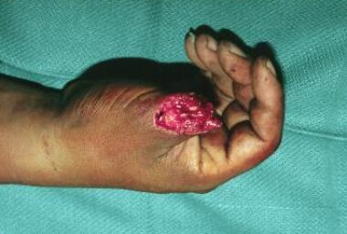
-
Thumb amputation.
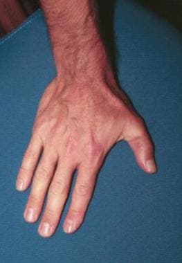
-
After thumb replantation.
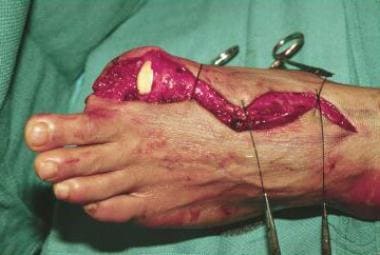
-
Amputation of left big toe.
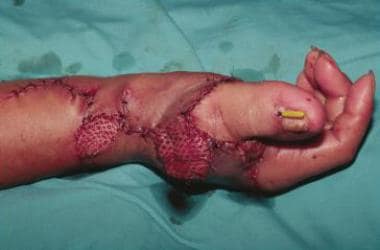
-
Toe-to-thumb transfer.


