Overview
Angiosarcoma is a vascular tumor, the cells of which manifest many of the morphologic and functional properties of normal epithelium. Angiosarcoma is rare, accounting for less than 2% of all soft-tissue sarcomas, and may vary from highly differentiated tumors to those with significant anaplasia, often making these tumors difficult to differentiate from melanoma or carcinoma. While it can occur in any location in the body, angiosarcoma is most commonly found in the skin and soft tissues. Previous radiotherapy and chronic lymphedema (Stewart-Treves syndrome) are known risk factors for cutaneous angiosarcoma. [1, 2]
The most common form of angiosarcoma is cutaneous angiosarcoma not associated with lymphedema. [3, 4] In contrast to the deep location of most soft-tissue sarcomas, angiosarcoma has a predilection for skin and superficial soft tissue on the scalps of elderly people, constituting less than 1% of all head and neck malignancies. [5] According to Aust et al, less than 5% of soft-tumor sarcoma occurs in the head and neck, with only approximately 10% being angiosarcoma. [6] For information on all types of skin cancer, visit Medscape’s Skin Cancer Resource Center.
This disease is primarily located on the head and neck of elderly males. In one of the largest series to date, Perez et al reported on 88 patients with cutaneous angiosarcoma divided into 4 groups: 38 head and neck, 30 radiotherapy-induced, 13 sporadic trunk/extremity, and 7 Stewart-Treves syndrome related. The median age at diagnosis for the entire cohort was 70 years (72.5 y for head and neck), with 95% being white. Of those with head and neck angiosarcoma, 82% were male. Wilson-Jones first distinguished a unique form of angiosarcoma developing in the face and scalp of elderly individuals, noting that it carries a relatively poorer prognosis than the other forms. This form of angiosarcoma is also known as senile angiosarcoma or malignant angioendothelioma. Currently, angiosarcoma of the face and scalp in elderly patients is recognized as a distinctive subgroup. The estimated ratio of male-to-female predominance is approximately 3:1.
Cutaneous angiosarcoma of the head and neck is a unique disease. It usually occurs in the dermis of the scalp and less often in the upper face. Typically, the disease manifests clinically with multiple separate foci, especially when it originates in the scalp (see images below). Angiosarcoma manifesting in the lower face is uncommon, and neck presentations are rare. According to Morrison et al, the rate of regional nodal involvement, reported to be 20-30%, is higher than that of most sarcomas. [7] However, in their review, Perez et al observed only 7% of patients to have regional nodal involvement. [1]
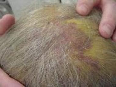 The most common presentation of a scalp angiosarcoma involves lesions that are single or multifocal; bluish or violaceous; nodules, plaques, or flat infiltrating areas; and occasionally may bleed or ulcerate. Most often, the tumors are on relatively exposed skin, either with no or very thin hair.
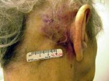 Cutaneous angiosarcomas primarily affect elderly persons and are usually located on the head and neck, particularly in the area of the scalp and upper forehead.
Etiology
The cause of angiosarcoma of the scalp is unknown, although several associations have been reported. Angiosarcoma developing in persons with chronic lymphedema is well described. However, lymph stasis is probably not involved significantly in angiosarcoma of the scalp. The most frequent association with angiosarcoma of the scalp is prior radiation for either a malignant or a benign condition. The mechanism for radiation-induced angiosarcoma is unknown. Radiation-induced angiosarcoma typically presents 5-10 years after irradiation, while radiation-induced tumors of the head and neck have shown greater latency periods. [8] Perez et al found radiation-induced angiosarcoma of the breast to occur at a median of 9 years, while head and neck angiosarcoma occurred at 15 years. [1] Interestingly, according to Lydiatt et al, the histologic appearance and clinical behavior in postirradiation tumors do not appear significantly different from those in tumors arising de novo. [9]
Other risk factors described for angiosarcoma at other locations include exposure to vinyl chloride and thorium dioxide (Thorotrast). Because most angiosarcoma occurs on the scalp and face of whites and usually in non–hair-bearing areas, ultraviolet light exposure has also been suggested as a contributing cause. However, Lydiatt et al investigated the prevalence of metachronous skin cancers as an expression of solar damage and found that only 3 of 18 patients had a history of basal or squamous skin cancer. [9] This is consistent with the prevalence in the general population for this age group and does not support solar exposure as a significant agent. Holden et al questioned sun exposure as a tumorigenic agent because many patients who develop angiosarcoma are women with a full head of hair. [10] Finally, Lydiatt et al suggested trauma as a causative agent, but trauma seems more likely to just be the reason the lesion is noticed by the patient. [9]
Clinical Presentation
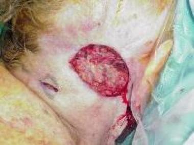
Most cutaneous angiosarcoma appears on the scalp or upper forehead. Angiosarcoma of the face and scalp is insidious, and its clinical manifestations vary widely. In the early stages, angiosarcoma of the face and scalp frequently appears clinically innocent. The lesions may be single or multifocal; bluish or violaceous; nodules, plaques, or flat infiltrating areas; and occasionally may bleed or ulcerate. Most early lesions begin as ill-defined, bruiselike areas with an indurated border. More advanced lesions can be elevated, nodular, or occasionally ulcerated. Grossly, the tumors may appear as ill-defined, hemorrhagic areas.
Extensive local growth is common, and margins are difficult to define surgically. Multifocality is noted in approximately half the patients. Metastasis to regional lymph nodes and to the lungs can occur, often after repeated surgical excisions of the primary growth. In these patients, the prognosis is poor. As reported by Weedon and Freedman, this may reflect the fact that clinical diagnosis is often delayed until the lesions are advanced. [11] Still, even very small tumors can extend microscopically far beyond the visible boundaries of the lesion.
Pathology
Histologically, early lesions may show benign, capillary, hemangiomalike structures. This pattern, however, is deceiving, because angiosarcoma usually has an aggressive course. Microscopically, angiosarcoma often shows extensive involvement of the dermis, with poorly differentiated tumors also invading deep structures such as fascia and subcutis. The histopathologic features of angiosarcoma are diverse. The 3 histologic patterns are vascular channels, sheets of cells, and cells of undifferentiated morphologic features. In some lesions, more than one pattern may be present.
Low-grade angiosarcoma is a well-differentiated lesion that retains some of the functional and morphologic properties of normal vascular endothelium. Well-differentiated lesions form distinct vascular channels, although these are often irregular in shape and size. Unlike benign hemangiomas, well-differentiated angiosarcoma forms vascular channels that create its own tissue planes as it dissects through the dermal collagen. In addition, unlike normal epithelium, angiosarcoma is characterized by cells with larger and more chromatic nuclei, with cells often piling up along the lumina, creating papillations. Low-grade tumors have infrequent mitoses.
In poorly differentiated (high-grade) tumors, sheets of pleomorphic cells may resemble a carcinoma. The vascular anastomosing channels typically observed in angiosarcoma are lined by atypical endothelium that can be either a single row of cells or multiple layers thick. In highly cellular tumors, the neoplastic process causes such close approximation of cells within vascular spaces that the tumor may appear solid. Higher-grade lesions have areas of hemorrhage, disordered architecture, and large cells with hyperchromatic, pleomorphic nuclei. Cells often display prominent mitotic activity. Occasionally, tumors may have well-differentiated areas, but often they are composed exclusively of poorly differentiated areas.
Prognostic Factors
Overall, the prognosis for cutaneous angiosarcoma is poor. Angiosarcoma has a tendency for metastasis via lymphatic or hematogenous routes, and late local recurrence and metastasis after years of apparent remission and successful local control are well documented. A number of large clinical series have assessed prognostic factors. [12, 13]
Tumor size may affect prognosis. Weiss and Goldblum report tumors less than 5 cm in diameter are associated with a significantly better prognosis than larger lesions. [14] Holden et al analyzed patients with tumors smaller than 5 cm, 5-10 cm, and larger than 10 cm and demonstrated a statistically significant correlation between tumor size and survival rate. [15] Multiple studies have compared tumors larger and smaller than 5 cm, all concluding that smaller tumors have a better overall prognosis for overall survival (OS) and/or disease-free survival (DFS). Perez et al, Maddox and Evans, and Pawlik et al report smaller (< 5 cm) tumors are associated with significantly longer OS and DFS. [8, 16, 1] In the most recent of these studies, Perez et al showed median 5-year OS of 48.4% for smaller than 5 cm versus 11.5% for larger than 5 cm. Thus, one of the most important factors in determining prognosis with cutaneous angiosarcoma appears to be initial lesion size.
Other factors have a less substantial effect on prognosis. Maddox and Evans found that patients with moderate or marked lymphocytic inflammatory responses in the tumor survived longer (P = .002). [8] Pawlik et al noted that patients with multifocal disease had decreased DFS compared with patients who had only one lesion. [16] In contrast, tumor grade did not appear to correlate with survival. Holden et al also could not correlate histopathologic grade with survival outcome. [15] Despite this, some researchers contend that low-grade tumors have a more indolent course and a better prognosis than high-grade lesions.
A retrospective study by Alqumber et al of 15 patients indicated that in facial and scalp angiosarcoma, treatment prognosis is not influenced by the reconstructive method. The investigators determined that reconstruction with a flap rather than a skin graft led to delayed median time to recurrence detection (8.75 vs 7.32 mo, respectively) and earlier median time to metastasis (3.75 vs 6.53 mo, respectively), but that both methods were associated with the same median overall survival period (16.7 mo). [17]
A study by Cassidy et al indicated that in patients with nonmetastatic scalp angiosarcoma, the overall survival rate is worse in those aged 65 years or older, in patients with a tumor size of 5 cm or greater, and in individuals who are not treated with definitive surgery. The investigators estimated that study patients who underwent definitive surgery had 1- and 5-year survival rates of 78.2% and 34.1%, respectively, compared with 68.0% and 18.0%, respectively, for patients treated not with definitive surgery but with definitive radiation or a combination of radiation and chemotherapy. [18]
A study by Bernstein et al indicated that survival rates associated with angiosarcoma of the scalp are worse than for angiosarcoma of the face. Scalp and facial angiosarcoma had 5-year locoregional control rates of 9% and 53%, respectively, as well as recurrence-free survival rates of 5% and 27%, respectively, and overall survival rates of 9% and 26%, respectively. In addition, scalp angiosarcomas tended to present at a larger size, possibly because they were less likely to be noticed until they were more advanced. [19]
Death from disease occurs by metastasis, local extension, or tumor invasion. Recurrences and metastases are usually noted within 2 years of diagnosis. Holden et al reported that only 12% of patients survived 5 years or longer, with half dying within 15 months of presentation. [15] In a single institutional review, Perez et al reported 5-year DFS and OS to be 32% and 35%, respectively. [1]
Standard Treatment
The optimum treatment of cutaneous angiosarcoma has not been defined. Generally, radical surgery and postoperative radiotherapy are advocated to treat patients with these tumors. [20] In many patients, surgery often is not feasible because of the multifocal nature and local spread pattern of these tumors. Achieving a negative surgical margin is frequently difficult in patients with scalp angiosarcoma because of the extensive microscopic spread common with this disease. To assist in achieving negative margins, intraoperative frozen specimens are often obtained to help guide the extent of the resection.
Pawlik et al, however, showed that analysis of frozen specimens is not accurate for evaluating the extent of disease at the surgical margins. [16] Others have reported that the use of Mohs surgery similarly does not improve the ability to accurately define tumor-free margins. Thus, even with multiple operations and resections, the goal of histologically negative margins remains elusive. Farhood et al, in a review of patients with head and neck sarcomas of various histologic types, reported that pathologic margins obtained by wide excision were positive in more than 50% of patients. [21] In the experience of Mark et al, only 1 of 12 patients had the disease locally controlled. [22] Holden et al reported one cure in 7 patients treated with surgery alone. [15]
Given the poor results obtained with surgery alone, radiation therapy has been offered as possible adjuvant therapy. Some authors report that radiation therapy provides no benefit. For example, a study by Zhang et al of 42 individuals with primary scalp angiosarcoma, including 39 who underwent surgery, either by itself, in combination with chemotherapy or radiation therapy, or in three-modality therapy, indicated that patients do not significantly benefit from a particular form of treatment with regard to overall and recurrence-free survival rates. [23] Other reports, however, have suggested that surgery combined with radiation therapy offers the best prognosis.
In contrast, the Cassidy study, as discussed above, found 1- and 5-year survival rates to be better in individuals treated with definitive surgery. [18] Similarly, a retrospective study by Oashi et al reported that curative-intent surgery led to improved overall survival in patients with primary cutaneous angiosarcoma of the scalp and face. Evidence indicated that such treatment is beneficial even when the lesion is over 5 cm in size, with negative margins and the use of combined modalities not significantly affecting overall survival in surgical patients. [24]
Mark et al reported improved disease-free survival (DFS) with the addition of radiation therapy to surgery. [22] Similarly, Hodgkinson et al from the Mayo Clinic reported that the only 2 survivors in their series underwent surgery and received radiation therapy. [25] Pawlik et al reported that a prior history of radiation therapy strongly impacted survival upon both univariate and multivariate analyses. In fact, patients who received radiation therapy had a median survival almost 4 times longer than patients who did not receive radiation therapy.
A literature review by Hwang et al indicated that in patients with angiosarcoma of the face and scalp, longer overall survival can be achieved with a combination of radiation therapy and chemotherapy (37.0 mo) than with either type of therapy alone (22.7 and 15.1 mo, respectively). [26]
A retrospective study by Patel et al of patients with angiosarcoma of the face or scalp also indicated that the disease responds better to multimodal therapy than to individual treatments alone. The study included 55 patients, 40 of whom underwent a combination of surgery, radiation treatment, and/or chemotherapy, with the rest treated with just one of these modalities (with the exception of one patient who underwent observation without treatment). The investigators determined that the multimodality patients had a better 5-year rate of locoregional disease control than did the others (20% vs 11%, respectively), as well as higher rates of recurrence-free survival (19% vs 10%, respectively) and overall survival (46% vs 16%, respectively). [27]
Morrison et al noted, however, that angiosarcoma often recurs at the radiation-field margins, sometimes at striking distances from clinically evident disease. [7] Additionally, the disease often recurs at regional and distant sites. For these reasons, the development of new approaches, including more effective local and systemic therapy, is gravely needed.
Reconstruction
The primary treatment for scalp sarcoma of all histologic types is wide surgical excision to histologically negative margins whenever possible. As noted above, achieving a negative margin is frequently difficult in angiosarcoma patients because of the extensive microscopic spread common with this disease. Therefore, in trying to achieve a negative margin, a wound is created that almost never can be closed primarily.
In this clinical setting, the reconstructive surgeon is faced with the dilemma of either (1) performing a primary reconstruction and potentially discovering later that further excision, possibly including sacrifice of the entire reconstruction, is necessary, or (2) performing a staged reconstruction after final confirmation of the margin status has been obtained. The authors prefer the latter approach, although it mandates a second surgical procedure for all patients.
The following are available options for reconstructive surgeons to use for initial temporary coverage and for definitive reconstruction:
-
Homograft skin/bovine collagen constructs
-
Autologous skin grafts
-
Rotation flaps
-
Free flaps
Each option is useful in different situations.
Staged reconstruction is the principal treatment of angiosarcoma of the scalp. Because most patients are elderly, the preferred reconstructive algorithm is skin grafts initially, followed by rotation flaps, and, finally, free-tissue transfer. (See image below).
 Although treatment of scalp angiosarcomas mandates excision to negative margins (when possible), plastic reconstruction should be delayed. Homograft is initially used to temporarily reconstruct the scalp. Homograft has the advantage of being a durable skin substitute for 5-7 days. Once the pathology report shows definitive clear margins, the patient is returned to the operating room for a staged reconstruction.
Typically, oncologic principles require that permanent sections confirm free margins. Therefore, the large defects created after resection must have some type of temporizing reconstruction. This aspect of the case is important because preserving the pericranium allows the use of skin grafts, the simplest form of reconstruction. Rarely does tumor removal require resection of the pericranium, thus preventing the use of a skin graft as the first option; previous radiotherapy is the only possible exception to using skin grafts initially.
Homograft/bovine collagen constructs
Regardless of the clinical scenario, the authors commonly use cadaveric homografts to temporarily reconstruct the scalp. Access to a skin bank with an available supply of homografts is necessary to reliably use this technique. Once the homograft is thawed, place it on the wound just like a skin graft and secure it in place with staples, Xeroform gauze, and a compressive type of head dressing such as self-adhering foam. Usually, the dressing remains on the wound for approximately 1 week, while the pathologist provides margins on the surgical specimens. Homograft has the advantage of being a durable skin substitute for 5-7 days before a purulent exudate develops beneath the graft. When the margins are free, the patient is returned to the operating room for a staged reconstruction.
Other authors have described the use of bovine collagen constructs for reconstruction of full-thickness scalp defects created after resection of angiosarcoma. In one study, 23 patients were treated with bovine constructs for defects with an average size of 51 cm. Histologic studies demonstrated persistence of the construct and infiltration of nascent fibroblasts. Skin grafting of the area was accomplished at an average of 30 days from the time of the initial reconstruction with bovine collagen construct.
Rotation flaps
Local rotation flaps derived from the patient's scalp are the standard when flaps are used to reconstruct the wound. The authors strongly prefer to take large scalp flaps based on single areas, rotate them into the defect, and then graft the donor site. This allows trimming of the skin flap to be placed, while providing the patient with a healed wound for adjuvant radiotherapy. The Crane principle, whereby the flap is used only temporally on the tumor defect, is never used in sarcoma reconstructions because of the potential spread of the tumor. The use of multiple skin flaps is uncommon because they open large areas of scalp to tumor spread.
Free flaps
Reserve free-tissue transfer for patients with angiosarcoma who have high-quality donor vessels for the free-tissue transfer. This problem is usually the limiting step for this operation and is often related to patient age. In the authors' institutions, the rectus muscle flap is the prime free-tissue transfer muscle. This may change with the advent of laparoscopic harvesting of the omentum. The rectus muscle is durable, and the patient's body position is such that the flap can be harvested while the ablative cancer procedure is being performed. Free-tissue transfer is the most complex reconstructive procedure and is the least used technique in elderly patients.
Innovative Treatments
The role of adjuvant chemotherapy in treating angiosarcoma is poorly defined. In a series from the University of California-Los Angeles, 4 of 6 patients who underwent surgery and received radiation and chemotherapy were disease free. [22] Other investigators have concluded that in patients with nonextremity soft-tissue sarcoma, adjuvant chemotherapy offers no statistically significant benefit for survival.
One agent that appears to have substantial activity is paclitaxel. In one study, a response rate of 89% was seen in patients with angiosarcoma of the scalp or face, even in patients previously treated with chemotherapy or radiation therapy. Additionally, a 2004 case report described that weekly administration of docetaxel induced complete remission in a patient with local recurrence and pulmonary metastasis. [10, 28]
Other investigators have examined the use of intra-arterial chemotherapy using pegylated liposomal doxorubicin in combination with intralesional injections of interferon alfa (IFN-alfa). [29] This treatment strategy may be a promising innovative therapeutic option for localized scalp angiosarcoma.
Evidence indicates that the poor prognosis in certain cancers may be associated with alterations in humoral and cellular immunity, and therapy with immunomodulators such as interferons and interleukins may be successful alternatives to chemotherapy. Ulrich et al reported on the long-term survival of an 88-year-old woman with angiosarcoma of the scalp using a combination of intralesional cytokines (ie, IFN-alfa and interleukin 2) and several courses of surface irradiation. [30] This combination therapy led to a 2-year remission of both the tumor and the ultrasonographically suspicious cervical lymph nodes in this patient, whose lesion was quite extensive (20 X 15 cm). Other investigators, however, have reported that tolerance of IFN-alfa is poor, and, when used alone, it does not appear to be an effective treatment of scalp angiosarcoma.
Conclusion
Angiosarcoma of the scalp is a rare, aggressive, and difficult to treat tumor that primarily affects older individuals. Surgeons need to be familiar with these tumors and must be aware of the extremely high risk of local recurrence despite aggressive treatment. Staged reconstructions should be considered in most patients so that definitive reconstruction can be performed after the wide excision margins are found to be negative on permanent section analysis.
Postoperative radiation, if used, should be given in a wide field, beyond the apparent edge of the tumor, and should not be applied in a "postage stamp" fashion. New treatment approaches are urgently needed.
Questions & Answers
Overview
What is angiosarcoma of the scalp?
What is the prevalence of angiosarcoma of the scalp?
Where does cutaneous angiosarcoma of the head and neck typically manifest?
What causes angiosarcoma of the scalp?
Which physical findings are characteristic of angiosarcoma of the scalp?
Which histologic findings are characteristics of angiosarcoma of the scalp?
What is the prognosis of cutaneous angiosarcoma of the scalp?
How is angiosarcoma of the scalp treated?
What is the role of reconstructive surgery in the treatment of angiosarcoma of the scalp?
What is the role of cadaveric homografts in reconstructive surgery for angiosarcoma of the scalp?
What is the role of rotation flaps in reconstructive surgery for angiosarcoma of the scalp?
What is the role of free flaps in reconstructive surgery for angiosarcoma of the scalp?
What is the role of chemotherapy in the treatment of angiosarcoma of the scalp?
What is the role of immunomodulators in the treatment of angiosarcoma of the scalp?
What are the advantages of staged reconstruction in the treatment of angiosarcoma of the scalp?
What is the role of postoperative radiation in the treatment of angiosarcoma of the scalp?
-
The most common presentation of a scalp angiosarcoma involves lesions that are single or multifocal; bluish or violaceous; nodules, plaques, or flat infiltrating areas; and occasionally may bleed or ulcerate. Most often, the tumors are on relatively exposed skin, either with no or very thin hair.
-
Cutaneous angiosarcomas primarily affect elderly persons and are usually located on the head and neck, particularly in the area of the scalp and upper forehead.
-
Although treatment of scalp angiosarcomas mandates excision to negative margins (when possible), plastic reconstruction should be delayed. Homograft is initially used to temporarily reconstruct the scalp. Homograft has the advantage of being a durable skin substitute for 5-7 days. Once the pathology report shows definitive clear margins, the patient is returned to the operating room for a staged reconstruction.


