Practice Essentials
Facial skeleton fractures can result from low-, medium-, or high-velocity trauma. Floor fractures may occur in combination with zygomatic arch fractures, Le Fort type II or III midface fractures, or fractures of other orbital bones.
The goal of treatment is to maintain or restore the best possible physiologic function and aesthetic appearance to the area of injury. [1] A conservative approach may be warranted in some instances, whereas more invasive intervention may be necessary in other situations. [2]
Signs and symptoms of orbital floor fractures (blowout)
Patients may describe the following after facial trauma:
-
Decreased visual acuity
-
Blepharoptosis
-
Binocular vertical or oblique diplopia (especially in upgaze)
-
Ipsilateral hypesthesia, dysesthesia, or hyperalgesia, in the distribution of the infraorbital nerve
Other signs and symptoms can include the following:
-
Pupillary dysfunction coupled with decreased visual acuity
-
Ocular misalignment
-
Hypotropia or hypertropia
-
Limitation of elevation ipsilateral to the fracture
-
Deepening of the supratarsal crease, along with narrowing of the palpebral fissure
Workup in orbital floor fractures (blowout)
Radiographs can be used for soft tissue but are limited by their lack of ability to detect differences in tissue density of less than 10%, making evaluation of soft tissue difficult at best. Anteroposterior views of the orbit usually are obtained with varying angulation of the x-ray beam vector. The most common views are the Caldwell and Waters projections.
Computed tomography (CT) scanning has supplanted radiographs in evaluation of midfacial trauma. Magnetic resonance imaging (MRI) enables multiplanar imaging and is excellent for evaluating soft tissue masses and optic nerve pathology. However, even though MRI provides exquisite detail of the orbital region, CT scanning remains the imaging modality of choice for evaluation of orbital trauma. Of note, intraocular ferromagnetic foreign bodies can add additional insult to the eye and surrounding structures secondary to the magnetic field of the MRI scan.
Management of orbital floor fractures (blowout)
Medical therapy
Medical treatment is warranted for patients for whom surgery is not indicated. This may include patients who present without significant enophthalmos (2 mm or more), a lack of marked hypo-ophthalmos, absence of an entrapped muscle or tissue, a fracture of less than 50% of the floor, or a lack of diplopia.
The patient can be treated with oral antibiotics on an empiric basis due to the disruption of the integrity of the orbit in communication with the maxillary sinus.
A short course of oral prednisone reduces edema of the orbit and muscle, allowing for a better assessment of enophthalmos or entrapment.
Surgery
The orbital floor can be accessed through a conjunctival approach, through cutaneous exposure, or through a transmaxillary approach. Access to this region allows for exploration and release of displaced or entrapped soft tissue, thereby correcting any extraocular motility disturbances. In addition, repair of the bony defect with removal or repositioning of bony fragments allows for restoration of the partition between the orbit and maxillary antrum, thereby preserving orbital volume and geometry and eliminating impingement of soft tissue structures.
Background
Orbital floor fractures may result when a blunt object, which is of equal or greater diameter than the orbital aperture, strikes the eye. The globe usually does not rupture, and the resultant force is transmitted throughout the orbit causing a fracture of the orbital floor. Signs and symptoms can be quite varied, ranging from asymptomatic with minimal bruising and swelling to diplopia, enophthalmos, hypo-ophthalmia (ie, hypoglobus), and hypoesthesia of the cheek and upper gum on the affected side. Treatment is titrated to the degree of injury. [5]
See the image below.
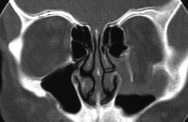 Coronal CT scan of orbits demonstrating loss of orbital floor on the left in contrast to the normal orbital floor on the right.
History of the procedure
According to Ng et al, orbital floor fractures were first described by MacKenzie in Paris in 1844. [6] In 1957, Smith and Regan described inferior rectus entrapment with decreased ocular motility in the setting of an orbital floor fracture and used the term blowout fracture. [7]
During the 1990s, rigid internal fixation became the most frequently used technique in repair of floor fractures. According to Patel and Hoffmann, materials employed for fixation reach back to the introduction of stainless steel wires by Dr. Buck in the 19th century. [8]
Plating has gained widespread acceptance, eclipsing stainless steel wiring in the repair of facial fractures. Refinement of plating for the repair of long bones, microplating systems, and biocompatible implants offer the surgeon several choices for restoration of normal bony architecture.
Problem
Orbital floor fractures can increase volume of the orbit with resultant hypoglobus and enophthalmos.
The inferior rectus muscle or orbital tissue can become entrapped within the fracture, resulting in tethering and restriction of gaze and diplopia.
Significant orbital emphysema from a communication with the maxillary sinus can occur. Orbital hemorrhage is possible with risk of a compressive optic neuropathy.
The globe can be ruptured or suffer less severe forms of trauma, resulting in hyphema, retinal edema, and profound visual loss. A study by Gaier et al indicated that the visual and ocular prognosis is worse in patients in whom open globe injury is concomitant with orbital fracture than in those with isolated open globe injury. The investigators reported, for example, that the presence of an orbital fracture is an independent risk factor for subsequent evisceration/enucleation, with an odds ratio of 4:6. Orbital floor fractures were the most common orbital fractures in the study. [9]
Pathophysiology
Etiology
Pure orbital floor fractures, referred to as isolated floor fractures, result from impact injury to the globe and upper eyelid. The object is usually large enough not to perforate the globe and small enough not to result in fracture of the orbital rim.
Pathophysiology
The orbit and its contents are affected by orbital floor fractures. Direct fractures of the orbital floor can extend from orbital rim fractures, whereas indirect fractures of the orbital floor may not involve the orbital rim. The cause of the fracture is thought to be from increased intraorbital pressure, which causes the orbital bones to break at their weakest point. This is usually the medial orbital floor. Another theory is that compression of the inferior orbital rim causes direct buckling of the orbital floor. In either case, if the intraorbital pressure is great enough at the time of injury, orbital contents can be forced into the fracture site and possibly into the maxillary sinus. [10]
Orbital floor fractures are secondary to a sudden increase in intraorbital hydraulic pressure. A high-velocity object that impacts the globe and upper eyelid transmits kinetic energy to the periocular structures. This energy results in pressure with a downward and medial vector usually targeting the infraorbital groove. Most fractures occur in the posterior medial region that is comprised of the thinnest bones. [11]
Another proposed mechanism that is less favored describes buckling of the orbital floor without displacement of orbital contents following high-velocity trauma.
Although most pure orbital fractures affect the region medial to the infraorbital groove, any fracture type, size, or geometry is possible.
Epidemiology
Frequency
Orbital floor fractures alone or in conjunction with other facial skeletal fractures are the most commonly encountered midfacial fractures, second only to nasal fractures.
The frequency of orbital floor fractures depends on demographics and socioeconomic conditions. Trauma centers and urban facilities encounter a higher prevalence of this injury type.
A retrospective, cross-sectional study by Iftikhar et al found that between 2001 and 2014, of an estimated 671,324 US inpatient admissions for ophthalmic disorders, orbital floor fractures were among the three most prevalent diagnoses (9.6%), together with orbital cellulitis (14.5%) and eyelid abscesses (6.0%). [12]
Mortality/Morbidity
With simple blowout fractures, there may be no morbidity at all, or the patient may complain of diplopia, enophthalmos, or hypoesthesia of the cheek and gum. Edema and ecchymosis of the eyelids and periorbital region usually are seen but are temporary. With any injury that involves a sinus, air may escape into the orbit or subcutaneous tissues. This is called orbital emphysema.
Vertical diplopia may be caused by entrapment of the perimuscular tissue surrounding the inferior rectus muscle in the fracture site. This results in limited upgaze and may cause pain on attempted upgaze as well. Damage to the third nerve branch to the inferior rectus muscle also may cause limited vertical motility. Severe pain with limited horizontal and vertical movements can be indicative of more severe orbital hemorrhage or edema. [13]
Enophthalmos may result when large orbital floor fractures occur and orbital contents prolapse into the maxillary sinus. If a medial wall fracture also has occurred, the enophthalmos may be compounded due to prolapse of orbital contents into the ethmoid sinus. Orbital edema that occurs at the time of injury initially may mask the enophthalmos, but the sunken eye appearance will become more apparent over the following 1-2 weeks as the edema subsides.
Fractures along the floor usually affect the infraorbital groove and therefore the infraorbital nerve. The resultant neuropraxia causes hypoesthesia of the cheek and upper gum on the affected side. This usually is temporary but can last up to 6 months or longer. In severe injuries, the hypoesthesia may be permanent.
Sex
Because the usual mechanism of injury is assault with a blunt object, the vast majority of cases occur in males. In a study of facial fractures in an urban population, 81% of the patients were males.
Age
Because of the nature of the injury and its etiology (eg, assault), most orbital floor fractures occur in teenagers or young adults. [14]
Prognosis
Most cases do well, and most patients obtain resolution of diplopia and correction of enophthalmos.
Successful repair of orbital blowout fractures may be complicated by persistent problems. Neuralgia in the distribution of the infraorbital nerve may worsen after surgery. Improvement of this problem, if any, may take 6 months or more.
More troubling is persistent diplopia. If isolated to extreme positions of gaze, it may go unnoticed or may not be bothersome to the patient. However, if the diplopia affects functional positions of gaze, corrective prisms can be tried. Ultimately, eye muscle surgery may be required to address this problem with repositioning of the extraocular muscles to allow for orthophoric fixation of images.
A study by Su et al of 83 pediatric patients with orbital blowout fractures found that the length of time for postoperative recovery from diplopia was associated with age, with the younger patients taking longer to recover than the older ones. [15]
Enophthalmos can worsen over time. Despite adequately repairing the fracture, atrophy of the orbital fat can occur, resulting in further enophthalmos.
Patient Education
Warn patients to avoid strenuous activity and to use common sense when determining their postoperative activity level.
Warn patients to avoid nose blowing for several weeks after the injury and repair. [3, 4]
Educate patients about nerve damage recovery. An injured motor nerve (third nerve branch) or sensory nerve (infraorbital nerve) can take weeks or months to return to normal. In some cases, the damage may be permanent.
-
Coronal CT scan of orbits demonstrating loss of orbital floor on the left in contrast to the normal orbital floor on the right.
-
Coronal CT scan (soft tissue window) showing right orbital floor fracture, vertical elongation of right orbit, reduction in size of right maxillary sinus, and soft tissue swelling of the right maxillary sinus mucosa.
-
Coronal CT scan showing orbital floor fracture posterior to the globe. A fracture of the lateral maxillary sinus wall also is present.
-
Coronal CT scan showing posterior extension of floor fracture.
-
Operative photo of fracture repair via transconjunctival approach.
-
The bones that contribute to the structure of the orbit.


