Background
The same goal exists for rhinoplasty performed on Asians as for rhinoplasty performed on Caucasians, which is to build a natural-appearing structure that blends harmoniously with the face. As a group, Asians require augmentation of the nose to achieve this result, in contrast to Caucasians who usually require reduction. As with other types of surgery performed on Asians, successful surgery results in a feature consistent with the patient's ethnic identity. Thus, the goal of surgery should be an attractive Asian nose, not the creation of an attractive Caucasian nose on an Asian face. [1, 2, 3, 4] For more information on aesthetic medicine, including news and CME activities, visit Medscape’s Aesthetic Medicine Resource Center.
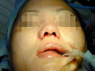
See the image below depicting completion of rhinoplasty procedure.
History of the Procedure
Previously, surgery has focused primarily on dorsal augmentation. Although still performed (especially by nonphysicians or those with limited training), injection of paraffin or liquid silicone has been replaced by alloplastic augmentation, most commonly silastic. Historically, the surgeon addressed the tip by augmenting it together with the dorsum in a one-piece, L-shaped implant with the bend of the L forming the new tip. Because extrusion at the tip remains an ongoing concern with implants of this type, the surgeon frequently protects the tip with cartilage from the ear, septum, or lower lateral cartilage.
The nasal tip and especially nasal tip lengthening vis-à-vis facial thirds remains the primary challenge of Asian rhinoplasty (see the image below).
 Lengthening procedure (different patient) for the short nose. A graft placed behind the toboggan graft holds it out to extension.
The popularity of open rhinoplasty in the United States has led to an increased interest in applying this method to Asian rhinoplasty.
Nevertheless, due to the poorer healing characteristics of Asian skin, some practitioners prefer the endonasal approach to Asian rhinoplasty.
For nasal dorsal augment, a recent surge of interest in autogenous reconstruction using autogenous cartilage has been observed. Costal cartilage is increasingly used in the United States. Recent attention has been drawn to diced conchal cartilage wrapped in Surgicel [5] or temporalis fascia. [6] The diced cartilage obviates the problems associated with warping, which can occur with rib. As much as 4 mm or more of dorsal augmentation can be achieved using conchal cartilage.
Problem
Address the problem as isolated to the dorsum, tip, alar base, vertical dimension, or all of the above. The Asian nose shares similar ideal dimensions with the Caucasian nose but with emphasis on subtleness: the dorsum requires less height, the tip less definition, the alar base less narrowness. As in Caucasians, ideally the radix begins at or slightly below the lash line. The length, measured from the idealized radix to the base of the columella (subnasale), occupies the central third of the face. The tilt of the columella measures 90-115° from the vertical plane, with higher angulation preferred for smaller women.
Southeast Asians (Malay, Filipino, southern Chinese) typically require the most dorsal augmentation (4 mm or more), while northeast Asians (Korean, Japanese, northern Chinese) require less (1.5-2.5 mm) or none. It may be necessary to better define the tip and increase its projection. A deficient premaxilla may need augmentation, as evidenced by a retracted columella with deficient columellar show from lateral view. The nose may require lengthening as measured from the radix to the tip or from the radix to the base of the columella.
The need for alar reduction is frequent in southeast Asians but much less so in northeast Asians. In most cases, both the flare and width need to be corrected; this necessitates an incision into the alar groove. [7]
Pathophysiology
Skin
Nasal skin's thickness better conceals the anatomic detail of the underlying nasal skeleton. This allows better blending of alloplastic or autogenous augmentation with native tissues. Nevertheless, do not use this as an opportunity for sloppiness in surgical technique, because in this patient population, expectations are exceedingly high.
Cartilage
The more delicate cartilaginous tissues of the lower lateral cartilage generally require reinforcement with autogenous cartilage from the ear or septum to obtain a desired result. Affecting a result with pure cartilage reshaping techniques is difficult and usually inadequate. Septal cartilage frequently requires two-layered reinforcement because of its thinness. When harvesting septal cartilage, preserve 1.5 cm of caudal and dorsal septum to prevent nasal dorsal collapse. In about 20% of cases, the septal cartilage is inadequate and additional cartilage from the ear is necessary.
Generally, the lower lateral cartilage is too soft and pliable to adequately support the tip. Such softness precludes the successful use of onlay grafts to the tip, except for the rare patient who has sufficiently strong cartilage (about 10% of patients). Currently, when the author performs an open rhinoplasty, preference is given to creating a columellar strut combined with a shield graft when the septal cartilage is sufficiently strong.
In many cases, septal cartilage as an anterior strut graft appears adequate for tip projection in closed rhinoplasty, if a minimum of 22 mm of cartilage can be obtained to act as both a strut and a shield graft. When an endonasal approach is used, a 1-layered graft appears adequate to maintain durable tip projection.
When tip grafting, defatting should be limited in order to decrease the likelihood of graft visibility. This is particularly true in the open approach, as the 2-layered graft combined with a columella incision can lead to compromised circulation at the tip area and more significant scarring.
More recently, Erol has reported on aggressive tip defatting when performed with closed rhinoplasty and a higher dose of triamcinolone (Kenalog 10/1 mL). [8]
Nasal bones
The shorter and more delicate nasal bones place the patient at higher risk for internal valve collapse; consider spreader grafts in the rare patient requiring isolated dorsal reduction. Fortunately, alloplastic dorsal augmentation functions as a spreader graft, precluding the need for this as a separate maneuver in many cases.
Because of the shorter height of the nasal bones, the author finds that a curved osteotome provides a more consistent result than the percutaneous approach.
Indications
The indication for Asian augmentation nasal surgery is a patient with realistic expectations and mental stability. Asian male rhinoplasty patients appear to have a higher rate of dissatisfaction from nasal surgery. Careful screening is recommended, especially with regard to outcome and the likely shortfalls of the operation.
Relevant Anatomy
First analyze the nasal dorsum, which begins at or slightly below the eyelash line. A straight line drawn from this point to the supratip area determines the appropriate dimensions of the nasal implant. Because of the high visibility of implants ending in the mid dorsum, it may be necessary to lower the height of the dorsum to accommodate a longer implant, even if the dorsal deficiency appears isolated to the radix.
Next, assess the nasal tip for three characteristics: the need for increased projection, tip definition, and/or length from radix to tip. Because of their interrelationship, the need for improvement in any one of these aspects impacts the other two.
The resilience of the lower lateral cartilage determines the approach used to correct the deficiency. According to Millard-Sheen, a well-developed firm nasal tip may require nothing more than a suture-reshaping technique or a graft isolated to the nasal tip identical to a Caucasian rhinoplasty. More typically, a tip grafting technique is required. As the tip becomes increasingly delicate, construct increasingly substantial tip cartilage.
As the nasal tip projection is increased with a graft, the nose rotates cephalically, shortening the radix-tip length. Several techniques can be used to offset the cephalic rotation. These include the extended septal strut with the a wider dorsal edge; a septal extension graft; and the backstopped shield graft or reversed shield graft (convex surface placed caudally). [9]
As mentioned above, when using an open approach, the author prefers conchal cartilage, made 2 layers thick, to serve as an anterior strut graft. Perhaps owing to circulatory disruption, septal cartilage appears to weaken or resorb over time, resulting in the loss of tip projection. The endonasal approach preserves better circulation to the tip area, and this may explain the better survivability of cartilage tip grafts.
Next, look for maxillary spine deficiency, as evidenced by a retracted columellar base and an acute nasolabial angle. The degree of deficiency may require a plumping graft of cartilaginous tissue. Finally, determine whether the alar base requires correction of width or flare. Increasing the projection of the nasal tip usually obviates the need for this in the northeast Asian population (Koreans, Japanese). More frequently, this procedure is performed on Southeast Asians such as Filipinos and Malay. When correcting the flare, the surgeon should avoid a pasted-on appearance of the nostril at all costs. This is done by preserving a slight curvature to the nostril when making the lateral incision.
A study by Wu et al indicated that in East Asian patients, bilateral accessory tip add-on grafts can be employed in augmentation rhinoplasty. Providing a bridge between the alae and the columellar strut, the crescent-shaped grafts can aid in tip support. Although the investigators did not recommend routine use of these add-on grafts, they stated that the grafts could be used for such indications as a severely short nose with insufficient interior soft tissue. [10]
Contraindications
Prior injections of liquid silicone or paraffin to the nasal dorsum predispose patients to infections when the nose is augmented with alloplastic material. The patient should accept the risk of infection rates, which border on 40% or more.
-
After completion of procedure.
-
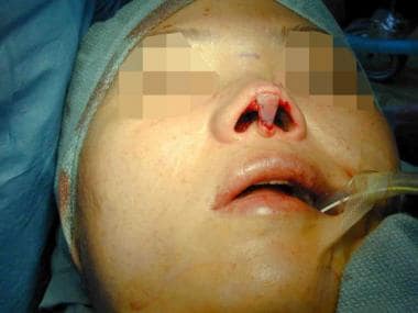
Placing an intercrural 6-0 Prolene taper needle suture to stabilize the framework.
-
A 2-mm osteotome placed percutaneously. Perform this step prior to tip work.
-
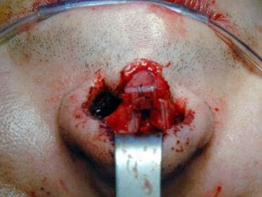
Septal cartilage graft secured to the columella.
-
Marking for alar base resection.
-
Lengthening procedure (different patient) for the short nose. A graft placed behind the toboggan graft holds it out to extension.


