Practice Essentials
Chronic leg or vascular ulcers typically manifest as arterial, neurotrophic, or venous ulcers. They are distinct with regard to their location, appearance, bleeding, and associated pain and findings.
Ulcers of the lower extremities, particularly in individuals older than 65 years, are a common cause for visits to the podiatrist, wound care specialist, primary care physician, vascular surgeon, or dermatologist. [1]
The great majority of vascular ulcers are chronic or recurrent. They cause a considerable amount of morbidity among patients with peripheral vascular disease, including work incapacity. The care of chronic vascular ulcers places a significant burden on the patient and the health care system. Additionally, these nonhealing ulcers place the patient at much higher risk for lower extremity amputation. [2]
Workup of vascular ulcers
Imaging studies
When noninvasive tests reveal unacceptable pedal perfusion, perform imaging studies of the lower extremity to identify the level of obstruction and to evaluate the distal runoff.
Perform angiography when visualization of the vessels of the lower extremities is desired. A femoral runoff analysis is the study of choice. Magnetic resonance angiography (MRA) can also be useful when evaluating lower extremity disease.
Doppler duplex scanning can detect venous reflux with a sensitivity greater than 75%, compared with approximately 40% for descending venography. Ascending venography also may be considered to obtain detailed anatomic information.
Other tests
Ankle-brachial indices (ABIs) and toe digital pressures with pulse volume recordings can provide good clues to the perfusion of the foot.
Xenon-133 clearance to measure blood flow can help to estimate the chance of wound healing.
Transcutaneous oxygen tension may be measured; however, a wide discrepancy exists with the minimal level below which wound healing does not occur. Most agree that a pressure of 30-35 mm Hg is sufficient for healing of more than 90% of wounds.
Management of vascular ulcers
Medical therapy
Research on wound care has resulted in increased use of interactive and active dressings rather than passive dressings that cover and absorb. Interactive hydrocolloid dressings provide a controlled microenvironment for wound healing. Active dressings deliver substances such as growth factors, which are important in the healing cascade.
Surgical therapy
When determining whether to perform surgical therapy for chronic vascular ulcers, consider which is more appropriate for the patient: (1) revascularization and/or coverage of the wound, (2) ligation of incompetent venous perforators, or (3) primary amputation and rehabilitation.
When the ulcer is caused by venous reflux in the superficial venous system, the problem can be addressed with minimally invasive procedures commonly practiced by vascular surgeons. These surgeries include saphenofemoral junction disconnection, stripping of the long saphenous vein to below the knee, calf varicosity avulsions, and saphenopopliteal junction disconnection. The rate of wound healing for those treated with surgery is not significantly higher than that of patients who are treated conservatively, but the resultant diminished rate of wound recurrence is a benefit. [3]
Ligation of superficial venous perforators has been shown to reduce the 4-year recurrence rate of vascular ulcers, from 56% in ulcers treated by compression alone to 31% in ulcers treated by compression plus surgery. [4]
Revision of the wound followed by split-thickness skin graft (STSG) has long been an option for chronic wound management. Often, however, the wound bed is not suitable for grafting or a structure such as a bone or tendon is exposed. Under these circumstances, consider pedicled or free flaps. Microvascular flap coverage of chronic ulcers has met with much success in the treatment of arterial ulcers.
Epidemiology
Frequency
In the United States, the prevalence of vascular ulcers in the general population is not known. However, as the obesity rate increases, the rate of vascular ulcers also increases because of the comorbidities that are associated with patients who are obese. In certain states, venous ulcers are seen in 2.5% of patients admitted to long-term care facilities. This rate is believed to be much higher than the overall population prevalence.
Internationally, studies performed in Ireland [5] and Australia estimate the prevalence of current chronic leg ulcers to be approximately 1%. Of these, most (approximately 80%) are thought to be caused by venous disease rather than arterial disease. A telephone survey performed in Sweden estimated the prevalence over time to be 9.8% for both healed and nonhealed ulcers in persons older than 70 years.
Etiology
Ulceration due to vascular causes is often multifactorial and can be caused by both arterial and venous disease. Hypertension and atherosclerosis of the peripheral vessels lead to arterial disease associated with ischemic ulcers. Chronic venous insufficiency and the resulting venous hypertension cause venous ulcers. Vasculitis such as Buerger disease (thromboangiitis obliterans) or Takayasu disease can also be associated with ulceration. The former tends to manifest with arterial or ischemic-type ulcers, while the latter manifests with cutaneous disease such as pyoderma gangrenosum or erythema nodosum.
When an ulcer does not respond to adequate medical and wound care, the potential for an underlying malignancy should be considered. Cutaneous malignancies that may masquerade as ulcers include nodulo-ulcerative basal cell carcinoma, squamous cell carcinoma, keratoacanthoma, nodular melanoma, tumor stage mycosis fungoides, lymphomatoid granulomatosis, lymphomatoid papulosis, angiosarcoma, and cutaneous metastases from internal malignancy. Healthcare providers must recognize these presentations and render appropriate therapeutic intervention. [6]
Labropoulos et al showed that most of these uncommon ulcers were located in the medial lower calf (n = 19). Specific causes revealed in the histology included neoplasia (5 patients), chronic inflammation (3 patients), sickle cell disease (2 patients), vasculitis (2 patients), rheumatoid arthritis (1 patient), pyoderma gangrenosum (1 patient), and ulcer due to hydroxyurea (1 patient). In 6 patients with ulcers, the histology did not reveal any specific cause. [7]
Pathophysiology
Arterial (or ischemic) ulceration can be caused by either progressive atherosclerosis or arterial embolization. Both lead to ischemia of the skin and ulceration.
Venous (or stasis) ulceration is initiated by venous hypertension that develops because of inadequate calf muscle pump action and after the onset of either primary (with no obvious underlying etiology) or secondary (as seen after deep venous thrombosis) valvular incompetence. Two hypotheses have been proposed to explain venous ulceration once venous hypertension develops.
The first states that distention of the capillary beds occurs because of increased stasis. This leads to leakage of fibrinogen into the surrounding dermis. Over time, a fibrinous pericapillary cuff is formed, impeding the delivery of oxygen and other nutrients or growth factors to the affected tissue. The resulting hypoxic injury leads to fibrosis and then ulceration.
The other hypothesis suggests that the endothelium is damaged by increased venous pressure and leukocyte activation. Proteolytic enzymes and free radicals are released, escape through the leaky vessel walls, and damage the surrounding tissue, leading to injury and ulceration.
In addition, studies have shown a relationship between obesity, chronic venous disease, and popliteal venous compression (PVC). [8] This syndrome may clarify the previously unexplained venous presentations. Other explanations of increased incidence of vascular ulcers in obese patients could be the direct result of intra-abdominal venous compression.
A study by Rasmussen et al using near-infrared fluorescence lymphatic imaging found impaired lymphatic function occurring early in the development of venous leg ulcers and revealed bilateral dermal backflow in the presence of chronic venous insufficiency, including in patients without ulcers in the contralateral limb. [9]
A study by Polak et al indicated that in patients with deep venous thrombosis, denser fibrin clots that are resistant to lysis not only increase the risk of postthrombotic syndrome but also raise the likelihood of postthrombotic venous ulcer development. [10]
Presentation
Chronic leg or vascular ulcers typically manifest as arterial, neurotrophic, or venous ulcers. They are distinct with regard to their location, appearance, bleeding, and associated pain and findings.
Arterial ulcers
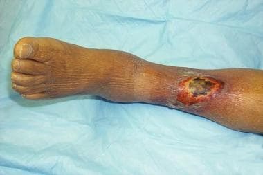
Arterial ulcers (see image below) are often located distally and on the dorsum of the foot or toes. Initially they have irregular edges, but they may progress to have a better-defined appearance. The ulcer base contains grayish, unhealthy-appearing granulation tissue. With manipulation, such as debriding, these ulcers bleed very little or not at all. The patient may report characteristic pain, especially at night when supine, which is relieved by dependency of the extremity. Upon examination, characteristic findings of chronic ischemia, such as hairlessness, pale skin, and absent pulses, are noted.
Neurotrophic ulcers
Neurotrophic ulcers are characterized by a punched-out appearance with a deep sinus. These are often seen underlying calluses or over pressure points (eg, plantar aspect of the first or fifth metatarsophalangeal joint). They are commonly surrounded by chronic inflammatory tissue. Probing or debriding may lead to brisk bleeding. Because these patients usually have a neuropathy leading to hypesthesia and diminished positional sense or 2-point discrimination, these ulcers are frequently painless.
Venous ulcers
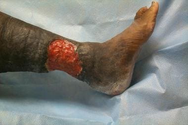
Venous ulceration (see image below) is commonly noted in the "gaiter" region of the legs. This region is located circumferentially around the lower leg from approximately mid calf to just below the medial and lateral malleoli. Larger but shallower than other ulcers, stasis ulcers have a moist granulating base and an irregular border. This base oozes venous blood when manipulated. The tissue surrounding these ulcers may exhibit signs of stasis dermatitis. Patients often report mild pain that is relieved by elevation.
Diabetic ulcers
Diabetic ulcerations occur as a result of various factors. Such factors include mechanical changes in conformation of the bony architecture of the foot or the combination of any of the ulcers mentioned previously. Nonenzymatic glycosylation predisposes ligaments to stiffness. Neuropathy causes loss of protective sensation and loss of coordination of muscle groups in the foot and leg, both of which increase mechanical stresses during ambulation.
Indications
Surgical therapy is an integral part of the treatment of nonhealing wounds. Wounds with necrosis or infection usually require debridement or incision of the affected tissue. The goal is to achieve a clean, granulating bed upon which a split-thickness skin graft (STSG) may be placed for closure. In other circumstances, the wound bed may be unable to support a skin graft or debridement or disease may have resulted in the exposure of a structure such as a joint or bone. Under these conditions, local or free flaps of tissue may be used to provide coverage of the wound. These flaps may be performed in concert with or independent of arterial revascularization or venous repair procedures. Revascularization often causes even moderately sized ulcers to heal primarily.
Contraindications
Surgical therapy of vascular ulcers may be accomplished by a number of methods; tailor the choice to fit both the patient's and surgeon's expectations. Primary coverage and/or revascularization may be most appropriate in one patient and amputation with rehabilitation most suitable in another. Evaluate contraindications to treating an ulcer with either an STSG or pedicled or free flap based on the likelihood of survival of the coverage tissue versus the risks of undergoing the procedure, each of which is associated with varying degrees of morbidity.
Factors to consider when evaluating an ulcer to determine the likelihood of successful coverage include existing infection or the likelihood of developing infection at the surgical site; the perfusion of the surgical site; the condition of the surrounding tissue, such as edema or ischemia; the rehabilitation potential of the patient; any existing comorbid conditions; or habits of the patient that preclude survival of the graft or flap.
-
Vascular ulcers. Arterial ulcer with characteristic features.
-
Vascular ulcers. Venous ulcer with characteristic features.


