Practice Essentials
Radiation ulcers are wounds caused by the acute or chronic effects of ionizing radiation. [1] The injury may involve the skin, underlying soft tissue, and even deep structures such as bone. The most common cause of radiation injury is an adverse effect of therapeutic radiation therapy. Other causes are occupational or environmental exposures. [2] See the image below.
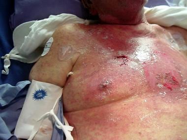 Case A. Cutaneous injury caused by irradiation of the chest wall to treat advanced lung cancer with metastases to the head and spine. This patient was transferred to a burn unit for adequate care of the burns and ulcerations caused by the radiation treatments.
Workup of radiation ulcers
Lab studies
Routine presurgical testing should be done, as indicated by the patient's age and comorbid conditions. Nutritional parameters, such as albumin, prealbumin, and ferritin levels, should be obtained if suboptimal nutrition is a possibility. Patients with chronic wounds are often debilitated, and they may have anemia due to chronic, minor blood loss.
Check the prealbumin and albumin levels, which indicate whether the patient's wound healing capability is optimized.
Imaging studies
Plain radiographs may be useful to look at the condition of the underlying bone and to screen for osteoradionecrosis. Computed tomography (CT) scanning or magnetic resonance imaging (MRI) may be useful in defining the extent of large, deep wounds and the involvement of underlying muscle and bone.
Biopsy
Biopsy of suspicious wounds should be done to rule out malignancy (Marjolin ulcer)
Management of radiation ulcers
Medical therapy
As a result of vascular changes and the resultant hypoxia, irradiated fields have a decreased capacity to fight infection. Impaired delivery of antibiotics also hinders the eradication of infection. As a result, use topical antibiotics, preferably those with tissue penetrance capabilities, to decontaminate irradiated wound beds.
Regarding acute radiation injury, radiation therapy, even when properly administered, may cause adverse skin effects. Treatment is supportive and includes protection from further trauma and use of topical antimicrobials (eg, silver sulfadiazine for partial-thickness skin losses). If frank, full-thickness ulcerations develop, they are unlikely to heal with purely medical intervention.
Chronic radiation injury decreases the ability of the body to tolerate bacterial contamination. When elective surgery is undertaken through radiated tissue, meticulous technique, gentle tissue handling, and antibiotic prophylaxis are essential.
Hyperbaric oxygen treatment is of value in healing of tissues of the head and neck, anus, and rectum. It can also be useful in preventing osteoradionecrosis of the mandible when dental work is needed after radiation.
Medical therapy with amifostine reduces the incidence of xerostomia.
Surgical therapy
Immediate, tension-free reconstruction should be performed at the time of ulcer excision since granulation tissue tends not to arise in irradiated beds. Because skin grafts typically fail, arterial-based flaps (free, locoregional, musculocutaneous, or fasciocutaneous) are the preferred means of reconstruction.
History of the Procedure
Wilhelm Konrad Roentgen discovered x-rays in 1895. The realization that radiation can cause tissue injury followed shortly thereafter. Many different forms of radiation have since been discovered, and applications have been developed for medical, industrial, and military use.
The common pathway of radiation injury to tissue, regardless of the source of the radiation, is interaction of the radiation energy with DNA that causes structural damage to the DNA. Depending on the precise area of injury in the cell, the damage may be repaired, cause cell death, or cause delayed effects. This damage can lead to both acute and chronic tissue effects.
Problem
Ionizing radiation may come from high-energy photons that can be the product of natural decay of radioactive material, such as gamma rays, or the product of artificial bombardment of electrons onto Tungsten, such as x-rays. Gamma rays and x-rays can be manipulated to control the amount of energy and the depth of penetration. The energy delivered to the tissues is measured in electron volts. Orthovoltage radiation is used in therapeutic radiation, and includes radiation at 80-400 keV. Supervoltage radiation delivers energy in excess of 1 million V.
Radiation can also be produced by high energy particles that are a product of radioactive decay. Alpha particles are helium nuclei emitted by various elements, including radium. Alpha particles penetrate poorly but can be taken up in local tissues. Beta particles are electrons. They penetrate the skin superficially and can be useful in treating relatively superficial skin conditions, such as mycosis fungoides.
Brachytherapy is radiation therapy delivered with a short distance between the radiation source and the target. Many radioactive isotopes are used, such as iodine-125, gold-198, and cesium-137. Needles filled with radioactive seeds, mechanical appliances, permanent seeds, and other modalities can be used.
Radiation delivery is measured by the amount of radiation absorbed by a gram of tissue. One rad is equal to 100 ergs of energy absorbed per gram of tissue. The most common unit of measurement is the gray, which is equal to 100 rad.
Epidemiology
Frequency
The true incidence of radiation injury to normal tissue is unknown. An acceptable short-term complication rate from radiation therapy is in the range of 5-15%. The long-term complication rate is unknown and, of course, affected by long-term survival rate. As cancer therapy becomes increasingly effective, patients are living long enough to have the adverse late effects of their radiation treatment.
In patients who underwent cardiac fluoroscopy for percutaneous coronary intervention, Wei et al, in a retrospective study, found the resulting incidence of radiation ulcers to be 0.42% (nine out of 2124 patients). [3]
Etiology
The acute effect of ionizing radiation is direct cell damage to DNA. This damage may cause immediate cell death, prevent proliferating cells from dividing, or induce apoptosis. The late effect is fibrosis. [4] The mechanism of fibrosis production is unclear, but it may involve proinflammatory and profibrotic cytokines, such as tumor necrosis factor-alpha. The fibrosis is progressive.
Pathophysiology
Tissues affected by acute high-dose radiation, as in industrial accidents, manifest progressive obliterative endarteritis culminating in tissue necrosis. [5] Long-term radiation injury results in fibrosis of the dermal and subcutaneous tissues. Elastin fibers are fragmented. The skin loses its rete pegs. The number of blood vessels may be normal; however, fibrosis around them impairs their ability to contract. Electron microscopy may show dehiscence of endothelial cells and microthrombi. [6]
Radiation damage to cells can also lead to future malignancies. These may arise in chronic wounds due to radiation damage. They can also be the cause of nonhealing wounds.
On a cellular level, radiation-induced damage can be direct or indirect. Direct damage results from the hits to, or radiation absorption by, the cells. In contrast, indirect damage occurs when the radiation causes cellular water to release free radicals, which in turn combine to make cytotoxic peroxides. [7] Direct effects of radiation can affect cellular DNA, which, when altered, may lead to cellular destruction or aberrant cellular replication and malignancy.
These direct and indirect cellular alterations also occur at the level of the fibroblast. In the development of soft tissue wounds and ulcerations in irradiated fields, extensive research implicates fibroblast dysfunction and depletion as culprits because of decreased collagen deposition in soft tissues. Although most investigators agree that vascular injury also inhibits wound healing, Rudolph et al question this notion. [8] They suggest that oxygen tension is not altered in irradiated tissues compared with nonirradiated fields and that the onus of impaired wound healing falls on this fibroblast dysfunction. [9] Some laboratory evidence indicates that local treatment of radiated wounds with transforming growth factor (TGF)-beta and fibroblast growth factor (FGF) improves tensile strength in acute radiated wound models. [10]
Presentation
The 3 types of radiation injury are acute, subacute, and chronic.
Acute injury refers to radiation exposure that often occurs in a setting such as an industrial accident. It is usually caused by orthovoltage radiation in the range of 5000-10,000 rads. The effects on the skin and exposed soft tissues are like those of a thermal burn, with alterations and destruction of the basal cells of the epidermis, though these effects are slower and more progressive than those due to thermal burns. Erythema results and may be recurrent. Along with erythema, the patient has pain, edema, itching, and eventual desquamation of the skin. This desquamation can progress to soft tissue necrosis and obliterative endarteritis. Superimposed infection also may complicate the clinical course.
Subacute injury differs from the acute form in that it is caused by recurrent exposures to radiation over time, as with therapeutic radiation. This type of radiation tends to be of lower energy than that observed in occupational or environmental exposures. It also causes cutaneous erythema and edema, but the erythema tends to be transient and necrosis is usually absent. Another characteristic finding after multiple radiation treatments is hyperpigmentation and a woody induration of the soft tissues. In terms of vascular effects, studies have demonstrated the following changes in irradiated vessels: decreased smooth muscle; increased collagen levels with fibrosis and thickening of the walls; hyaline degeneration and breakdown of elastic lamina; dehiscence of endothelial cells; and fibrinoid necrosis and microthrombi in the lumina leading to ineffective delivery of oxygen, other vital nutrients, and antibiotics.
Chronic injury results from long-term exposures and typically results from repeated occupational exposures (eg, in radiology technicians). The injuries are similar to those noted above but tend to be more pronounced. This is particularly true of vascular damage. Fibrosis and thrombosis progress to an obliterative endarteritis and its attendant ischemia.
Given the progressive nature of radiation damage, a soft-tissue ulceration may develop at any time after radiation exposure. This ulceration may be large, or it may initially manifest as a draining sinus.
In patients who have undergone breast cancer surgery, Ma et al classified chronic radiation-induced ulcers of the chest wall as follows [11] :
-
Mild - Less than 10 cm 2, ulceration to the subcutaneous layer, ulcer base containing relatively healthy granulation tissue, and no disorders of upper limb movement
-
Moderate - Over 10 cm 2, ulcer base containing partially exposed bone and no healthy granulation tissue, and partial sensory and motor disorders of the upper limb
-
Severe - Over 100 cm 2, ulcer base revealing obvious collarbone and/or rib osteonecrosis, and total loss of upper limb function
Indications
Indications for treatment of radiation ulcers include control of infection and prevention of sepsis, pain control, management of the malignant potential, and closure of wounds.
Relevant Anatomy
Radiation ulceration may affect skin, underlying soft tissues, and bone. The effects of injury may be seen beyond the area of treatment. Before reconstructive surgery is undertaken, the physician must thoroughly evaluate the probable extent of injury and the effect of the chosen reconstructive method.
Contraindications
Radiation injury of deep tissues may be underestimated. Radiated tissue heals poorly by secondary intention. Therefore, reconstructive methods that lead to primary wound healing are strongly preferred. Exposure of vital structures should be avoided unless the reconstructive plan allows for their immediate coverage.
-
Case A. Cutaneous injury caused by irradiation of the chest wall to treat advanced lung cancer with metastases to the head and spine. This patient was transferred to a burn unit for adequate care of the burns and ulcerations caused by the radiation treatments.
-
Case A. Cutaneous injury caused by irradiation to the chest wall to treat advanced lung cancer with metastases to the head and spine.
-
Case A. Cutaneous injury caused by irradiation to treat advanced lung cancer with metastases to the head and spine. View illustrates radiation burns to the head and neck region. Note the residual silver sulfadiazine and mafenide acetate cream on the patient's face and ears, which was applied to treat the injury and prevent infectious complications.
-
Case A. Cutaneous injury caused by irradiation to treat advanced lung cancer with metastases to the head and spine.
-
Case A. Cutaneous injury caused by irradiation to treat advanced lung cancer with metastases to the head and spine.
-
Case A. Cutaneous injury caused by irradiation to treat advanced lung cancer with metastases to the head and spine. View shows the posterior aspects of the patient's ears and neck.
-
Case B. This patient presented to the plastic surgeon with complaints of a small opening along her mid sternum. She was receiving follow-up care from her primary physician, who had been treating the wound with parenteral antibiotics, with no improvement. The patient's history was noteworthy for previous left radical mastectomy followed by cobalt radiation approximately 20 years ago. At first glance, the wound appeared to be a small, draining sinus surrounded by the erythema typically seen with radiation-damaged skin.
-
Case B. The patient was scheduled for debridement of the affected area. Use of a myocutaneous flap was planned because a large area of underlying osteonecrosis was suspected. Image depicts the extensively débrided chest wall. Most of the sternum and numerous costochondral cartilages were excised.
-
Case B. Photograph obtained after a right-sided pectoralis major myocutaneous flap was used to close the resultant defect. The pectoralis muscle was disinserted at the shoulder to facilitate movement of the flap across the midline.


