History of the Procedure
The history of chest wall reconstruction illustrates the challenges associated with this type of repair. In 1778, Aimar resected the first osteosarcoma of the ribs. In 1820, Cittadini reported a case of bony chest wall tumor resection. Parham, in 1899, was the first in the United States to report resection of a bony chest wall tumor involving 3 ribs. This apparently caused a pneumothorax, which was controlled with soft tissue coverage. In the early 1900s, Fell and O'Dwyer described intubation techniques and positive-pressure ventilation. [1, 2]
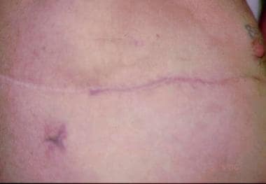
The image below is a postoperative photo of chest wall reconstruction.
In 1906, Tansini used the latissimus dorsi myocutaneous flap, apparently for the first time, for coverage of radical mastectomy defects. [3] Hutchins and Campbell shared this approach. [4, 5] Graham and Singer were the first to successfully perform a pneumonectomy in the early 1930s. [6] In the 1940s, Watson and James used the fascia lata for closure of skeletal wound defects. [7] Bisgard and Swenson described the use of ribs for closure of sternectomies. [8]
Pickrell offered techniques in chest wall resection for breast cancer, [9] and Maier described his use of cutaneous flaps for patients with breast cancer postresection. [10] The 1950s and 1960s included refinement of the reconstructive techniques and the implementation of multistaged procedures. Other pioneers of mention include Arnold and Pairolero, whose studies concluded that chest wall reconstruction is safe, durable, and associated with long-term survival. [11] For the past 25 years, chest wall reconstruction has undergone a vast growth in technique and alternatives. Flaps often used for this task are the latissimus dorsi, pectoralis major, serratus anterior, rectus abdominis, external oblique, and omentum.
The congenital defect of the thorax, Poland syndrome, was described by Sir Alfred Poland in 1841. [12] He noted restricted musculature on one side of the thorax on a single autopsy. In his report entitled "Deficiency of the pectoralis muscle," he described absence of the sternocostal portion of the pectoralis major, an absent pectoralis minor, and a severely hypoplastic serratus anterior and external oblique. [12] de Haan associated the defects of Poland syndrome to the overlooked concomitant deformities of the ipsilateral upper extremity and hand. [13]
Etiology
One of the most common acquired chest wall deformities is sequela from infection. This may be the result of mediastinitis, trauma, or empyema. The resulting defects, from débridement of the chest wall or the pleural space and its contents, may require fill procedures with flaps of thoracic or abdominal origin, sterilization procedures, or collapse procedures as in thoracoplasty. Tumor radiation injury promoting scar and nonfunctional tissue also may require débridement and reconstructive measures. Resection of large chest wall, pulmonary, or mediastinal tumors, as well as defects created by trauma, may merit chest wall reconstruction. [14]
The etiology of Poland syndrome, a congenital defect of the chest wall, is unclear, yet the current theory describes hypoplasia of the ipsilateral subclavian artery in utero. The subclavian artery supply disruption sequence (SASDS) described by Parker et al illustrates the kinking of the upper extremity artery as the ribs grow forward and medially. [15] The reduction in lumen diameter and thus flow impedes distal growth, which supports the theory that more proximal blocked flow results in more severe deformity. The incidence of Poland syndrome is 1 in 30,000. The right side in Poland syndrome is affected twice as often as the left and it is considered to be autosomal dominant with low penetrance.
Möbius syndrome involves the anomalies observed in Poland syndrome in addition to bilateral facial paralysis and the inability to abduct the eyes. Möbius syndrome is observed in 1 individual per 500,000.
The etiologies of pectus excavatum and pectus carinatum are unknown. Pectus excavatum is the most common congenital anomaly of the chest (90%). The male-to-female ratio is 3:1.
Pathophysiology
The muscles of inspiration, an active action, involve primarily the diaphragm, which contracts inferiorly and creates a negative intrapleural pressure, thus inducing inhalation. Secondary muscles involved in inspiration are termed accessory muscles and are the sternocleidomastoids, which aid in raising the sternum superiorly and outward; the scalene muscles, which elevate the upper ribs; and the external intercostal muscles, which elevate all the ribs.
Expiration is a passive process. The intrinsic elasticity of the lung and musculature promotes exhalation. The muscles mentioned above relax and initiate the expiratory phase of breathing. Pulmonary function tests that measure forced expiratory volume in 1 second (FEV1), tidal volume, and the ratio of FEV1 to forced vital capacity ratio also are beneficial, yet these values are not critical in the face of mandatory surgical intervention. Lung disease takes on two broad categories, obstructive and restrictive. With obstructive disease, expiration is impeded by proximal obstruction of the bronchioles and bronchi, causing air trapping, increased functional residual capacity and residual volume, and decreased FEV1 and vital capacity.
Restrictive lung disease is an interstitial process that causes lung tissue to be less compliant, thus reducing the ability of the lung to expand. This promotes reduced lung volumes. Flail chest refers to a segment of chest wall, usually 5 cm in diameter, which loses continuity with the surrounding chest wall, resulting in a paradoxic respiratory pattern and inefficient ventilation. Adequate fixation of this segment is necessary to correct this phenomenon and restore proper respiratory physiology and ventilation.
The size of the defect above which bony stabilization is required is not clear. Two-rib segmental loss may be repaired with soft tissue reconstruction. While Dingman cautions that a 4-rib loss results in flail, [16] Arnold argues that complete sternectomy or resection of 4-6 ribs at the cartilage level does not result in flail or respiratory instability. McCormack and Picciocchi et al agree that defects less than 5 cm in diameter or resection of 3 ribs or fewer do not merit skeletal stabilization. [17, 18]
Indications
Chest wall defects are grouped into 2 general categories, acquired and congenital defects. Acquired defects include tumor (primary or recurrent), infection, radiation injury, and trauma. [19, 20] This group entails most cases that require an operative plan that balances function, durability, and aesthetics in the reconstructive effort. [21]
Congenital defects, although less common, also can be a reconstructive challenge. This article focuses on the congenital defects of Poland syndrome, pectus excavatum, and pectus carinatum.
Remember that prior to undertaking the challenge of chest wall reconstruction, the status of the pleural cavity, the requirement for skeletal support, and the extent of the soft tissue defect must be understood.
Relevant Anatomy
The paired internal thoracic arteries, the deep epigastric systems, provide the main blood supply to the ventral aspect of the chest. This system connects the major vessels of the neck to those in the groin. Many flaps are based on understanding this vascular supply. Collateral blood supply from the acromiothoracic axis is also important to recognize.
-
Latissimus dorsi muscle: The thoracodorsal artery and vein supply the anterior two thirds of the latissimus dorsi muscle (LDM). Posteriorly, the LDM relies on perforators. The thoracodorsal nerve is the only nerve supply. The LDM is the largest flat muscle, extending from the lower 6 thoracic vertebrae posteromedially, the crest of the ileum inferiorly, wrapping around anterosuperiorly with the teres major muscle to form the posterior axillary fold, and attaching to the intertubercular sulcus of the humerus.
-
Pectoralis major muscle: The pectoralis major muscle (PMM) is a fan-shaped muscle that covers the anterior superior portion of the chest and forms the anterior axillary fold. The proximal attachments are the medial half of the clavicle, the anterior surface of the sternum, the superior 6 costal cartilages, and the aponeurosis of the external oblique muscle. The distal attachments include the lateral lip of the intertubercular groove of the humerus. Blood supply includes perforators from the internal thoracic artery, intercostal arteries, and from the thoracoacromial artery. Innervation is supplied by the lateral and medial pectoral nerves.
-
Serratus anterior muscle: Located in the lateral portion of the thorax overlying the intercostals, the proximal attachments of the serratus anterior muscle (SAM) include the lateral external surfaces of ribs 1-8; the distal attachment is the anterior surface of the medial border of the scapula. Innervation and blood supply are provided via the long thoracic nerve and artery.
-
Rectus abdominis muscle: This long, broad, strap muscle is the principal vertical muscle of the anterior abdominal wall. Its origin is at the pubic symphysis and crest and its insertion is the xiphoid process and fifth to seventh costal cartilages. The innervation to the rectus abdominis muscle (RAM) includes the ventral rami of the inferior 6 thoracic nerves. The arterial supply is mainly from the inferior and superior deep epigastric arteries, supplemented by branches of the subcostal arteries.
-
External oblique muscle: The external oblique muscle (EOM) is located in the anterolateral portion of the abdominal wall. Its origin is the external surfaces of ribs 5-12 and its insertion is the linea alba, pubic tubercle, and the anterior half of the iliac crest. Its innervation is from the inferior 6 thoracic nerves, including the subcostal. The blood supply primarily involves the small arteries that arise from anterior and collateral branches of the posterior intercostal arteries in the 10th and 11th intercostal spaces and from anterior branches of the subcostal arteries.
-
Intercostal muscles: Comprising the external, internal, and innermost layers, the intercostal muscles, located between ribs, are used for inspiration and expiration. The innervation and blood supply to these muscles involve the intercostal nerves and arteries.
-
Omentum: Located intra-abdominally, the omentum drapes off of the greater curvature of the stomach. The omentum is a highly vascular and versatile sheet of adipose supplied by the gastroepiploic arteries.
Poland syndrome is characterized by abnormalities of the costal cartilages and anterior rib ends and total absence of the anterolateral ribs, resulting in herniation of the lung and deformities of the chest wall and musculature. This may manifest as absence of the sternal head of the pectoralis major (pectoralis minor may be absent), hypoplasia or aplasia of the nipple or breast, lack of subcutaneous fat and axillary hair, and shortening of the ipsilateral upper extremity along with brachysyndactyly and potential complete absence of the middle phalanges.
Pectus excavatum, also known as funnel chest, is characterized by a depressed sternum, which is held posteriorly and sunken by rib cartilage overgrowth. This explains the worsening of the condition as the child grows. Rounded sloping shoulders and mild kyphosis also are evident as well as the cardiopulmonary dysfunction that is associated with this deformity.
Pectus carinatum is due to overgrowth of the rib cartilages. This results in a protrusion of the lower sternum and xiphoid in the chondroglandular type and of the upper sternum and manubrium in the chondromanubrial type.
Contraindications
The procedure is for reconstruction, thus the contraindications are related to operative risk and flap design. The former is discussed in a separate article titled Flaps, Muscle and Musculocutaneous Flaps, and the latter is discussed in the Surgical Therapy section.
-
Right lateral photo of a patient who underwent pneumonectomy, which was complicated by a bronchopleural cutaneous fistula. The photograph is of the fistula tract.
-
CT scan showing large right-sided empyema.
-
Fistulogram.
-
Omental flap for obliteration of the empyema cavity.
-
Postoperative photo, chest wall reconstruction.
-
Infected and open sternal wound after coronary artery bypass graft.
-
Sternal wound debridement with vacuum-assisted closure therapy.
-
Sternal wound with omental flap.
-
Healed sternal wound appearance after omental flap and split-thickness skin graft.