Background
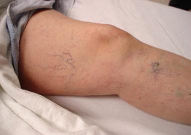
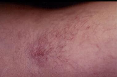
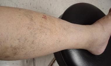
Sclerotherapy, in which a solution is injected into a vein, causing it to collapse, scar, and fade, remains the primary treatment for small-vessel varicose disease of the lower extremities. [1, 2] These small vessels include telangiectasias, venulectasias, and reticular ectasias. Telangiectasias are flat red vessels smaller than 1 mm in diameter. Venulectasias are blue, sometimes distended above the skin surface, and smaller than 2 mm in diameter. Reticular veins have a cyanotic hue and are 2-4 mm in diameter. Large varicosities do not respond as well as small varicosities to sclerotherapy. [3, 4] See the images below.
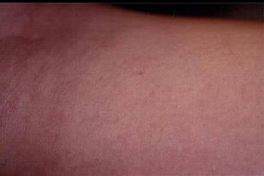
Treatment of telangiectasias, venulectasias, and reticular veins may greatly improve their appearance (see the image below). Treatment may also improve the associated painful symptoms. These vascular abnormalities are common. Telangiectasias are present in up to 28.9% of men and 40.9% of women. [5]
A single-center, prospective study by Cuffolo et al indicated that foam sclerotherapy is effective against venous ulcers. In varicose vein patients with venous ulceration, 3% sodium tetradecyl sulfate was administered to occlude incompetent superficial veins of over 3 mm, with compression treatment subsequently applied. The investigators found at 6-week follow-up that in 21% and 46.1% of patients, respectively, ulcers had either completely healed or undergone clinical improvement (with no additional venous incompetence). Among patients followed up at 1 year, healing had been maintained in 77.1% of ulcers. [6]
Etiology
Genetics and individual behavior patterns are important factors in the development venous disorders. Familial inheritance is reported in 15-40% of cases. Caucasians are most commonly affected. Pregnancy, prolonged standing, and prolonged walking also predispose people to venous disease. [7, 8]
The presence of clusters of reticular veins and telangiectasias on the lateral thigh area is called the lateral subdermic plexus of Albanese and is considered to be a remnant of embryonic development. The presence of clusters of telangiectatic veins on the medial or the lateral aspects of the ankle region is likely the result of incompetence in the great saphenous vein (medial) or the small saphenous vein (lateral). Finding a collection of telangiectatic veins along the medial thigh or knee areas should generate suspicion about an underlying incompetence in the great saphenous vein. Any concern about an underlying saphenous vessel insufficiency should warrant an investigation of the lower extremities by duplex ultrasonography.
Indications
The major indications for sclerotherapy are to improve cosmetic appearance and to reduce the associated symptoms such as pain and burning. Sclerotherapy can also be used to treatment any remnant tributaries after endovenous laser ablation of a saphenous or truncal vessel.
Visual sclerotherapy refers to the process of injecting a sclerosant into target veins without the aid of ultrasonography, whereas duplex-guided sclerotherapy (endovenous chemical ablation) is performed using duplex ultrasonography to guide the injections. This article discusses visual sclerotherapy only.
Relevant Anatomy
A thorough review the lower extremity venous system is essential before treatment is administered. Venous anatomy is very variable in some parts of the lower extremities but more constant in other parts. The lower extremity has both a superficial and a deep venous system. The deep venous system includes the femoral, popliteal, anterior tibial, posterior tibial, peroneal veins, and others. The superficial system is tremendously complex and extremely variable; it includes the great and short saphenous systems and other unnamed veins. The great and short saphenous veins occasionally connect by intersaphenous veins, such as the Giacomini vein. Several communicating vessels, called perforating veins, are present between the 2 superficial and deep systems. Occasionally, telangiectasias may communicate directly with the deep system.
Contraindications
Contraindications to sclerotherapy include pregnancy, thrombophlebitis, pulmonary emboli, hypercoagulable states, and allergy to the sclerosing agents.
-
Telangiectasias.
-
Reticular veins.
-
Venulectasias.
-
Venulectasias after sclerotherapy treatment.


