Overview
Wound healing is a complex and dynamic process of replacing devitalized and missing cellular structures and tissue layers. [1] The human adult wound healing process can be divided into 3 or 4 distinct phases. Earlier authors referred to 3 phases—inflammatory, fibroblastic, and maturation, [2] which has also been denoted as inflammatory, proliferation, and remodeling—and this is maintained by some authors. [3] In the 4-phases concept, there are the hemostasis phase, the inflammatory phase, the proliferation phase, and the remodeling phase. In the 3-phases approach, the hemostasis phase is contained within the inflammatory phase.
Not only do authors vary the number of phases, they also denote differences in the phase descriptors used; they may designate phases as the hemostasis phase, inflammatory phase, proliferation phase, and remodeling phase, or they may refer to the hemostasis phase, inflammatory phase, granulation phase, and maturation phase. [4] Therefore, certain phases have more than one name, such as remodeling or maturation and proliferation or granulation. [5] As our understanding of wound healing progresses, further phases and subphases may well be delineated.
Within these broad phases are a complex and coordinated series of events that includes chemotaxis, phagocytosis, neocollagenesis, collagen degradation, and collagen remodeling. In addition, angiogenesis, epithelization, and the production of new glycosaminoglycans (GAGs) and proteoglycans are vital to the wound healing milieu. The culmination of these biological processes results in the replacement of normal skin structures with fibroblastic mediated scar tissue. For more information on wound healing, visit Medscape’s Wound Management Resource Center.
This process can go awry and produce an exuberance of fibroblastic proliferation with a resultant hypertrophic scar, which by definition is confined to the wound site. Further exuberance can result in keloid formation (see image below), in which scar production extends beyond the area of the original insult. The collagen is thicker, more irregularly arranged, and more often causes pain. In a hypertrophic scar, the collagen is thinner and arranged more parallel to the wound. Furthermore, hypertrophic scars occur in all races, although less so in young and elderly persons. Hormonal changes may have an impact. Keloid scarring is more often seen in nonwhite persons. [6]
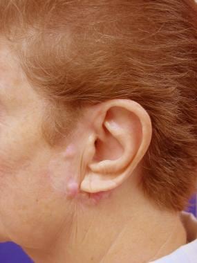 A patient referred for keloid formation after excision of facial cancer and reconstruction.
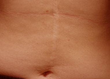
Conversely, insufficient healing can result in a hypotrophic or atrophic scar formation (see image below). All wounds in adult skin heal with a scar. The degree of inflammation has a direct impact on the ultimate scar formation. [7]
Types of Wound Healing
Although various categories of wound healing have been described, the ultimate outcome of any healing process is repair of a tissue defect.
Primary healing, delayed primary healing, and healing by secondary intention are the 3 main categories of wound healing. Even though different categories exist, the interactions of cellular and extracellular constituents are similar.
A fourth category is healing that transpires with wounds that are only partial skin thickness. [8]
Categories of Wound Healing
Category 1
Primary wound healing, or healing by first intention, occurs within hours of repairing a full-thickness surgical incision. This surgical insult results in the mortality of a minimal number of cellular constituents.
Category 2
If the wound edges are not reapproximated immediately, delayed primary wound healing transpires. This type of healing may be desired in the case of contaminated wounds. By the fourth day, phagocytosis of contaminated tissues is well underway, and the processes of epithelization, collagen deposition, and maturation are occurring. Foreign materials are walled off by macrophages that may metamorphose into epithelioid cells, which are encircled by mononuclear leukocytes, forming granulomas. Usually the wound is closed surgically at this juncture, and if the "cleansing" of the wound is incomplete, chronic inflammation can ensue, resulting in prominent scarring.
Category 3
A third type of healing is known as secondary healing, or healing by secondary intention. In this type of healing, a full-thickness wound is allowed to close and heal. Secondary healing results in an inflammatory response that is more intense than with primary wound healing. In addition, a larger quantity of granulomatous tissue is fabricated because of the need for wound closure. Secondary healing results in pronounced contraction of wounds. Fibroblastic differentiation into myofibroblasts, which resemble contractile smooth muscle, is believed to contribute to wound contraction. These myofibroblasts are maximally present in the wound from the 10th-21st days.
Category 4
Epithelialization is the process by which epithelial cells migrate and replicate via mitosis and traverse the wound. This occurs as part of the phases of wound healing, which are discussed in Sequence of Events in Wound Healing. In wounds that are partial thickness, involving only the epidermis and superficial dermis, epithelization is the predominant method by which healing occurs. Wound contracture is not a common component of this process if only the epidermis or epidermis and superficial dermis are involved.
Overview of Wound Healing
The amalgam of coordinated events that constitute the process of wound healing is quite complex. The steps in the procession of wound healing include inflammation, the fibroblastic phase, scar maturation, and wound contracture. [9, 10] Wound contracture is a process that occurs throughout the healing process, commencing in the fibroblastic stage. [9]
The inflammatory phase occurs immediately following the injury and lasts approximately 6 days. The fibroblastic phase occurs at the termination of the inflammatory phase and can last up to 4 weeks. Scar maturation begins at the fourth week and can last for years. [9]
An analogous system depicts the 4 phases as hemostasis, inflammation, granulation, and remodeling in a continuous symbiotic process. [11] This is the phase system used in this text.
Sequence of Events in Wound Healing
Following tissue injury via an incision, the initial response is usually bleeding. The cascade of vasoconstriction and coagulation commences with clotted blood immediately impregnating the wound, leading to hemostasis, and with dehydration, a scab forms. An influx of inflammatory cells follows, with the release of cellular substances and mediators. Angiogenesis and reepithelialization occur and the deposition of new cellular and extracellular components ensues.
Initial phase - Hemostasis
The initial injury results in an outflow of blood and lymphatic fluid. This is also the process during which the initial reparative coagulum is created. Both the intrinsic and extrinsic clotting mechanisms are activated. The intrinsic mechanism is enjoined from the thrombocytes and the extrinsic mechanism from the injured tissues. Following vasoconstriction, platelets adhere to damaged endothelium and discharge adenosine diphosphate (ADP), promoting thrombocyte clumping, which dams the wound. With the short-lived vasoconstriction complete, the vessels dilate allowing the influx of more thrombocytes and other blood cells.
At this stage, one can think of the commencement of the inflammatory phase. Although some speak of a separate inflammatory phase, it commences during the hemostasis phase, again providing evidence of the overlapping nature of the healing compendium. These thrombocytes, as well as the recruited white blood cells, release numerous factors to ramp up the healing process. Alpha-granules liberate platelet-derived growth factor (PDGF), platelet factor IV, and transforming growth factor (TGF)–β). The processes of inflammation, collagen degradation and collagenogenesis, myoblastic creation from transformed fibroblasts, growth of new blood vessels, and reepithelialization have all commenced.
These processes are mediated by a host of cytokines and growth factors. The interleukins strongly influence the inflammatory process. Vascular endothelial growth factor (VEGF) and other factors enhance blood vessel formation, and some have multiple roles such as fibroblast growth factor (FGF)–2, which affects not only the process of angiogenesis but also that of reepithelialization. Vasoactive amines such as histamine and serotonin are released from dense bodies found in thrombocytes. PDGF is chemotactic for fibroblasts and, along with TGF-β, is a potent modulator of fibroblastic mitosis, leading to prolific collagen fibril construction in later phases. Fibrinogen is cleaved into fibrin, and the framework for completion of the coagulation process is formed. Fibrin provides the structural support for cellular constituents of inflammation. This process starts immediately after the insult and may continue for a few days.
Second phase - Inflammation
As the hemostasis phase can be construed to consist of an early and a late phase, the early phase being bleeding and hemostasis and the late phase being coagulation, so is it also with inflammation. While the inflammatory phase commences during the hemostasis phase, the early component of the inflammatory phase is predominated by the influx of the polymorphonuclear leukocytes (PMNs) and the later component predominated by monocytes/macrophages.
Within the first 6-8 hours, the next phase of the healing process is underway, PMNs engorging the wound. TGF-β facilitates PMN migration from surrounding blood vessels, where they extrude themselves from these vessels. These cells cleanse the wound, clearing it of debris. The PMNs attain their maximal numbers in 24-48 hours and commence their departure by hour 72. Other chemotactic agents are released, including FGF, TGF-β and TGF-α, PDGF, and plasma-activated complements C3a and C5a (anaphylactic toxins). They are sequestered by macrophages or interred within the scab or eschar. [12]
As the process continues, monocytes also exude from the vessels. These are termed macrophages once they leave the vessel. The macrophages continue the cleansing process and manufacture various growth factors during days 3-4. The macrophages orchestrate the multiplication of endothelial cells with the sprouting of new blood vessels, the duplication of smooth muscle cells, and the creation of the milieu created by the fibroblast. Many factors influencing the wound healing process are secreted by macrophages. These include TGFs, cytokines and interleukin 1 (IL-1), tumor necrosis factor (TNF), and PDGF.
Third phase - Granulation/proliferation
This phase consists of different subphases. These subphases do not happen in discrete time frames but constitute an overall and ongoing process. The subphases are fibroplasia, matrix deposition, angiogenesis and reepithelialization. [11]
In days 5-7, fibroblasts have migrated into the wound, laying down new collagen of the subtypes I and III. Early in normal wound healing, type III collagen predominates but is later replaced by type I collagen.
Tropocollagen is the precursor of all collagen types and is transformed within the cell's rough endoplasmic reticulum, where proline and lysine are hydroxylated. Disulfide bonds are established, allowing 3 tropocollagen strands to form a triple left-handed triple helix, termed procollagen. As the procollagen is secreted into the extracellular space, peptidases in the cell wall cleave terminal peptide chains, creating true collagen fibrils.
The wound is suffused with GAGs and fibronectin produced by fibroblasts. These GAGs include heparan sulfate, hyaluronic acid, chondroitin sulfate, and keratan sulfate. Proteoglycans are GAGs that are bonded covalently to a protein core and contribute to matrix deposition.
Angiogenesis is the product of parent vessel offshoots. The formation of new vasculature requires extracellular matrix and basement membrane degradation followed by migration, mitosis, and maturation of endothelial cells. Basic FGF and vascular endothelial growth factor are believed to modulate angiogenesis.
Reepithelization occurs with the migration of cells from the periphery of the wound and adnexal structures. This process commences with the spreading of cells within 24 hours. Division of peripheral cells occurs in hours 48-72, resulting in a thin epithelial cell layer, which bridges the wound. Epidermal growth factors are believed to play a key role in this aspect of wound healing.
This succession of subphases can last up to 4 weeks in the clean and uncontaminated wound.
Fourth phase - Remodeling/maturation
After the third week, the wound undergoes constant alterations, known as remodeling, which can last for years after the initial injury occurred. Collagen is degraded and deposited in an equilibrium-producing fashion, resulting in no change in the amount of collagen present in the wound. The collagen deposition in normal wound healing reaches a peak by the third week after the wound is created. Contraction of the wound is an ongoing process resulting in part from the proliferation of the specialized fibroblasts termed myofibroblasts, which resemble contractile smooth muscle cells. Wound contraction occurs to a greater extent with secondary healing than with primary healing. Maximal tensile strength of the wound is achieved by the 12th week, and the ultimate resultant scar has only 80% of the tensile strength of the original skin that it has replaced.
Summary
The process of trying to understand wound healing traces back to ancient times [6] and has continued to be investigated. Interest grew in the 1900s, and, by 1960, it was understood that wound healing time could be decreased up to 50% if appropriate dose-dependent settings are created. [13] Continuing from that time, there has been an ongoing expansion to not only understand the vast array of intrinsic and extrinsic factors of wound healing, but also the intracellular, extracellular, molecular, and biochemical processes and interactions that facilitate healing.
The process of wound healing constitutes an array of interrelated and concomitant events. Understanding of these processes and effectors on these processes continues to expand.
Future and Controversies
Future advances in wound healing will focus on affecting the agents that influence the processes involved in the repair of damaged tissue. Laser techniques, nonlaser techniques, and other modalities are being explored to enhance the proliferation of cells, the migration of cells, and the acceleration of the healing of wounds. [14, 15, 16]
Human cell–conditioned media developed in embryologiclike conditions has been shown to improve healing times in postlaser facial skin. [17] Fetal tissue can heal scarless due to the unique characteristics of fetal epithelial and mesenchymal cells and the functioning of the fetal immune system. [18] The inclusion of transforming growth factor (TGF)–β3 during the healing of wounds in adults can be beneficial. [19] TGFs -β1, -β2, and -β3 all have significant roles in wound healing, and the simple addition, subtraction, or ratio of these growth factors may not be fully explanatory for scarless healing. [20]
Hyperbaric oxygen has also been used to promote healing. [21]
Agents such as platelet-rich plasma (PRP) and erythropoietin (EPO) are modulators that have a positive effect on tissue regeneration and have been used successfully to enhance the healing of wounds. [22, 23, 24] In a study of wound healing in patients who had undergone maxillofacial surgery, Menchisheva et al found that individuals to whom PRP was applied during surgery demonstrated an earlier appearance and increase in fibroblasts, macrophages, and collagen fibers than did controls. Moreover, on postoperative days 1 and 5, wound fluid in the treatment group had a greater concentration of IL-1β and TNF-α than did that of the control group, a manifestation, according to the investigators, of PRP’s effects on healing’s inflammatory and granulation phases. [25]
A study by Misiura et al suggests that PRP promotes wound healing by activating a complex of growth factors and adhesion receptors, so that cell proliferation, migration, and collagen biosynthesis are stimulated. According to the study’s findings, activation of the epidermal growth factor receptor (EGFR) via PRP brings about peptidase-D (PEPD)–dependent proliferation of human keratinocytes. [26]
A study by Hoeferlin et al suggested that although peptide growth factors are considered essential to the wound-healing properties of PRP, the lipid fraction of PRP also plays an important role in this, by aiding in the proliferation and migration of primary adult human dermal fibroblasts and overcoming the suppression of fibroblast proliferation by chronic wound fluid. [27]
Nutritional factors are also critical for proper wound healing. Improvement in the nutritional status of adults correlates with enhanced wound healing. [28, 29, 30]
A literature review by Seth et al looking at the effect of nutrition on surgical wound healing reported that enhanced wound healing and immune function are associated with intake of omega-3 fatty acids and specific amino acids, including glutamine. Moreover, wound-healing stages were found to be positively impacted by vitamins A, B, and C and by zinc, while results concerning the effects of vitamin E were variable. It was further indicated that polyphenolic compounds can aid recovery through their anti-inflammatory function. The investigators also stated that increased postoperative complications and infections could be linked to malnutrition and that “preoperative nutritional support correlated with reduced hospital stays and complications.” [31]
Honey has been shown to be less beneficial in wound healing, despite its use since early times, and has even caused delayed healing in certain types of wounds. [32] A literature review by Jull et al found evidence that treatment with honey causes partial-thickness burns to heal more quickly than they would with conventional management and that infected postoperative wounds may heal more quickly with honey than with antiseptics and gauze, but the investigators determined evidence for honey’s effects on other wounds to be of low or very low quality. [33]
A literature review by Beitz, utilizing narrative reviews, clinical trials, and animal studies, indicated that medications with the greatest risk of negatively affecting wound healing and skin integrity include antibiotics, anticonvulsants, angiogenesis inhibitors, steroids, and nonsteroidal anti-inflammatory drugs. Drugs that may aid wound healing, on the other hand, include ferrous sulfate, insulin, thyroid hormones, and vitamins. [34]
In a rat model, pine bark from the Turkish pine (Pinus brutia) increased the rate of wound healing. [35] Aloe arborescens has enhanced healing properties when compared to Aloe vera. [36]
Appropriate neurologic stimulation is also important in the healing of wounds. In a 2013 report, it was shown that capsaicin-induced nerve damage resulted in small-fiber neuropathy and was associated with slower healing in shallow wounds not deep wounds. [37]
Reparative strategies involving engineered tissue matrices, either exogenous or endogenous, have also been used. [22]
Stem cells continue to be a new frontier of research in the armamentarium of wound healing strategies. [38] There still exists controversies with the use of fetal stem cells. Stem cells, in particular adipose-derived stem cells, have been shown to ameliorate wound healing, and continued research in these areas appears promising. [39, 40] Exogenous mesenchymal–derived stem cells, commonly obtained from bone marrow, but available from other sources, are being used in the setting of nonhealing inflammatory wounds. [3] Third trimester human fetal placental chorionic stem cells did not compare favorably with first semester ones in promoting wound healing. [41] . However, first-, [42] and more recently mid-, [43] trimester amniotic stem cells, when treated using valproic acid, obtained pluripotentialism.
-
A long-standing hypotrophic scar. Patient had abdominal surgery as a child.
-
A patient referred for keloid formation after excision of facial cancer and reconstruction.