Practice Essentials
Of all tumors in the hand, 1-2% are malignant. These can be divided into primary and metastatic tumors. Primary malignant tumors are subdivided further into skin tumors, musculoskeletal tumors, and soft tissue tumors. While malignant hand tumors are relatively uncommon, the incidence of metastatic tumors is exceedingly rare. [1] An incidence rate of 0.0016-0.5% of neoplastic metastasis to the skin of the hand has been reported.
In order of decreasing incidence, squamous cell carcinoma (SCC), basal cell carcinoma (BCC), basosquamous cell carcinoma, and melanoma account for 90% of primary malignant tumors of the hand. Other malignant skin lesions include dermatofibrosarcoma protuberans, Kaposi sarcoma, sweat gland tumors, and Merkel cell carcinoma.
Bone and soft tissue malignancies of the hand are much less common than cutaneous malignant tumors. Malignant bone tumors that occur in the hand include chondrosarcomas, osteogenic sarcomas, and Ewing sarcoma. Additional soft tissue malignant tumors include epithelioid sarcoma, synovial sarcoma, liposarcoma, fibrosarcoma, malignant fibrous histiocytoma, malignant schwannoma, rhabdomyosarcoma, leiomyosarcoma, vascular leiomyosarcoma, angiosarcoma, lymphangiosarcoma, and clear cell sarcoma.
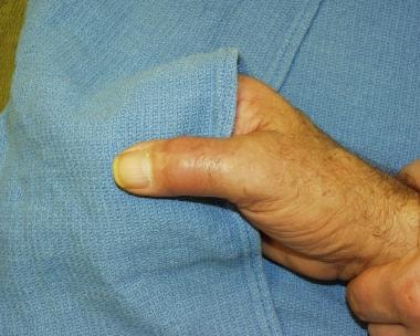
Lumps and bumps at the hands are quite common. Most of the time, such growths are benign tumors; however, a high index of suspicion is always imperative to avoid the misdiagnosis of a malignant tumor (see image below). [2, 3]
A retrospective study by Cavit et al found that out of 402 cases of hand tumors, 10 (2.5%) were malignant, including 6 out of 361 soft tissue tumors (1.7%). Five of the soft tissue malignancies were squamous cell tumors (83.3%), and one was a synovial sarcoma (16.7%). The malignant osseous tumors consisted of two primary lesions (an osteosarcoma and a chondrosarcoma) and two that had metastasized to the hand (one from a lung carcinoma and the other from a chondrosarcoma). [4]
Workup in malignant hand tumors
A complete history and physical examination with special attention to the affected limb, supplemented by radiologic studies and biopsies, are fundamental to obtaining the correct diagnosis of and and administering the proper treatment for malignant hand tumors. [5]
Cutaneous lesions
Evaluating cutaneous lesions may include radiographic studies of the chest, computed tomography (CT) or magnetic resonance imaging (MRI) scans, and evaluation of other anatomic structures.
Laboratory studies may include alkaline phosphatase, calcium, and liver function tests.
Tissue biopsy is the next step in diagnosis.
Malignant soft tissue tumors
Laboratory studies may include measuring serum calcium, phosphorous, and alkaline phosphatase levels to assess for possible bony metastasis. Other helpful laboratory studies include a hematology profile if considering an infectious etiology, a rheumatoid panel if an arthritide is suggested, and an erythrocyte sedimentation rate to assess for inflammation.
Images from plain radiographic studies offer a good degree of bony definition with possible bony involvement of a lesion. If findings of bony invasion are equivocal on plain radiographs, CT scanning is the next logical step in assessing the lesion.
MRI can be used to determine the area of gross involvement when assessed in various views (eg, coronal, axial, sagittal), to better define lesions prior to operative intervention.
Bone scans can be useful in finding hot spots or areas of increased radioisotope uptake.
Ultrasonography is effective in determining if a lesion is solid or cystic. It can assist in defining the type of biopsy that may be pursued on a lesion.
Plan carefully prior to biopsy because an excisional biopsy may lead to a cure. [6]
Malignant bone tumors
These include the following:
-
Chondrosarcoma - After a well-planned incisional biopsy, patients must be staged appropriately to determine if adjunctive therapy is indicated
-
Osteogenic sarcoma - Plain radiographs illustrate intramedullary lesions with lytic and sclerotic components; a well-planned incisional biopsy precedes treatment
-
Ewing sarcoma - MRI studies can assist in defining bone and soft tissue involvement
Management of malignant hand tumors
Adequate resection is the key for treatment of malignant skin neoplasms. Clearly, minimizing the deformity created is important, but the goal must be removal of the malignant tissue, which may require later stages of reconstruction. Achieve complete resection of the lesion with clear histologic margins in normal surrounding tissue.
Classification
Staging is an important aspect in evaluating a malignant tumor in order to describe the degree of extension of the disease. The tumor can be staged once all the clinical, radiographic, and histologic data have been gathered. The stage of the tumor determines the extent of resection. Basically, the criteria for staging of malignant tumors includes anatomic location (T), lymph nodes involvement (N), presence of metastases (M), and histologic grade (G) in case of sarcomas.
Primary Malignant Tumors - Cutaneous
The skin is by far the most common site for malignant tumors of the hand. In order of decreasing incidence, squamous cell carcinoma (SCC), basal cell carcinoma (BCC), basosquamous cell carcinoma, and melanoma account for 90% of primary malignant tumors of the hand. Other malignant skin lesions include dermatofibrosarcoma protuberans, Kaposi sarcoma, sweat gland tumors, and Merkel cell carcinoma. For more information on these and other skin cancers, see Medscape’s Skin Cancer Resource Center.
Squamous cell carcinoma
Squamous cell carcinoma (SCC) is the most common primary malignant tumor of the hand, accounting for 75-90% of hand malignancies. Risk factors include solar radiation, ionizing radiation, chronic inflammation, immunosuppression, xeroderma pigmentosa, Bowen disease, leukoplakia, human papillomavirus, and smoking.
Clinical presentation ranges from small hyperkeratotic, erythematous to ulcerated exophytic large tumors. Tumors often manifest as small firm nodules with indistinct margins or plaques. The surface may have various irregularities ranging from smooth to verruciform to ulcerated. Skin coloration is often brown to tan. Scaling, bleeding, and crusting may also occur. Typically, SCC is locally invasive; however, metastatic rates of up to 20% have been reported in more aggressive lesions such as in radiation wounds and burn scars (Marjolin ulcers).
A single-institution, retrospective cohort study by Mull et al found that among patients with cutaneous SCC, the cancer occurred on the hand more commonly in immunosuppressed individuals than in immunocompetent persons (38% vs 24% of cases, respectively). [7]
Surgical excision is the standard treatment. For invasive SCC less than 2 cm, a 4 mm surgical margin is recommended; for those greater than 2 cm, a 6 mm margin is recommended. A 95% cure rate has been reported with these margins. Because of the low incidence of lymph node metastasis (2-5%), an elective lymph node dissection is not recommended unless the axillary nodes are clinically enlarged.
A study by Park et al suggested that arterialized venous free flaps are effective in the management of skin cancer of the digits. Of 12 patients who received the flaps—following excision of SCC (7 patients) or malignant melanoma (5 patients)—complete flap survival occurred in nine cases and partial necrosis in three patients, although only one of the necrosis cases required skin grafting. [8]
A study by Askari et al indicated that despite a low metastasis rate, SCC of the hand has a high local recurrence rate. The study, which involved 86 patients with SCC tumors of the hand and a mean follow-up period of 6.4 years, found that the 5- and 10-year recurrence-free survival rates were 67% and 50%, respectively, while the metastasis rate was 4%. Risk factors for recurrence included web space location, the presence of bilateral or multiple tumors, and a prior history of SCC. [9]
Basal cell carcinoma
Basal cell carcinoma (BCC) accounts for 3-12% of hand malignancies, second to SCC. Usually, they are slow-growing tumors that may present as areas of skin atrophy, pink to reddish discoloration, telangiectatic changes, and, ultimately, ulceration with an elevated pearly border. Risk factors are similar to those for SCC and include chronic solar radiation, fair skin, immunosuppression, inorganic arsenic exposure, and Gorlin syndrome.
Several variants of BCC have been described. Nodular BCC is the most common form. Other variants, such as superficial, infiltrating, morpheic, pigmented, basal-squamous, adenoid, cystic, keratotic, and fibroepithelioma are less common. Metastasis occurs at a rate of 0.0028-0.1%.
A study by Subramaniam et al found that for BCC, the relative tumor density (“the proportion of tumors occurring at a specified site” divided by “the proportion of skin area of that site”) on the hand is low, while being high on areas receiving less sun exposure. According to the investigators, this suggests that the relationship between sun exposure and the development of BCC is complex. [10]
Similarly, Loh et al stated that the fact that BCC occurs only rarely on the dorsal hand, even though this area receives intense sun exposure, suggests that the pathogenesis of BCC is affected by factors besides ultraviolet radiation. [11]
The standard treatment involves surgical excision. At least a 4 mm margin should be included. Mohs microdermographic surgery is rarely indicated in the hand. Other treatment options include curettage with electrodesiccation, cryosurgery, and radiation.
Melanoma
Melanoma accounts for approximately 3% of primary malignant hand tumors. The increase in incidence appears to be greater than any other malignancy. Risk factors include solar radiation, previous dysplastic nevi, fair skin, family history of melanoma, and congenital nevi.
Various subtypes exist, including lentigo maligna melanoma, intraepidermal malignant melanoma, superficial spreading malignant melanoma, acral lentiginous malignant melanoma, nodular malignant melanoma, and desmoplastic melanoma. Lesions with increased growth, that change in color or shape, that have irregular borders, and that are greater than 0.5 cm are suggestive of melanoma. Any pigmented lesion under the nail plate with no previous history of trauma requires a biopsy because of the possibility of a subungual melanoma. This usually affects the thumb or the great toe.
Survival is related to the thickness of the lesion (in mm) (Breslow thickness; also see Skin Malignancies, Melanoma) in malignant melanoma without metastasis. Breslow classification has 4 groups. Tumors in group 1 have a depth of invasion of less than 0.76 mm. Group 2 tumors have a depth of invasion of 0.76 to 1.5 mm. Those in group 3 have a depth of invasion of 1.51 to 4 mm. Group 4 tumors have a depth of invasion greater than 4 mm. Because of the close proximity of the nail matrix to the distal phalanx, this system is not reliable for subungual melanoma.
The optimal management of melanoma requires a multidisciplinary approach. The principal treatment is surgical excision or amputation, especially in subungual melanoma. The tumor thickness is used to determine the surgical margins. For in situ lesions, a 5-mm margin is adequate. For invasive lesions less than 1 mm in thickness, a 1 cm margin is recommended. For thickness of 1-2 cm, a 1-2 cm margin is recommended, while for those over 2 cm in thickness, a 2-3 cm margin is recommended. For clinically palpable regional lymphadenopathy, an elective lymph node dissection should be provided. Otherwise, a sentinel lymph node dissection can be performed, especially for melanoma of intermediate thickness (1-4 mm).
Dermatofibrosarcoma protuberans
Dermatofibrosarcoma protuberans (DFSP) is an uncommon low-grade malignant tumor that arises in the dermis. Most often presents as a painless violet-red plaque or nodule. May present for several years and history of a traumatic injury is common. Treatment consists of surgical excision with 3 cm margin including the deep fascia to minimize the risk of recurrence.
Kaposi sarcoma
Kaposi sarcoma is a vascular malignancy of lymphatic endothelial cells. The lesion manifests as a painless, violet nodule or plaque. Often, edema is associated with this lesion. The classic patient population is of Eastern European Jewish descent; however, with the rise in HIV infection, these lesions are encountered in many other groups. Patients who are HIV positive often have more aggressive clinical disease with widespread lesions and internal organ involvement. Therapy consists of radiotherapy, cryosurgery, laser surgery, or primary excision.
Eccrine sweat gland malignant tumors
Eccrine sweat gland malignant tumors in the hand are rare. They manifest as slow-growing painless lesions. Typically, the patient is elderly and the lesion can range from nodule to plaque or ulcerated tumor. The lesion is usually locally invasive, and the risk of metastasis is based on the histologic grade. Treatment consists of wide local excision with a margin of normal tissue. These tumors are not responsive to radiation or chemotherapy.
Merkel cell carcinoma
Merkel cell carcinoma is a rare and aggressive cutaneous neoplasm observed in sun-exposed areas. Older patients with long-term solar radiation exposure are at greatest risk. The tumor originates in the basal layer of the epidermis and it is described as a rapidly growing, painless, bluish-red intracutaneous nodule. Treatment is wide surgical excision with regional lymphadenectomy. Postoperative radiation and chemotherapy should be considered because of the high incidence of metastasis and recurrence. [12]
A study by Broida et al indicated that even with aggressive local treatment and the use of adjuvant radiotherapy to the primary tumor site and regional lymph nodes, Merkel cell carcinoma of the hand and wrist has, within 5 years post diagnosis, high recurrence and mortality rates. Five-year recurrence and disease-specific mortality rates in the study were approximately 45% and 33%, respectively, among patients with hand and/or wrist Merkel cell carcinoma, despite wide local incision in 22 of the cohort’s 25 patients (including one amputation). Seventeen patients received adjuvant radiation to the primary site, and six patients (with evidence of nodal disease), to the regional lymph node basin. Three patients underwent adjuvant chemotherapy. [13]
Primary Malignant Tumors - Bone and Soft Tissue
Bone and soft tissue malignancies of the hand are much less common than cutaneous malignant tumors. Malignant bone tumors that occur in the hand include chondrosarcomas, osteogenic sarcomas, and Ewing sarcoma. Additional soft tissue malignant tumors include epithelioid sarcoma, synovial sarcoma, liposarcoma, fibrosarcoma, malignant fibrous histiocytoma, malignant schwannoma, rhabdomyosarcoma, leiomyosarcoma, vascular leiomyosarcoma, angiosarcoma, lymphangiosarcoma, and clear cell sarcoma.
A study by Ahmed et al of patients with hand/wrist and foot/ankle sarcomas who were treated with a combination of limb-sparing surgery and radiation therapy reported a 5-year local tumor control rate of 88%. Functional outcomes were considered acceptable among survivors aged 18 years or older who did not undergo subsequent amputation, with the mean score on the Michigan Hand Outcomes Questionnaire (MHQ) being 72.8. [14]
Chondrosarcoma
Chondrosarcoma is the most common primary malignant bone tumor of the hand. This tumor originates from the cartilage cells in joints and may arise in a previously benign lesion, such as enchondroma or osteochondroma. [15] Men are affected more often than women (male-to-female ratio of 2:1), and it is observed most frequently in individuals older than 60 years.
The metacarpals and proximal phalanges are affected most commonly. [16] Clinically, chondrosarcoma manifests as a slow-growing, firm, and painful mass. Radiographically, it is characterized by poorly defined lesions with stippled calcifications at the matrix and areas of lysis. Most chondrosarcomas are locally invasive, but rarely metastasize with a risk of 10%. After a well-planned incisional biopsy, patients must be staged appropriately to determine if adjunctive therapy is indicated. Most commonly, it metastasizes to the lungs. [17] Treatment consists of surgical excision or amputation. Chemotherapy or radiation therapy has no role in the treatment of this tumor.
Osteogenic sarcoma
Osteogenic sarcoma (osteosarcoma) is a highly malignant bone tumor characterized by the formation of neoplastic bone tissue. It is the most common childhood malignant bone tumor but is rarely observed in the hand. Osteogenic sarcoma in the hand often manifests in patients older than 40 years, and, typically, the proximal phalanges and metacarpals are involved.
These lesions may manifest as rapidly enlarging painful masses. Plain radiographs illustrate intramedullary lesions with lytic and sclerotic components. Periosteal elevation with new bone formation and soft tissue involvement is a common finding. After a well-planned incisional biopsy, treatment consists of wide local excision or amputation. Adjuvant chemotherapy, which improves survival, can be performed prior to or following surgical treatment. External-beam radiation has no role in the treatment of these lesions.
Ewing sarcoma
Ewing sarcoma is an uncommon aggressive malignant bone tumor composed of small, round cells of uncertain origin. The tumor more often arises in long bones of children and is rarely found in the hand. The most common areas of involvement in the hand are the metacarpals or phalanges. It has a consistent chromosomal abnormality of reciprocal translocation of chromosomes 11 and 12. In addition to pain and swelling, this tumor can cause fever and leukocytosis. Radiographs reveal an "onion skin" pattern, representing a circumferential layer of aggressive periosteal reaction with an associated lytic lesion of the cortex. All these findings can lead to the misdiagnosis of osteomyelitis. Magnetic resonance imaging (MRI) studies can assist in defining bone and soft tissue involvement.
Treatment consists of induction chemotherapy, wide surgical excision, and then maintenance chemotherapy. [18] Multiagent chemotherapy has improved survival rates from less than 10% to more than 50%. These tumors are radiosensitive; however, this modality is not appealing in young patients.
Epithelioid sarcoma
Epithelioid sarcoma is the most common soft tissue sarcoma in the hand. This lesion frequently manifests as a painless nodule, which may ulcerate, on the volar aspect of the hand. It is often misdiagnosed as an infection or Dupuytren nodule. Epithelioid sarcoma tends to spread proximally from its origin and carries a high risk of nodal metastases. Wide local excision or amputation followed by radiation therapy offers the best chance for cure. Some authors have suggested elective lymph node dissection vs. sentinel node biopsy even without clinical nodal disease. Chemotherapy should be considered for those with recurrence, involvement of the lymph nodes, or metastasis.
Synovial sarcoma
Synovial sarcoma is a highly malignant soft tissue tumor that arises in the region of a joint, tendon, or bursae. The tumor typically manifests as a painful or tender mass near a joint, often in the carpus. The lesion may have a long latent period with slow growth or may have a rapid growth phase. Plain radiographs often demonstrate soft tissue calcifications. This tumor metastasizes via lymphatic spread, which occurs in 25% of patients. Treat synovial sarcoma with wide local excision or amputation followed by radiation therapy, which decreases local recurrence.
Liposarcoma
Liposarcoma is a soft tissue sarcoma that is rarely observed in the hand. Histologic subtypes include well-differentiated, myxoid, pleomorphic, round cell, and dedifferentiated subtypes. These lesions can be confused clinically with a benign lipoma, but more often they are painful and grow more rapidly. After a well-planned tissue biopsy, wide excision or amputation followed by adjuvant radiation is recommended. Chemotherapy may be considered for high-grade lesions or metastasis.
Fibrosarcoma
Fibrosarcoma is a malignant tumor of fibroblasts. These arise from connective tissue such as fascia, periosteum, or tendons. Clinically, this lesion appears as a soft tissue mass that exerts local effects by compression of adjacent structures. Treatment for fibrosarcomas is similar to that for other soft tissue malignancies of the hand and includes wide excision and radiation therapy.
Malignant fibrous histiocytoma
Malignant fibrous histiocytoma is a malignant soft tissue tumor with foci of macrophage differentiation. Few reports exist of this lesion occurring in the hand. Treatment is similar to that for the tumors described above, with wide excision or amputation and adjuvant radiation therapy.
Malignant peripheral nerve sheath tumors
Malignant peripheral nerve sheath tumors include those tumors of neural origin. Of these tumors, 50% occur in individuals with neurofibromatosis. This tumor grows along and within the peripheral nerve. The clinical manifestation is a soft tissue mass that often is painful to palpation. Treatment consists of wide excision. [19] A high incidence of local recurrence is typical.
Rhabdomyosarcoma
Rhabdomyosarcoma is rare in the upper extremities, but it is the most common soft tissue sarcoma in children. Rhabdomyosarcoma arises from skeletal muscle and is divided into 4 subtypes, embryonal, botryoid, alveolar, and pleomorphic. Alveolar rhabdomyosarcoma is the most common in the hand. Clinically, this manifests as an enlarging, painful mass. Associated paresthesias can occur secondary to local nerve compression. Often, the patient has a history of associated trauma. Treatment has improved in recent years, although survival rates have been reported to be 6% in 4 years. Local and regional lymphatic spread is common, and metastasis to the heart, skeletal bone, lungs, pancreas, liver, and kidneys can occur. Early diagnosis with appropriate surgical resection or amputation followed by radiation and chemotherapy improves outcome.
Leiomyosarcoma
Leiomyosarcoma is a malignant tumor that arises from smooth muscle cells. These lesions are rarely observed in the extremities. They reportedly appear as a solitary soft tissue mass, although they may be lobulated. Epidermal changes with discoloration or ulceration may be present. After appropriate staging, treatment involves excision with adjuvant chemotherapy and radiation therapy. Reportedly, 5-year survival rates are 70-80%, with metastatic disease observed most commonly in the lungs.
Vascular leiomyosarcoma
Vascular leiomyosarcoma arises from large arteries or veins and has been reported in the hand. This lesion manifests as an edematous mass, and mass effect can cause local symptoms. Treatment consists of wide local excision. Reportedly, 50% of patients have metastatic disease at the time of surgery.
Angiosarcoma
Angiosarcoma is an aggressive, highly malignant tumor composed of neoplastic endothelial cells. No sex predominance is recognized, and they can occur in persons of any age. Most tumors undergo slow growth with central necrosis, which reveals overlying skin changes. Treatment consists of wide local excision.
Lymphangiosarcoma
Lymphangiosarcoma is a rare malignancy that reportedly develops in 0.1% of patients with lymphedema of the arm following radical mastectomy. Clinically, this manifests as multiple purplish nodules in the affected brawny skin. Wide local excision is the best treatment, but this tumor is associated with a poor prognosis.
Clear cell sarcoma
Clear cell sarcoma is an uncommon soft tissue sarcoma that reportedly occurs near tendons and aponeurosis. It has been characterized as "the malignant melanoma of soft parts" and may be misdiagnosed as metastatic melanoma. This tumor is aggressive, with regional lymphatic invasion commonly observed. For this reason, perform lymph node biopsies on these patients. Treatment, as with most of these sarcomas, consists of wide local excision.
Evaluation - Cutaneous Lesion
History
Obtain a thorough history, including changes in color, size, pain at the site, bleeding, scaling, or anything else the patient may have noticed. Question the patient about habits in regard to solar exposure, the use of sunscreens, and exposure to ionizing radiation. Importantly, determine if the patient has had other cutaneous malignancies excised. Also obtain a family history of malignancies.
Physical
Focus the physical examination on the lesion. Determine the size, color, and any border irregularity, and assess adjacent skin and structures. Magnification of the lesion under excellent lighting can assist in the evaluation. Palpate adjacent lymph nodes to assess for lymphadenopathy, possibly indicating spread of the malignancy.
Workup
Beyond a careful history and physical examination, evaluating cutaneous lesions may include radiographic studies of the chest, computed tomography (CT) or MRI scans, and evaluation of other anatomic structures. Laboratory studies may include alkaline phosphatase, calcium, and liver function tests.
Biopsy
Tissue biopsy is the next step in diagnosis. Individualize the type of biopsy specimen obtained (eg, incisional, excisional, shave, punch) for each lesion. The goal is to deliver to the pathologist a sample of tissue that represents the pathology of the lesion. When possible, pursue complete excisional biopsy.
Treatment - Cutaneous Lesion
Adequate resection is the key for treatment of malignant skin neoplasms. Clearly, minimizing the deformity created is important, but the goal must be removal of the malignant tissue, which may require later stages of reconstruction. Achieve complete resection of the lesion with clear histologic margins in normal surrounding tissue. [20]
Malignant Soft Tissue Tumors
Malignant soft tissue tumors of the hand (excluding skin) are exceedingly rare; however, a high index of suspicion is necessary to prevent misdiagnosis.
History and physical examination
As with the evaluation of a cutaneous lesion, obtain a thorough history when evaluating a soft tissue mass. Pertinent issues to pursue when taking the history include time of onset, change in the lesion, skin changes, pain, bleeding, and a history of trauma.
A complete and thorough physical examination is imperative for the evaluation and diagnosis of a malignant hand lesion. Certainly, most soft tissue lesions in the hand are benign, but use a systematic approach when evaluating a soft tissue mass. First, inspect the lesion under an adequate light source. Consider the anatomic structures surrounding the lesion and possible etiologies for the mass. Next, palpate the lesion. Determine the size, shape, and contour. Determine if the lesion is fixed or mobile, ie, whether it is attached to surrounding structures. Ask the patient to flex and extend the fingers and/or wrist to determine if the mass is arising from a tendon or joint space. Also, do not ignore the remainder of the hand. Inspect the hand for lesions other than the one in question. Following a focused examination of the hand, complete the evaluation of the extremity for possible lymphadenopathy or other suggestive lesions.
Workup
Laboratory studies may include measuring serum calcium, phosphorous, and alkaline phosphatase levels to assess for possible bony metastasis. Other helpful laboratory studies include a hematology profile if considering an infectious etiology, a rheumatoid panel if an arthritide is suggested, and an erythrocyte sedimentation rate to assess for inflammation.
Multiple modalities are available for diagnostic imaging of potentially malignant hand lesions. Images from plain radiographic studies offer a good degree of bony definition with possible bony involvement of a lesion (see image below). CT scan images have better overall and bony resolution than plain radiograph images. If findings of bony invasion are equivocal on plain radiograph images, CT is the next logical step in assessing the lesion.
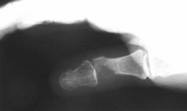 X-ray image demonstrating bony lucency at the distal portion of the proximal phalanx.
MRI has a distinct advantage over CT scanning, with improved resolution in soft tissue imaging. MRI can be used to determine the area of gross involvement when assessed in various views (eg, coronal, axial, sagittal), to better define lesions prior to operative intervention.
Bone scans can be useful in finding hot spots or areas of increased radioisotope uptake. This technique is quite sensitive to pathology; however, the findings are nonspecific. Bone scans are currently used more often when assessing for bony metastasis of a primary malignancy.
At times, ultrasonography can be helpful when assessing a soft tissue lesion. Ultrasonography is inexpensive, quite readily available, and effective in determining if a lesion is solid or cystic. It can assist in defining the type of biopsy that may be pursued on a lesion.
Biopsy
The next step in assessing a soft tissue mass is biopsy. The type of biopsy performed depends on the outcome of previous diagnostic studies. Plan carefully prior to biopsy because an excisional biopsy may lead to a cure. [6] Excisional biopsy is the goal; however, various other biopsies may be implemented. Needle biopsy can be performed on a lesion, although it is suited best to a lesion with a cystic component. Incisional biopsy may be performed on a lesion that is too large to be excised without compromising vital surrounding structures.
Remember that the goal is to deliver a representative sample of the lesion to the pathologist. Whenever possible, pursue complete excision; however, weigh the benefits of complete excision (with possible loss of function and cosmetic result) against an incomplete excision. This is often a function of experience and an index of suspicion that the lesion is malignant. All biopsies should be performed by an experienced hand surgeon because the type of incision should be based on sound techniques that minimize contamination of surrounding tissue and allow for additional resections to be performed without undue functional loss.
Metastatic Lesions
Metastatic tumors to the hand are rare entities, with only a handful reported in the literature, often as case reports. The lungs, kidneys, head, and neck are the most common sites of metastatic source to the hand, with the lungs accounting for 40% of reported primary tumors metastatic to the hand. Presentation of a metastatic lesion to the hand can occur in an individual with an unknown primary tumor or with a known source of metastasis. Often, the diagnosis is delayed because the presentation (ie, erythema, pain, swelling) can be confused with infection.
A retrospective study by El Abiad et al of 28 patients with osseous metastases to the hands or feet found lung carcinoma to be the primary cancer (32.1%) most commonly associated with these lesions. Of the 12 patients with hand metastases, the majority of lesions were in the metacarpals and phalanges. [21]
Typically, the prognosis is grim in patients with metastatic hand tumors; however, long disease-free intervals after appropriate excision have been reported.The El Abiad study found that the median period of survival following diagnosis of osseous metastases to the hands or feet was 9.7 months. [21]
Treatment of metastatic lesions entails therapy for the primary tumor along with adjuvant appropriate resection of the area of metastasis with clear margins. Exercise caution and treat each patient on an individual basis according to the degree of metastatic disease.
-
Patient with metastatic adenocarcinoma to the proximal phalanx.
-
X-ray image demonstrating bony lucency at the distal portion of the proximal phalanx.