Overview
Facial paralysis is a debilitating condition that is often associated with dramatic functional, psychological, and cosmetic sequelae. Varied functional deficits pose significant physiologic challenges. The inability to express oneself with spontaneous facial expression or intelligible speech can have extraordinary psychological ramifications, and facial asymmetry can scar a patient’s self-image, rendering him or her less secure in everyday interactions with the world. [1, 2]
The surgical team has an armamentarium of surgical strategies for facial reanimation. These procedures are categorized as either dynamic or static. [3] (See also Facial Nerve Paralysis, Dynamic Reconstruction.) The surgeon must decide on the most appropriate method of reconstruction on the basis of a detailed medical assessment of the patient, a thorough evaluation of the injury, and sound clinical judgment. The following should be determined:
-
The location, extent, and degree of paralysis
-
The etiology of the nerve injury
-
The duration of paralysis
-
The time delay between injury and presentation
-
The patient’s age, wishes, expectations, and overall health
Manifestations of facial nerve paralysis include the following:
-
Facial laxity
-
Asymmetric smile
-
Lower lip asymmetry at rest
-
Droopy oral commissure (from the weakened major and minor zygomatic muscles)
-
Inspiratory nasal collapse
-
Oral incompetence (difficulty with mastication and speech)
-
Lower-eyelid ectropion or laxity
-
Lagophthalmos
-
A sense of disfigurement
Therefore, the goals of reconstruction of the paralyzed face are as follows:
-
Facial symmetry at rest
-
Oral competence and eye closure
-
Voluntary facial movements with spontaneous facial expression
-
Minimal to absent synkinesis and mass movement
Dynamic procedures aim to reanimate the face by local muscle transfer or by nerve grafting and free muscle transfer; they should be considered in every patient with facial nerve paralysis. Although such procedures provide the best functional and cosmetic results for the paralyzed face, they may not be suitable for a patient who is debilitated or terminally ill.
Static techniques are employed to suspend the soft tissue structures of the face, but they do not provide facial reanimation. They are often adjunctive maneuvers performed in conjunction with dynamic techniques to enhance facial symmetry, particularly in treating lagophthalmos and lower-lid laxity. [4, 5]
However, static procedures may also be performed alone for patients who are not candidates for dynamic reanimation procedures (because of physical debilitation, advanced age, increased time delay from injury to repair, [6] or poor health) but who would still benefit from the restoration of facial symmetry.
Plastic surgeons, along with other specialists, must incorporate static procedures with advanced dynamic muscle transfers into their arsenal of facial nerve reconstruction techniques.
Indications
At times (eg, in elderly patients), dynamic facial reanimation is not possible or indicated, and static reconstruction is performed. The goals of static suspension procedures are to protect the cornea by restoring eyelid competence, to enhance mastication and speech production through commissure elevation, and to achieve cosmetic improvement by restoring facial symmetry at rest. Static procedures include the following:
-
The use of injectables or implants (eg, ENDURAGen [Stryker Craniomaxillofacial, Kalamazoo, MI] or AlloDerm [LifeCell, Branchburg, NJ]) [7]
-
Rejuvenation techniques – These include brow lift, open and endoscopic [8, 9] ; facelift; and blepharoplasty
-
Ocular care – This includes lower-lid shortening or tightening (Kuhnt-Szymanowski procedure), gold weight implantation, tarsorrhaphy, canthoplasty, and canthopexy
-
Upper- and lower-lip shortening or thickening with commissure preservation
-
Repositioning of the nasal alar base
-
Facial musculature plication or shortening [10]
-
Nasolabial fold recreation or lifting
Management of the eye is one of the most problematic issues in treating a patient with facial paralysis. The ocular sequelae of facial palsy include lagophthalmos with corneal exposure, [12] lower-lid ectropion, brow ptosis, and decreased tear production. Inadequate corneal protection can cause exposure keratitis, corneal ulceration, and blindness. Most dynamic procedures do not provide adequate reanimation and protection of the eye. However, several static techniques can adequately address this issue.
Not every patient is a suitable candidate for a dynamic procedure for facial reanimation. Patients who are severely debilitated or elderly may not be able to endure the lengthy operations required by dynamic reconstructions, nor can they wait for the delayed results generated by dynamic modalities (which sometimes take as long as 2-3 years to develop), given that their life expectancies are limited by advanced age or terminal illness.
For these patients, static suspension of the lower face with autologous or alloplastic materials can provide symmetry at rest and may improve oral incompetence and nasal collapse. These improvements in function enhance quality of life despite life expectancy.
Static procedures can be performed alone or in combination with other reanimation procedures. Adjunctive cosmetic procedures included brow lift, blepharoplasty, and rhytidectomy. Utilization of these techniques depends on the extent of facial asymmetry, brow ptosis, dermatochalasia, and skin laxity.
Clinical Presentation
Evaluation of a patient with facial paralysis commences with a thorough and detailed history and physical examination.
Patient history
Etiology is the most important factor in determining the timing and choice of reconstructive technique. Bell palsy is an idiopathic form of facial paralysis and is a diagnosis of exclusion. Trauma is the second most common cause of facial paralysis. Iatrogenic injury from mastoid surgery, parotid surgery, or pontine angle surgery for schwannoma may render a patient paralyzed. Ramsay-Hunt Syndrome type II, from herpes zoster reactivation is a possible presentation.
A thorough history includes the onset of paralysis, initial degree of paralysis, duration of paralysis, and associated symptoms. These details can often help identify the etiology, which, as noted, may affect the reconstructive method employed. Therefore, facial nerve injuries from Bell palsy, trauma, or malignant neoplasm must be distinguished from each other. The etiology of the denervation also dictates the timing of surgical treatment, if any is to be done.
If reconstructive efforts and interventions are to be tailored appropriately, a patient’s paralysis must be assessed for the possibility of spontaneous recovery, such as often occurs in a patient with Bell palsy. An estimated 56% of patients with Bell palsy will make a full recovery. [13] In these cases, an irreversible technique to reanimate the face may not be the best choice.
The patient’s overall health, psychological stability, and life expectancy are significant considerations. Patients with significant health risks and medical problems are not appropriate candidates for invasive reconstructive operations in which results do not manifest for 2-3 years postoperatively.
The surgeon must uncover the psychological impact of the paralysis on the patient and discuss the patient’s expectations. During this conversation, patient education is paramount. The physician should establish realistic expectations and determine if the patient can expend the time and finances required for the multiple procedures and potential revisions that may be necessary to increase the likelihood of successful, rewarding results.
Physical examination
Although electromyographic (EMG) assessment may evaluate the degree of muscle atrophy and thereby predict the potential for successful outcomes with dynamic restoration procedures, [14] the surgeon must perform a comprehensive physical examination of the patient with facial paralysis.
Scrutinizing the face at rest and during voluntary and reflex emotional movements, determining the involvement of unilateral or bilateral facial nerves, and mapping the nature and degree of facial asymmetries are important components of the physical examination. The degree of brow ptosis, ectropion, and lid laxity—as well as oral laxity, skin laxity, or commissure incompetence—must also be noted. The surgeon must identify the presence of other cranial, facial, or neurologic deficits or anomalies.
The condition of the ipsilateral eye must be carefully inspected to assess for excursion and ability to fully close, ectropion, lower-lid laxity, corneal irritation or ulceration, and tear production. The cornea can be inspected by using fluorescein dye and a Wood lamp to identify exposure keratitis or corneal ulcerations. The surgical team must record objective measures of facial motion and movement with digital images (either photographs or video recordings). This assists with preoperative evaluation and postoperative assessment of outcomes.
Management of Eye in Facial Paralysis
Paralysis of the upper branches of the facial nerve results in disorders of eyelid and lacrimal function. Sequelae include incomplete closure of the eye with corneal exposure, lower-lid ectropion with epiphora, decreased tear production, and loss of the corneal “squeegee effect.” These factors contribute to inadequate corneal protection, which can result in exposure keratitis, corneal ulceration, and blindness.
Eyelid and lacrimal function
Orbicularis oculi
The orbicularis oculi is a concentric muscle that is innervated by the frontal and zygomatic branches of the facial nerve. It provides tone to the upper and lower eyelids, promoting normal lid position and eyelid closure. Contraction of the pretarsal portion of the orbicularis oculi serves as a pump mechanism on the lacrimal sac to induce tear drainage. Normal function of this muscle is essential for lacrimal function, protection of the cornea, and preservation of vision.
Upper lid
The upper lid is a dynamic anatomic structure that is controlled by the opposing forces of the orbicularis oculi and levator muscles. The oculomotor nerve innervates the levator, which is responsible for lid opening and retraction. While a person is awake, the orbicularis and levator muscles are in a state of equilibrium, but the levator predominates.
Inhibition of the oculomotor nerve produces eyelid ptosis, eyelid closure, or both. Paralysis of the orbicularis oculi results in incomplete closure of the lid and lid retraction during wakefulness caused by the unopposed levator tone.
Lower lid
The orbicularis oculi provides tone and movement to the lower lid. Upward movement of 1 mm completes lid closure and induces tear drainage. The normal position of the lower lid is vital for eyelid closure and tear drainage. Paralysis of the orbicularis results in lower-lid ectropion with conjunctival exposure and incomplete lid closure and in inadequate tear drainage with possible pooling and epiphora.
Lacrimal gland
Appropriate lacrimal function depends on tear production, distribution, and drainage. The lacrimal gland is innervated by parasympathetic fibers that travel with the facial nerve. Disruption of these fibers may result in a decreased basal rate of tear production. Blinking of the eyelids distributes tear film uniformly across the corneal surface. Properly positioned upper- and lower-lid puncta, a functional lacrimal sac, and a patent nasolacrimal duct are essential for normal tear drainage.
Management strategy
Supportive therapy
Management of the eye in a patient with facial paralysis centers on corneal protection. The patient should use artificial tears during the day and lubricating ointment at night. Taping the eyelids can assist with eye closure. Patching is not recommended, because it does not protect the cornea from trauma or ulceration. The surgeon must examine the cornea frequently to rule out injury and irritation.
Tarsorrhaphy
Tarsorrhaphy is a popular and effective method of eye protection in facial paralysis. A central tarsorrhaphy completely impairs vision and is not cosmetically acceptable as a permanent procedure. It should be used only as a tool for temporary eye protection.
A lateral tarsorrhaphy is preferred, but it also limits peripheral vision and does not provide a cosmetically acceptable long-term result. Permanent tarsorrhaphy should never be entertained; moreover, tarsorrhaphy should never be a first line of treatment because of the functional and aesthetic downfalls and drawbacks.
Permanent lateral tarsorrhaphy is not generally recommended, but it can be used for eye protection in the severely debilitated patient who is not a candidate for other procedures. In this approach, the lateral aspects of the upper and lower lid are deepithelialized and then approximated with sutures.
Gold weight lid loading
Gold weight lid loading is an invaluable technique for the treatment of the paralyzed upper eyelid. A weight in the upper eyelid causes greater gravitational pull, passively closing the lid. [15] Gold is the material of choice for lid weighting because of its high density, relative inertness, and color, which blends with most skin tones.
Commercially manufactured gold implants are available in a wide range of weights. The most suitable weight is determined by taping different weights to the patient’s upper lid to assess which weight provides the most suitable eye closure in upright and supine positions.
Placement of a gold weight lid load is a simple procedure that is performed with local anesthesia. A supratarsal crease incision is made, and dissection is carried down to the tarsus. Disruption of the levator aponeurosis must be avoided. A pretarsal pocket is created, in which the selected gold weight is centered and secured to the tarsus with an absorbable suture.
May reported a 90% success rate in 482 gold weight lid loading procedures, with a 5% rate of persistent lagophthalmos. [16] Complication rates are generally low. Potential complications include incomplete closure, displacement or migration of the weight, foreign body reaction, cosmetic lid deformity, shifts in the astigmatic axis of refraction, and extrusion. Careful attention to pocket size and to securing the implant to the tarsus can minimize complications of migration. Closing the orbicularis and subcutaneous tissue over the implant reduces the risk of extrusion.
If necessary, revision procedures can be performed to reposition the implant or replace it with a different weight. Removal of the implant is simple, and postremoval sequelae have not been described.
Early use of gold weight lid loading is recommended; the procedure can be performed at the time of the initial facial nerve injury. In situations of nerve repair or grafting, recovery of facial nerve function may take several months. Lid loading provides corneal protection during the recovery period, and when facial nerve function returns, the lid load is easily removed. For surgeons who prefer autologous material, conchal cartilage grafts are an alternative treatment for lagophthalmos.
Lower-lid procedures
Loss of orbicularis tone in the lower lid results in ectropion and problems of lid closure and lacrimal drainage. Techniques to reanimate the lower lid include canthoplasty, lid-tightening procedures, and lid suspension.
A lid-shortening procedure does not adequately address medial canthal laxity. The classic technique, the Collin medial canthoplasty, involves exposure of the canthal tendon through upper and lower incisions just medial to the puncta. The surgeon must approximate the 2 arms of the tendon with a mattress suture in order to tighten it. This technique is appropriate for treatment of mild-to-moderate medial canthal laxity.
Crawford et al reported that 90% of patients with paralytic medial ectropion treated in this fashion experienced complete relief of symptoms. [17] A potential complication of the medial canthoplasty is inferior canaliculus scarring with inexorable epiphora.
Lateral lid laxity can be addressed by means of lid-shortening or lateral canthoplasty procedures. Lid shortening is accomplished by a full-thickness wedge resection of the lower lid through a subciliary skin incision. A full-thickness incision is made at the lateral limbus, followed by overlapping of the cut ends and wedge excision. Overcorrection should be done, and the tarsal plate is reapproximated with a nonabsorbable suture.
The lateral canthoplasty corrects canthal tendon laxity and shortens the lower lid. The tarsal strip procedure is a powerful procedure, whereby a lateral canthotomy is executed and the inferior portion of the lateral canthal tendon is released from its insertion at the lateral orbital wall. The tarsal strip is deepithelialized, elevated, and suspended to the periosteum of the orbital wall to produce sufficient lower-lid tightening.
Ellis described using a sling to suspend the medial lower lid. In this technique, a Gore-Tex strip (WL Gore and Associates, Newark, DE) is tunneled subcutaneously from the anterior lacrimal crest to the zygomatic process. [18] Tension on the sling elevates the lid and positions the punctum against the globe. Excess lid laxity often must be addressed with a lid-shortening procedure in conjunction with the sling.
Lower-lid sagging can recur after lid-shortening and lid-tightening procedures because of poor orbicularis oculi tone. Numerous grafts (eg, septal and conchal cartilage, hard palate mucosa, or contralateral tarsal plate), secured to the lower tarsal border, can bolster the lower lid. Cartilage is harvested easily from either the septum or the fossa triangularis. Conchal cartilage is thinner and more elastic than septal cartilage, lending itself to more facile molding and shaping.
The cartilage graft is tailored to fit the convexity of the globe and the inferior border of the tarsus. After a subciliary or transconjunctival incision, the depressors or lower-lid retractors are released, the lower edge of the tarsus is identified, and a pocket is created. The graft is sutured and secured within this pocket to the lower tarsal border.
Li and Shorr, describing their experience with AlloDerm versus hard-palate graft for lower-lid retraction, reported equal success for the 2 materials in treating ectropion and elevating the lower lid. [19] In any case, this procedure is easily combined with other lower-lid procedures, including the lateral tarsal strip.
May implanted the lower lids of 51 patients with auricular cartilage and reported improvement in lid position in 100%; there were no extrusions and only 2 cases of implant migration. [20]
Static Reconstruction of Lower Face
Static techniques generally are unsatisfactory as a single modality for rehabilitation of the paralyzed lower face and thus should not be used as a primary modality of reconstruction. Static procedures are most appropriate for debilitated patients who are unable or unwilling to endure the extensive operations of dynamic reanimation or those who are not expected to have a life expectancy beyond the nerve and muscle recovery period following dynamic strategies. They can also enhance dynamic reanimation by augmenting facial symmetry.
Most static procedures involve suspension of a part of the face by a sling. The most commonly used materials are fascia lata and the palmaris longus tendon. [21, 22] Both grafts are easily harvested and afford adequate length and strength; the fascia lata is preferred because multiple strips can be acquired. Initial overcorrection is necessary to compensate for the stretching that occurs with autologous grafts.
Alloplastic materials for facial suspension include polypropylene mesh, polytetrafluoroethylene (PTFE) patch, and acellular dermis. The advantages of the mesh and patch alloplasts include elimination of donor-site morbidity and minimal stretching and laxity. However, because alloplasts are foreign material, they have higher complication rates as a result of infection and extrusion.
Another option is acellular dermis, which has tensile strength similar to that of alloplasts but does not evoke any of the foreign body reactions. Acellular slings have shown significant improvements in oral commissure position and oral competence. Unlike Gore-Tex grafts, acellular slings can also be used in patients who are undergoing radiation therapy.
Oral commissure and lip suspension
Drooping of the oral commissure secondary to facial paralysis can be aesthetically and functionally problematic. Static suspension of the commissure can reestablish symmetry and enhance oral competence. The sling involves suspension of autologous or alloplastic materials from the orbicularis oris muscle to either the zygomatic arch or the orbital rim. [23]
Sundry surgical approaches and incisions are used in facial suspension. A standard rhytidectomy incision and dissection provide excellent exposure to the entire hemiface. Moreover, exposure of the oral commissure can be achieved via incisions at the vermillion border of the upper and lower lip or at the nasolabial fold. An extended subciliary incision or a vertical incision anterior to the sideburn provides exposure to the orbital rim and zygomatic arch.
The sling is sutured to the modiolus or split into 2 tongues and fixed to orbicular fibers of the upper and lower lip. The suspension vector is determined by analyzing the position of the mouth on the unaffected side, and the free end of the sling is suspended and fixed to the zygomatic arch or infraorbital rim with a permanent suture, Mitek screw (Mitek Surgical Products, Westwood, MA), or miniplate. [24]
Multiple strips of sling material can be used to create different vectors of suspension for the upper and lower lip. Some degree of overcorrection is necessary to account for postoperative relaxation and laxity, especially when autologous material (eg, fascia lata) is being used.
A 2011 article described a well-camouflaged incision that allows minimally invasive access to the temporalis tendon for its transposition to the modiolus. [25] The authors treated 17 consecutive patients, and their early results suggest that this technique may prove to be a promising addition to the surgical armamentarium. The following key elements from this article should be seen as “take-home points”:
-
A single incision is used, either well hidden in the nasolabial fold or in the buccal sulcus, and provides direct access to the most inferior aspect of the temporalis tendon as it converges and inserts upon the anterior mandibular ramus; the natural glide plane beneath the zygomatic arch is undisturbed, and the previous anatomic deformities (ie, zygomatic fullness and temporal donor site depression) are avoided
-
In all 17 patients, primary tendon-to-modiolus suturing was achieved without the use of additional incisions or other materials, either autogenous or synthetic, for lengthening
-
Physical therapy and patient motivation are indispensable components of a successful reanimation procedure using the temporalis tendon transfer; the goal is a “Mona Lisa” (or spontaneous temporal) smile, which is achieved through a rigorous 3-phase rehabilitation program, supplemented by video self-modeling and implementation intentions
Nasal lateralization
Buccal branch denervation induces paralysis of nasalis muscles and subsequent nostril collapse. Patients may experience unilateral nasal airway obstruction and internal valve collapse. This can be corrected with a lateralization procedure in which a sling of fascia or alloplast is secured to the deep tissue of the lateral alar base and suspended lateral to the ascending maxillary buttress with a nonabsorbable suture, an anchoring suture, or a titanium plate or screw.
Other anchoring techniques
Out of dissatisfaction with autologous fascial slings (because of resorption, scarring, and laxity) and alloplasts (because of complications and foreign body reactions), some surgeons have developed other stratagems for anchoring. Surgisis ES (Cook Biotech, Lafayette, IN), a xenograft made from porcine small intestinal submucosa, showed promising initial results in a pilot study of 6 patients with facial paralysis. [26]
Seeley and To described a system in which they suspended the face, commissure, and midface in multiple vectors. [27] They developed a static, multivector, bone-anchored suspension system with braided sutures that provides depression of the lower lip with an anchor to the mentum, elevation of the lower face with an anchor to the angle of the mandible, and suspension of the lip and nasolabial region and midface with an anchor to the lateral canthal region.
This multivector suspension system restored nasal breathing, improved drooling, restored normal speech, and enhanced cosmetic results through symmetry, all with minimal operative time and morbidity. [27]
Similarly, Horlock and Sanders achieved improved oral competence and oral asymmetry at rest and movement by performing a sub–orbicularis oculi fat (SOOF) lift and a subperiosteal midface lift. [28] This approach yields improved resting symmetrical tone and spontaneous synchronization. However, it generally is not indicated for patients with poor mouth excursion or with severe static asymmetry identified preoperatively.
Cheiloplasty
The corner of the mouth can be suspended by means of either dynamic or static techniques, but residual lip asymmetry with loss of tone and gapping often occurs. In cheiloplasty, the redundant paralyzed lip tissue is resected and exchanged for normal orbicularis oris and lip from the contralateral unaffected side.
The lip resection should be achieved with a full-thickness V or W wedge. As much as one third of both the upper and lower lip can be excised and closed primarily. The goal of this rotation and transfer of normal tissue is to reestablish a dynamic sphincter. Cheiloplasty can improve speech, eating, commissure competence, and appearance.
Illustrative case
The case illustrated below depicts a patient with facial nerve paralysis of all 5 major branches as a consequence of surgical sacrifice during total parotidectomy.
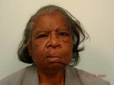 Facial nerve paralysis after total parotidectomy and modified radical neck dissection. Note left-side descent of soft tissues, oral commissure asymmetry, absent nasolabial fold, and accentuated nasojugal groove. On close inspection, scleral irritation resulting from lower-lid incompetence can be appreciated.
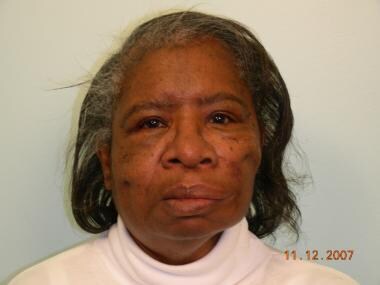 Correction of facial palsy via temporalis muscle transfer/Gore-Tex facial sling, midface lift, and formation of nasolabial fold. Medial and lateral canthoplasty was done on left eye, and endoscopic forehead lift was performed. Note improvement in resting position of lower lip, oral commissure symmetry, nasolabial fold creation, and restoration of lower lid margin to more anatomic position. Overall harmony and facial balance that is restored can be appreciated.
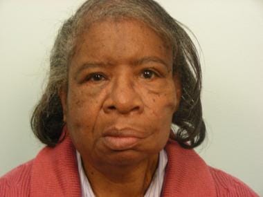 Same patient in January 2009 (> 1 y after surgery). Symmetry persists as soft tissues settle into more natural look.
The reconstruction combined a temporalis muscle transfer used as a facial sling with Gore-Tex suspension proximally and multitongue fixation distally. Adjunctive static procedures for restoration of ocular, nasal, and oral competence included medial and lateral canthoplasty of the left eye, formation of the nasolabial fold, endoscopic forehead lift, and a left-side midface lift. Moreover, the symmetry attained via static maneuvers allowed refinement of the overall aesthetic appearance.
Adjunctive Procedures
Soft tissue descent and ptosis may not be apparent until well after 9-12 months after the onset of facial nerve denervation. The surgeon should wait at least 12 months before considering any mode of cosmetic suspension or rehabilitation. The cosmetic or adjunctive techniques should be postponed until all necessary reconstruction has been performed and muscle and nerve recovery realized. This is paramount for free muscle transfer for facial reanimation; restoration of neurotization and muscle function can take 2-3 years.
Brow lift, blepharoplasty, and rhytidectomy procedures can be used in various configurations to battle soft tissue changes and descent that occur in the paralyzed face.
-
Facial nerve paralysis after total parotidectomy and modified radical neck dissection. Note left-side descent of soft tissues, oral commissure asymmetry, absent nasolabial fold, and accentuated nasojugal groove. On close inspection, scleral irritation resulting from lower-lid incompetence can be appreciated.
-
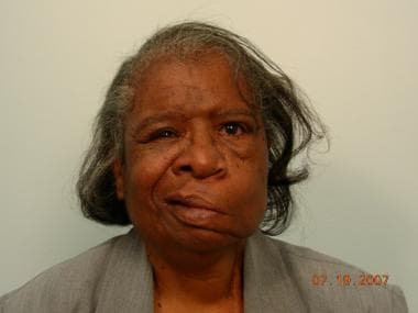
Patient during smile attempt.
-
Correction of facial palsy via temporalis muscle transfer/Gore-Tex facial sling, midface lift, and formation of nasolabial fold. Medial and lateral canthoplasty was done on left eye, and endoscopic forehead lift was performed. Note improvement in resting position of lower lip, oral commissure symmetry, nasolabial fold creation, and restoration of lower lid margin to more anatomic position. Overall harmony and facial balance that is restored can be appreciated.
-
Same patient in January 2009 (> 1 y after surgery). Symmetry persists as soft tissues settle into more natural look.


