Overview
Burn rehabilitation is an undeniably difficult and time consuming effort that, to attain the objective of optimal long-term function, must begin at the outset of burn care. Treatment goals and strategies vary, depending on the patient's injury, stage of treatment, age, and comorbidities. Goals range from minimizing loss of range of motion (ROM) in the critically ill patient to establishing a work-hardening program in recovered patients. See the image below.
See Thermal Injuries: A Matter of Degree, a Critical Images slideshow, to help identify and treat various types and degrees of burn injuries.
Survival was once the only gauge of success in managing serious burn cases. Today, however, the overriding objective of burn care has become reintegration of the patient into the home and community. This goal has extended the traditional role of the burn care team beyond acute wound closure.
Three broad aspects are involved in this effort: rehabilitation, reconstruction, and reintegration. The importance of early and active focus on long-term rehabilitation goals cannot be overemphasized.
Modern burn care may be divided into the following 4 general phases:
-
First phase - Initial evaluation and resuscitation; occurs on days 1-3 and requires an accurate fluid resuscitation and thorough evaluation for other injuries and comorbid conditions
-
Second phase - Initial wound excision and biologic closure; includes the maneuver that changes the natural history of the disease; this is accomplished typically by a series of staged operations that are completed during the first few days after injury
-
Third phase - Definitive wound closure; involves replacement of temporary wound covers with a definitive cover; there is also closure and acute reconstruction of areas with small surface area but high complexity, such as the face and hands.
-
Fourth phase - Rehabilitation, reconstruction, and reintegration; although this begins during the resuscitation period, it becomes time consuming and involved toward the end of the acute hospital stay
A study by Holavanahalli et al of over 90 patients who survived large burns found that an average of 17 years following the burn injury, patients continued to report joint pain and stiffness, as well as difficulty in walking or running, fatigue, and weakness in the arms and hands. Moreover, 68 out of 93 patients (73%) had limited motion, including in the neck (47%), hands (45%), and axilla (38%). [1]
Treatment Goals and Planning
As previously stated, burn rehabilitation is undeniably difficult and time consuming, but the time spent on outlining short-term and long-term treatment goals and modalities is worthwhile. Realistic therapeutic goals, as well as an appropriate plan of care, should be devised by the treatment team, including the patient and family.
Goals and daily schedules ideally are posted where the patient and family can review them easily, thereby reinforcing the expectation that the goals be met. Treatment goals and strategies vary, depending on the patient's injury, stage of treatment, age, and comorbidities. Goals range from minimizing ROM loss in the patient whom is critically ill to establishing a work-hardening program in recovered patients.
In critically ill patients, goals are to limit loss of ROM, reduce edema, and prevent predictable contractures through positioning and splinting. This process generally involves twice-a-day therapy sessions, which take advantage of planned anesthetics to allow more aggressive joint ROM.
In patients who have recovered from critical illness but still are hospitalized, treatment is much more time consuming, as well as physically and emotionally demanding of the patient and therapist. Appropriate therapist time must be budgeted.
Prior to hospital discharge, appropriate functional goals for the patient should include the ability to stand, ambulate, feed, and toilet. Regular meetings to discuss progress are appreciated by adults and children.
Acute Rehabilitation
Critically ill burn patients
To attain the objective of optimal long-term function, rehabilitation efforts must begin at the outset of burn care. Physical and occupational therapists play essential roles in the acute treatment of all burn patients, including critically ill patients and even during the resuscitation of patients with large injuries.
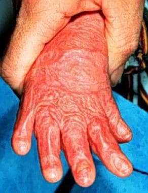
If a body part is left immobile for a protracted period of time, capsular contraction and shortening of tendon and muscle groups (which cross the joints) occur, as seen in the image below.
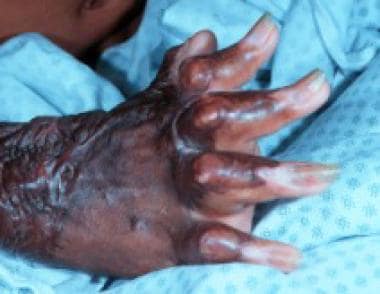 Contractures develop rapidly in patients with burns if proper range of motion and splinting are not performed from the outset of acute care.
Contractures are especially likely to develop if wounds are not closed promptly (see the image below). Moreover, if prevention strategies for contractures are not part of routine care, they can develop at astonishing speed. [2, 3] A study by Godleski et al, using the Burn Model Systems National Database, found that of the joint motions studied, adult burn patients who suffered contractures had a mean absolute loss of 20-65° in normal ROM, an 18-45% reduction in normal movement. [4]
The following are the 3 principal priorities for the burn therapist in the acute setting:
-
Performing passive ROM
-
Splinting and antideformity positioning
-
Establishing a long-term relationship with the patient and family members to ensure compliance with therapy goals and to increase the patient's morale for recovery
Passive ROM
Passive ROM is best performed twice daily, with the therapist taking all joints through a full ROM. The therapist must be sensitive to the patient's pain, anxiety, wound status, and extremity perfusion. ROM procedures should be performed in coordination with the intensive care unit (ICU) staff, with attention to the security of endotracheal tubes, nasogastric tubes, and arterial and central venous catheters being paramount, as unexpected loss of these devices can contribute to morbidity and mortality. Routine in-service training of therapists facilitates adherence to necessary precautions.
Although these procedures are important, they cannot be accomplished effectively (or humanely) if they cause excessive pain and anxiety. Medicating the patients before therapy sessions is often useful to increase treatment efficacy and decrease discomfort. Performing ROM often can be timed to coincide with dressing changes and wound cleansing, thereby minimizing the need for medication.
Antideformity positioning
Proper antideformity positioning minimizes the shortening of tendons, collateral ligaments, and joint capsules and reduces extremity and facial edema. Although splints are used less frequently, there are several predictable contractures that occur in patients with burns that can be prevented by a proper ROM, positioning, and splinting program (demonstrated in the chart below). These contractures generally are associated with the flexed position of comfort, except in the hands. (See the chart below.)
Splinting
Flexion neck deformities
Flexion deformities of the neck can be minimized with thermoplastic neck splints, conformers, and split mattresses. In critically ill patients, positioning the neck in slight extension is often all that can be done. Do not allow the ventilator tubing to pull the head so that a contracture develops; without proper care, a rotary contracture can develop, generally with the patient turned toward the ventilator. (See the image below.)
 If proper care is not taken, a rotary contracture can develop, generally with the patient turned toward the ventilator.
Axillary adduction contractures
Axillary adduction contractures can be prevented by placing the patient’s shoulders in a widely abducted position using axillary splints, padded hanging troughs of thermoplastic material, or a variety of support devices mounted to the bed.
Elbow flexion contractures
Elbow flexion contractures are minimized by statically splinting the elbow in extension. Elbow splints can be alternated with flexion splints to help retain a full ROM.
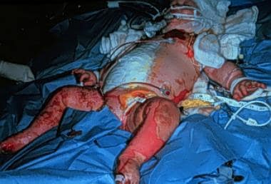
Hip and knee flexion contractures
Flexion contractures of the hips and knees are particularly common in young children but can be prevented by careful positioning and ROM. Prevention of contractures is important even in infants, as these contractures can interfere with subsequent ambulation. Prone positioning, although poorly tolerated by some, can assist in minimizing hip flexion contractures; knee immobilizers can minimize knee flexion contractures.
Equinus deformity
The equinus deformity, in which the ankle is plantar flexed and the foot is in a varus position, is a serious problem that can occur even if the ankles are not burned. It can be prevented, however, with static splinting of the ankles in the neutral position and the performance of ROM twice daily. Splints designed for this purpose can cause pressure injury over the metatarsal heads or calcaneus if improperly designed, but these injuries can be prevented by using padding to distribute pressure evenly across the metatarsal heads and by extending the footplate of the splint beyond the heel and cutting out the area around the calcaneus (as seen in the image below).
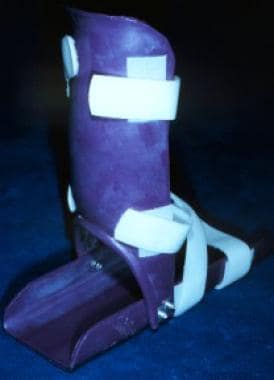 Pressure to the calcaneus can be prevented by extending the footplate of the splint beyond the heel and cutting out the area around the calcaneus.
Monitoring
Inspect all splints at least twice daily for evidence of poor fit or pressure injury. An in-service nursing staff minimizes splint-related skin injury. Positioning affected extremities just above the level of the heart reduces edema, which is another important aspect of antideformity positioning.
Resistance training
A study by Gittings et al indicated that the addition of high-intensity resistance training to standard rehabilitation may improve function in adults with burn injuries of the upper limbs. While all 48 of the study’s patients received standard physical therapy, they also underwent four weeks of either high-intensity or sham resistance training, 3 days per week, starting within 72 hours of the burn injury. The investigators found, over a 6-month follow-up period, that total quality of life scores and lower limb disability outcomes did not differ between the groups. However, the resistance training group showed functional improvement in upper limbs with burn injuries, with improved outcomes seen in the functional domain of the Burn Specific Health Scale-Brief and the Quick Disabilities of Arm, Shoulder and Hand score. [5]
Patient education
The burn therapist's initial assessment and care of patients with serious burns is the beginning of a long-term relationship. The therapist should make sure that the patient and his or her family know who the therapist is and understand the essential role of the therapist in their care. Everyone is grateful for regular communication and updates as to progress made and problems encountered. This information helps to ensure compliance with therapy goals. It also fuels the expectation that the patient will again become active and strong upon recovery.
Recovering Burn Patient
As critical illness abates and wounds progressively close, the roles of the physical and occupational therapists (as well as the demands on the patient) expand and become more difficult. Patients become more aware of what has happened to them, and they can become fearful of the therapist and the associated, potentially uncomfortable rehabilitation procedures.
The principal components of burn therapy that characterize this period include the following:
-
Continued passive ROM
-
Increasing active ROM and strengthening
-
Minimization of edema
-
Training in activities of daily living (ADL)
-
Initial scar management
-
Preparation for work, play, or school
A long-term favorable outcome requires hard work during this period, but it is important for the therapist not to push too hard. An early program of passive ROM greatly facilitates successful retention of normal ROM during this period. Intraoperative ROM also can be useful; in coordination with the operating room team, passive ROM can be performed between induction of anesthesia and preparation of the surgical site. Other maneuvers that can be used to increase the patient's tolerance for passive ROM include the following:
-
Timing of the ROM session with medication for dressing changes
-
Administration of opiates or benzodiazepines
-
Gentle conversation and encouragement
-
An unhurried approach to therapy sessions
Edema
Burned and grafted extremities commonly have lingering edema that can contribute to joint stiffness. Reducing this edema facilitates rehabilitation efforts.
The use of custom-fitted elastic garments this early after injury is expensive, as they frequently need to be downsized as the edema resolves; however, simply wrapping the fingers with self-adherent elastic helps to reduce digital edema. Tubular elastic dressings, elastic wrap dressings, elevation, and retrograde massage also help to reduce extremity edema.
In addition, topical silicone may have a favorable influence on selected evolving hypertrophic scars.
Focus of rehabilitation
As definitive wound closure nears and hospital discharge approaches, the focus of rehabilitation efforts becomes practical. Performance of ADL and the impending return to play/school/work are important considerations. Resisted ROM, isometric exercises, active strengthening, and gait training are important objectives.
When treating children, it is important to use developmentally appropriate play to facilitate rehabilitation goals. For example, children with serious hand burns are ideally engaged in play that requires the use of their hands at a motor level that is consistent with their development. [6]
Outpatient rehabilitation goals
For many burn patients, the first 18 months after discharge are more difficult than the acute stay. The principal rehabilitation goals at this time include the following:
-
Progressive ROM and strengthening
-
Evaluation of evolving problem areas
-
Specific postoperative therapy after reconstructive operations
-
Scar management
Ideally, the same therapist who worked with the patient during the acute inpatient hospitalization continues with him/her through the outpatient setting. Not only does this continuity enhance the patient's experience, it also helps the therapist to monitor burn recovery. If, for reasons of distance or managed care, it is not possible to maintain this relationship, regular contact at each clinic visit back at the burn unit can achieve this goal indirectly.
Unfortunately, it is not uncommon for ROM and strength to be lost during the first months after discharge. This is particularly true if there is inadequate outpatient rehabilitation. The burn unit team should monitor the quality of outpatient rehabilitation services during routine clinic visits at the burn unit. If the patient is losing substantial ROM and strength due to inadequate therapy, readmission for focused rehabilitation efforts is appropriate.
The realities of distance, transportation, and managed care often cause patients to work with inexperienced therapists. Therapists should visit the burn unit prior to the patient's discharge, videotape therapy sessions (with the patient's written permission), and maintain frequent telephone contact. Family education and involvement with rehabilitation plans may facilitate early identification of evolving problems and improve rehabilitation efforts. [7]
Burn therapists play a central role in planning and performing reconstructive procedures in the months and years after acute discharge. They help to identify needed operations, plan sequencing of operations, and educate patients and families about perioperative care. Planning appropriate postoperative rehabilitation activities helps patients to optimize surgical outcome.
Rehabilitation facilities
A study by Weissman et al of patients admitted to burn units over a 7-year period found that hospitals chose which of these patients to transfer to a rehabilitation facility based primarily on the following factors (listed in order of importance) [8] :
-
Performance of a surgical procedure during acute care
-
Occurrence of an inhalation injury
-
Length of hospitalization period
The study included data from 974 patients with second-degree burns or worse, covering 20% of each patient’s total body surface area.
Upper Extremity Burn Rehabilitation
High-quality acute burn care minimizes early upper extremity reconstructive needs, but problems regularly occur. Perhaps the most common upper extremity deformities are dorsal hand and web-space contractures. [9, 10]
Dorsal hand contractures
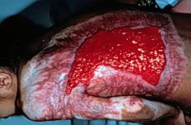
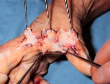
Dorsal hand contractures are prevented ideally by attention to proper positioning presurgically and postsurgically. If the initial surgical excision is performed tangentially rather than at the level of the fascia (ie, there is some remnant dorsal subcutaneous fat), the release is likely to slide and accept a large piece of skin (see the image below). The release must result in complete, resistance-free ROM of the metacarpophalangeal joints.
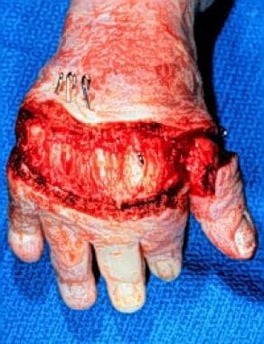 Successful release of a dorsal hand contracture substantially improves hand function. Do not delay this procedure, because it is functionally significant.
Web-space contractures
Although web-space contractures are common deformities that require correction, they can be minimized by proper early surgery and compressive gloves supplemented with web-space conformers. In the normal web space, the leading edge of the volar aspect of the web is distal to the dorsal aspect; in the typical dorsal web-space contracture, this pattern is reversed (the syndactyly is usually a dorsal deformity). When severe (ie, limiting digital abduction), it should be corrected. The typically normal leading palmar edge of the web space must not be compromised.
Elbow contractures and heterotopic ossification
Very deep burns of the elbow are associated commonly with difficulty in maintaining a complete ROM. Normal elbow ROM is required for performance of ADL such as feeding and toileting.
Limited elbow extension is commonly a volar soft tissue problem that responds to simple release. Heterotopic ossification is a sometime contributing factor, with bone forming in the soft tissues around the triceps tendon (as in the image below). This leads to a mechanical problem; the range of the elbow joint is compromised when components of the joint abut the abnormal bone.
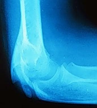 Heterotopic ossification may contribute to limited elbow motion and should be excluded by plain radiographs. This condition most commonly presents when bone forms in the soft tissues around the triceps tendon.
Although the ossification may resolve spontaneously over the course of years, it should be managed surgically if it interferes significantly with recovery. [11] A careful dissection is required, with bone being removed so that the elbow joint is not blocked. It is important to visualize and protect the ulnar nerve during this dissection.
Axillary contractures
Axillary contracture is not uncommon and can interfere with important upper extremity functions, such as feeding. Axillary release should encompass the entire rotational axis of the shoulder to facilitate complete ROM; the defect is closed with a sheet autograft. Postoperatively, abduction splints should maximize the ROM without creating traction or pressure on the brachial plexus or vessels.
Lower Extremity Burn Rehabilitation
Patients who have been supine for protracted periods often tolerate immediate upright positioning poorly. Prior to initial efforts at assisted standing, such patients benefit from tilt-table training and graduated sitting. Lower extremity edema, which can hinder recovery, is prevented best by using gentle elastic wraps prior to placing the patient in an upright position. [9]
The most common lower extremity deformities that require correction in patients who have sustained burns are dorsal foot extension contractures, popliteal flexion contractures, and hip flexion contractures. The latter 2 are particularly common in infants and very young children; they spend long periods of time with the hips and knees flexed and are particularly difficult to splint and range.
A deep dorsal foot burn may result in a contracture of the metatarsophalangeal joints, so that the toes are brought off the ground, causing the patient to have an abnormal gait. When the abnormal gait is severe enough to interfere with ambulation, surgery is required. An incisional release accepts a large piece of split thickness skin, particularly if the initial operation was performed in a layered fashion so that viable subcutaneous fat remains.
Flexion contractures of the popliteal fossa also interfere with ambulation. Correction generally requires incisional release and grafting, with directed postoperative efforts to maintain knee extension. Avoiding injury to the relatively superficial underlying neurovascular structures of the popliteal fossa is important.
Flexion contractures at the hips are common in infants and very young children who spend little time with the hips in extension. The contracted position of comfort is with the hip in flexion. This deformity interferes with ambulation and should be addressed early in recovery. Avoid injury to the femoral vessels and nerve, as the overlying contracted tissues may distort the normal anatomy.
Scar Management
Hypertrophic scarring is a difficult problem for burn patients, and scar management is an essential aspect of outpatient burn therapy.
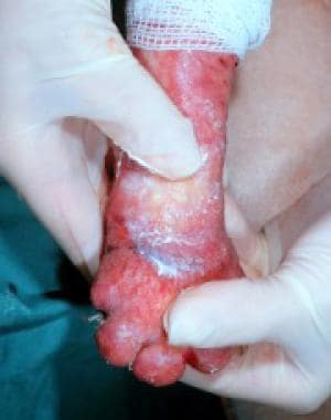
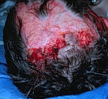
Perhaps the most virulent hypertrophic scarring is seen in deep dermal burns that heal spontaneously in 3 or more weeks; this seems especially true in areas of highly elastic skin (eg, the lower face, submental triangle, anterior chest and neck). The wound hyperemia seen universally following burn wound healing should begin to resolve about 9 weeks after epithelialization. In wounds destined to become hypertrophic, increased neovascularization occurs with increasing (rather than decreasing) erythema after 9 weeks. (See the images below.) [12]
 Despite the ubiquitous occurrence of hypertrophic scarring, its physiology is not understood. Perhaps the most virulent hypertrophic scarring is seen in deep dermal burns that heal spontaneously over the course of 3 or more weeks, particularly in highly elastic skin such as that covering the lower face, submental triangle, and anterior neck and chest.
 Despite its ubiquity, the physiology of hypertrophic scarring is not understood. Perhaps the most virulent hypertrophic scarring is seen in deep dermal burns that heal spontaneously over the course of 3 or more weeks, particularly in highly elastic skin (eg, the lower face, submental triangle, anterior neck, chest).
Available tools to modify the progression of hypertrophic scar formation are limited in number and effectiveness. These tools include the following:
-
Scar massage
-
Compression garments
-
Topical silicone
-
Steroid injections
-
Surgery
-
Serial casting - Useful in some contractures over major joints
Scar massage
Conscientious scar massage can be effective in limited areas of scarring; it is falso convenient, because it can be performed by family members. Ideally, this technique is performed several times each day. Bland moisturizers, which minimize drying of recently healed burns and skin grafts, are applied. Evolving hypertrophic areas then are massaged in a firm and slow manner. (See the image below.)
 Scar massage optimally is performed several times each day. Use firm, slow pressure on evolving hypertrophic areas after applying bland skin emollients.
Compression garments
Despite the controversy over their use, compression garments seem to improve control of broad areas of hypertrophic scarring, particularly in young children in whom this process seems to be more severe.
Compression garments should be worn 23 hours per day until wound erythema begins to abate, usually about 12-18 months after injury.
In growing and young children, frequent refitting and replacement of compression garments are required. Garment fit must be verified after manufacture, as a poorly fitting garment is less effective and can be uncomfortable.
Topical silicone
Topical silicone, applied to the healed wound as a sheet, is effective when used on small areas of a troublesome hypertrophic scar. Having the silicone in place 24 hours a day is ideal, except during bathing.
Some children develop a rash beneath the topical silicone, but this quickly resolves with the silicone’s removal. In these patients, 12-hour or every-other-day application seems to help.
Silicone sheets can be placed beneath compression garments or can be held in place by one of several elastic devices. Firm pressure is not required for the silicone to be effective.
Steroid Injections
For localized, extremely symptomatic areas of early hypertrophic scars, especially those in highly cosmetic areas or where extreme pruritus has developed, direct steroid injection (seen below) can be useful. The total dose, however, should be limited so that systemic effects do not occur.
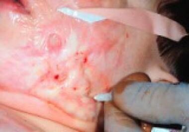 Steroid injections directly into localized, early hypertrophic scars can be useful, especially in highly cosmetic locations or in scars that cause extreme pruritus.
These injections are painful, as they require high pressure to infiltrate the dense hypertrophic scars; in children, general anesthesia usually is required.
Surgery
Surgical excision or incision and autografting are useful procedures when routine scar management tools are ineffective.
Extracorporeal shock wave therapy
A study by Moortgat et al suggested that extracorporeal shock wave therapy (ESWT) may increase hypertrophic burn scar elasticity. Forty patients were divided into two groups, those who received low-energy ESWT and those who underwent placebo therapy. Treatment was performed once weekly for 10 weeks, with follow-up over a 6-month period. The ESWT group was found to have better improvement in vertical elasticity of the hypertrophic burn scars by a statistically significant margin. [13]
Extreme Pruritus
Extreme pruritus is a frequent part of burn wound healing. It typically begins shortly after the wound has healed, peaks in intensity 4-6 months after injury, and then gradually subsides in most patients. Pruritus can be especially troubling at night. [14, 15]
In most patients, it is adequately treated with massage, moisturizers, and oral antihistamines at night. Alternative approaches are available, although none of them works reliably for everyone.
In patients particularly troubled by pruritus, a sequential therapeutic trial of each management option often identifies one particularly helpful method, such as any of the following:
-
Topical creams containing vitamin E
-
Topical antihistamine creams
-
Topical cold compresses
-
Frequent application of moisturizing creams
-
Colloidal baths
-
Steroid injection - Localized, highly pruritic scars often respond to this
A study by Goutos et al suggested that the administration of gabapentin alone or in combination with 2 antihistamines is more effective against acute burn pruritus than is the employment of the antihistamine chlorpheniramine alone or with 2 other antihistamines. [16] The authors also found that a combination of pharmacologic agents were needed to effectively treat study patients with higher initial itch scores. In addition, a linear regression model indicated that the lower a patient's age or the larger the total body surface area of an individual's burns, the greater the likelihood that monotherapy would fail.
Excoriation management
In rare cases, pruritus becomes so intense that excoriations develop. These wounds can become superinfected with Staphylococcus aureus, which further exacerbates the pruritus. To allow healing of excoriated areas, some patients require admission for wound care and antibiotics to control the pruritus and infection.
Acute Reconstruction
Proper acute burn care minimizes the need for burn reconstruction. Even in optimal circumstances, however, a predictable set of reconstructive operations commonly is required during the first postinjury years. A reconstructive plan is made best collaboratively with the patient and family, the patient's burn therapist, and the surgeon. One should not rush these procedures; however, waiting until all scars have matured completely for over 2 years prior to embarking on any reconstructive operations may prolong recovery unnecessarily.
The physical and emotional trauma of surgery must be balanced against the patient's functional and cosmetic needs. These plans are never easy to develop and must be considered carefully and individualized. Imagination and patience are important components of planning staged burn reconstruction.
Most burn reconstructive procedures can be performed using a combination of some basic techniques, as follows:
-
Incisional release and grafting
-
Excisional release and grafting
-
Z-plasty
-
Random flaps
Tissue expansion and free flaps are needed less commonly, but they can be useful in selected patients.
Incisional and excisional release
Most burn reconstructive operations can be effectively performed with an incisional or excisional release or with a common combined release, with the resulting wound being closed via a split thickness autograft. The contracture is placed under tension, and the release is performed sharply. (See the image below.)
 Most burn deformities can be corrected with release and grafting procedures. Any contracture around the mouth or neck that makes airway access difficult assumes a high priority in early reconstruction.
Adjacent areas of hypertrophic scar can be excised if donor sites are adequate to close the larger wound. Full-thickness skin grafts are less likely to contract than are thin split-thickness grafts; the former is the closure graft of choice in selected circumstances, such as the development of flexion contractures of the digits.
Site availability for full-thickness grafts generally is more limited than it is for split-thickness grafts, and thicker split-thickness grafts are adequate in most situations. In those patients with limited donor site availability, thin split-thickness grafts can be placed over acellular allogenic dermis to enhance results obtainable with thin split-thickness grafts alone.
Z-plasty
Although simple in concept, properly planned and executed Z-plasties are powerful reconstructive tools. [17, 18] The basic steps involved in constructing a Z-plasty include the following:
-
Defining the line(s) of tension that need to be modified
-
Planning the central limb of the Z-plasty(ies) on this line
-
Designing the lateral lines, if possible, so that they fall along natural skin lines (Langer lines) after transposition
-
Designing the angle between the central and lateral lines of the Z-plasty to be less than 90° with the lateral limbs curved and no longer than the central limb
Within these limits, a tremendous amount of variety is possible through modification of the blood supply of flaps and local tissue elasticity. Indeed, the utility of the Z-plasty is limited more by the surgeon's imagination than by the elasticity of adjacent available tissues. (See the image below.)
 Although simple in concept, properly planned and executed Z-plasties are powerful reconstructive tools, limited more by imagination than by their inherent efficacy.
A 5-flap Z-plasty can be constructed by placing 2 Z-plasties along the same band, oriented so that they are mirror images of each other. This results in a fifth "dog-ear" flap that can be inset to insert additional elastic tissue into the band. Multiple Z-plasties can be used in series along a band for excellent effect.
Tissue expanders and flaps
Local flaps, tissue expanders, and free flaps have a more limited, but nonetheless important, role in burn reconstruction. Thin, random flaps can be raised on the chest wall to cover small fourth-degree wounds of the hands in selected cases; the flap is divided at 3 weeks.
Groin flaps are more commonly used. These have earned an important role in reconstructing defects, particularly volar wrist defects associated with high-voltage electrical injury.
Tissue expanders are useful, particularly in the head and neck. Perhaps most useful are tissue expanders to correct burn-associated alopecia. (See the image below.)
 Tissue expanders are particularly useful in the head and neck and are perhaps most useful in the correction of burn-associated alopecia.
Like tissue expanders, free flaps offer an important option in selected, difficult wounds (eg, those associated with high-voltage injury [19] and extensive soft tissue loss of the distal lower extremity).
Head and neck reconstruction
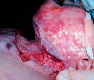
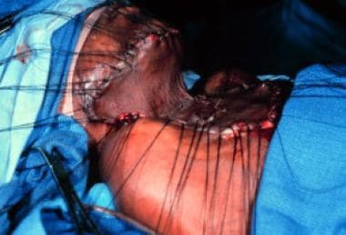
Usually, few reconstructive procedures are necessary in the face, head, and neck during the initial year after injury if deep facial burns have been resurfaced in cosmetic units with thick sheet grafts. The 2 common exceptions are the ocular adnexa and the neck. The pliant nature of the tissue around the eyes and mouth, combined with the important functions related to their normal position, render these areas at extreme risk for early problems related to tissue contraction. Any contracture that may impede access to the airway assumes a high priority in initial reconstruction. (See the image below.)
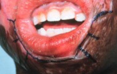 Early reconstruction should be a high priority for contractures around the mouth or neck that interfere with airway access.
Other predictable needs relate to lip eversion, microstomia, thickened nasolabial bands, and obstruction or distortion of the nares that occur with progressive contraction and thickening, particularly of deep dermal burns that heal over a protracted period of time.
Perhaps the most mobile structures of the face are the eyelids. Therefore, ectropion commonly occurs in the months following injury. Typically, only the skin and subcutaneous tissue are contracted, rolling the other structures away from the globe. Lid elevation is compromised if muscle is injured, unsightly protrusion of periorbital fat occurs if the orbital septum is violated, and coverage of the globe can be threatened if the tarsal plate is damaged. This anatomy must be understood prior to embarking on lid release; otherwise these normal, albeit distorted, structures may be injured. (See the image below.)
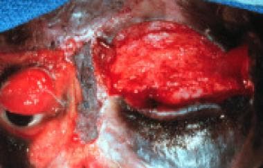 The ocular adnexa are very mobile; therefore, they are exquisitely vulnerable to the contractile forces of nearby or overlying wounds. Correct deformities of the ocular adnexa as soon as they occur.
The skin of the anterior neck is thin and elastic. Full-thickness burns in this area ideally are replaced with thick-sheet grafts early in the acute course. However, even with the best short-term surgical effort, optimal long-term results are difficult to produce.
Moreover, even with diligent use of conformers and neck splints, contractures are common, with a loss of the normal concavity between the tip of the chin and the sternum. When this becomes functionally important, neck release is indicated. Most patients have a satisfying result with release and split-thickness sheet autografting, although local flaps and tissue expanders provide additional options in selected patients and certain anatomic circumstances.
A study by Clayton et al found that in patients with partial-thickness orofacial burns (229 individuals), exercise and stretching begun within 48 hours of admission and conducted over an average of 30.7 days for contracture management led to improvements in horizontal and vertical mouth opening. However, only horizontal opening was statistically comparable to controls, with vertical mouth opening remaining significantly smaller. [20]
Psychiatric Aspects of Recovery
Attitude and psychological well-being play powerful roles (either helpful or destructive) in a patient’s physical recovery from burn injuries. The importance of understanding this concept cannot be overemphasized. Every member of the burn team can have a strong and favorable impact by considering these 2 factors during day-to-day patient interactions. [21, 22]
Indeed, a literature review by Spronk et al reported that the following factors were found, through multivariable analyses, to be associated with a poorer health-related quality of life following burn injury: postburn depression, posttraumatic stress symptoms, avoidance coping, a lesser degree of emotional or social support, a greater degree of neuroticism, and postburn unemployment. [23]
Various authors have described the following 3 basic stages of burn recovery, each with unique psychological implications:
-
Critical illness stage of recovery
-
Acute recovery phase of care
-
Final stage of psychological recovery
Critical illness stage of recovery
In this stage, survival often is in doubt, and immediate psychiatric issues dominate, including anxiety, fear, pain, delirium, sleep deprivation, and confusion. These problems ideally are addressed by the ICU team and psychiatric consultants
Acute recovery phase of care
Patients enter this phase after survival is assured and the intensities of surgery and intensive care diminish. This phase typically encompasses the noncritical remainder of acute hospitalization and is characterized by intensive physical and occupational therapy, fewer smaller surgical procedures, and the patient’s growing awareness of the impact and long-term implications of his/her burn injuries.
Patients often become depressed, and up to 30% of them experience symptoms of posttraumatic stress disorder (PTSD), such as hyperarousal, fearfulness, and sleep disturbances. Focused pharmacotherapy and individual counseling can be helpful
Final stage of psychological recovery
The final stage of psychological recovery encompasses the 1-2 years after initial hospital discharge. This time is often emotionally difficult, as patients adjust to new limitations at home and at work while experiencing waning PTSD symptoms.
Moderate depression can be expected in many patients and may be magnified if optimal recovery potential has not been reached because of inexpert therapy.
Recovery can be facilitated by a long-term therapeutic relationship. In many patients, participating in peer support groups, such as the Phoenix Society, is beneficial.
Reintegration
A few years ago, the goal of the burn team was survival of the patient; discharge was the measurement of success. Burn care, however, does not stop with wound closure.
Rehabilitation and reconstruction of the seriously burned patient is part of acute care. A burn ICU with a separate reconstructive surgery unit does not offer optimal care. As currently defined, successful burn care requires commitment by a focused, multidisciplinary team over the continuum of care from resuscitation through reconstruction, rehabilitation, and reintegration.
The ultimate goal of all burn care is reintegration of the patient into society, and it is important not to lose sight of this goal.
PTSD is common in burn patients, so be vigilant of symptoms (eg, hyperalertness, nightmares, chronic fearfulness). Ignoring this problem compromises recovery.
Ideally, patients return to their families, schoolmates, and communities as if the injury had never occurred. Consider this goal when planning the timing and type of reconstructive operations.
The stress on families of burn patients is enormous. Family counseling and support services are important. Moreover, any help afforded to these families indirectly can aid the patient.
-
Contractures develop rapidly in patients with burns if proper range of motion and splinting are not performed from the outset of acute care.
-
The set of predictable burn contractures can be minimized through focused and early intervention.
-
If proper care is not taken, a rotary contracture can develop, generally with the patient turned toward the ventilator.
-
If wounds are not closed promptly, contractures can rapidly occur.
-
Despite the ubiquitous occurrence of hypertrophic scarring, its physiology is not understood. Perhaps the most virulent hypertrophic scarring is seen in deep dermal burns that heal spontaneously over the course of 3 or more weeks, particularly in highly elastic skin such as that covering the lower face, submental triangle, and anterior neck and chest.
-
Pressure to the calcaneus can be prevented by extending the footplate of the splint beyond the heel and cutting out the area around the calcaneus.
-
Successful release of a dorsal hand contracture substantially improves hand function. Do not delay this procedure, because it is functionally significant.
-
Although simple in concept, properly planned and executed Z-plasties are powerful reconstructive tools, limited more by imagination than by their inherent efficacy.
-
Heterotopic ossification may contribute to limited elbow motion and should be excluded by plain radiographs. This condition most commonly presents when bone forms in the soft tissues around the triceps tendon.
-
Tissue expanders are particularly useful in the head and neck and are perhaps most useful in the correction of burn-associated alopecia.
-
Despite its ubiquity, the physiology of hypertrophic scarring is not understood. Perhaps the most virulent hypertrophic scarring is seen in deep dermal burns that heal spontaneously over the course of 3 or more weeks, particularly in highly elastic skin (eg, the lower face, submental triangle, anterior neck, chest).
-
Scar massage optimally is performed several times each day. Use firm, slow pressure on evolving hypertrophic areas after applying bland skin emollients.
-
The ocular adnexa are very mobile; therefore, they are exquisitely vulnerable to the contractile forces of nearby or overlying wounds. Correct deformities of the ocular adnexa as soon as they occur.
-
Steroid injections directly into localized, early hypertrophic scars can be useful, especially in highly cosmetic locations or in scars that cause extreme pruritus.
-
Most burn deformities can be corrected with release and grafting procedures. Any contracture around the mouth or neck that makes airway access difficult assumes a high priority in early reconstruction.
-
Early reconstruction should be a high priority for contractures around the mouth or neck that interfere with airway access.


