Practice Essentials
Approximately 5% of the population has some sort of ear malformation. Protruding ear and external ear microtia (or a variant) are the two most frequently encountered congenital ear problems in plastic surgery. The former condition is commonly treated by many practitioners, while the latter has become the bailiwick of just a few surgeons. Most of this discussion focuses on prominent ears because of their common occurrence. Otoplasty has undergone important developments, with numerous techniques being presented in the surgical literature. Congenital ear microtia and atresia is treated in only a few centers by surgeons with an established reputation, similar to the way some centers specialize in craniofacial osteotomy surgery.
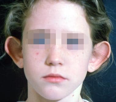
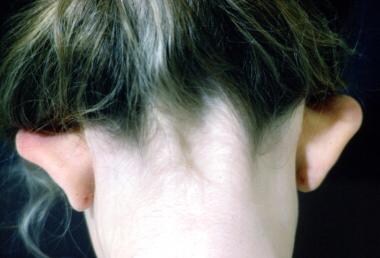
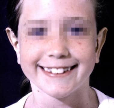
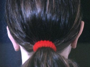
Examples of preoperative and postoperative otoplasty are shown in the images below.
An observational study by Litschel et al indicated that when viewing a face with protruding ears, observers tend to have a longer visual fixation time on the ears than they do when viewing an individual with nonprotruding ears but that protruding ears do not significantly affect the observer’s opinion of an individual’s personality with regard to assiduousness, intelligence, and likeability. The study involved 20 observers who viewed photos of children with either protruding or nonprotruding ears. [1]
Management
Historically, prominent or protruding ears have been treated surgically. However, nonsurgical techniques have emerged to treat neonates immediately after delivery. [2] The posterior helical rim is taped to the posterior retroauricular region with surgical tape. Tubular elastic net bandage or some type of ear wrap is used for reinforcement. To achieve the desired result, such techniques must begin in the first few weeks of life and take several weeks or months of constant and vigilant therapy.
Surgical techniques employed in the correction of prominent or protruding ears include the following:
-
Skin excision
-
Radial placed mattress sutures
-
Conchomastoid sutures
-
Excision of conchal cartilage
-
Laser-assisted cartilage reshaping
History of the Procedure
Many different approaches to setback otoplasty have been developed. Originally, the first operations were merely resection of skin from the posterior sulcus. Approximately 200 different techniques have now been described for setback otoplasty; each technique has strengths and weaknesses. Over the years, an evolution of operations has occurred, including those with and without sutures, with and without resection of cartilage, and with or without scoring of cartilage. With this many variations, no single "right" technique exists.
Microtia as a modern operation was first pioneered by Tanzer from Dartmouth Medical School. Tanzer was the first to develop the technique of using a whole piece of rib cartilage to simulate the cartilaginous structure of the external ear. Burt Brent from Stanford, Calif, expounded upon Tanzer's work and has written extensively about microtia reconstruction.
Problem
The expression "beauty is in the eye of the beholder" is often quoted and still quite relevant. What constitutes a prominent ear? Ray Elliot stated in his 1990 review article in Clinics of Plastic Surgery, "the esthetic ear protrudes less than 2 cm when measured from the surface of the helix to the mastoid scalp at the midpoint of the ear's length." [5] However, once this measurement achieves a distance of less than 1.2 cm, the ear has an equally displeasing "pinned back" appearance. The ear protrudes more at the lower pole and less so at the upper pole because of the shape of the skull. The scaphoconchal angle should have a natural soft roll and should not block the view of the helix anywhere along its course when viewed anteriorly.
Epidemiology
Frequency
No statistics are available on the prevalence of protruding or prominent ears. Heredity plays a role in many deformities of the external ear.
Etiology
Most embryologic studies of the ear focus on the development of the 6 ear hillocks. These hillocks appear around the fundus of the first branchial groove by 38 days of gestation. As the groove closes and the first and second arches come together, the primitive ear is formed by day 50 of gestation. The first 3 hillocks come from the first branchial arch and the second 3 from the second branchial arch. Absence of hillocks 2-5 produces a frequent and typical microtia. Malformations of the ear can appear anywhere during this development.
Pathophysiology
Features seen in the patient with prominent or protruding ears (in decreasing order of importance) include the following:
-
Absent antihelical fold
-
Obtuse scaphoconchal angle
-
Increased distance of helical rim to scalp
-
Deep conchal bowl
Address some or all of these problems in the planning and execution of the operation to correct the abnormality.
Auricular anatomy in children varies only slightly from that in adults. By the third year, 85% of ear growth and development occurs, and little growth occurs after 10 years. The ear's height may increase gradually into adulthood but its distance from the scalp changes little after 10 years. Because of this, setback otoplasty can safely be performed on children as young as 5-6 years.
Presentation
Most parents, while emotionally distressed when their baby is born with a portion of the external auricle missing or severely distorted, are unconcerned about protruding ears at birth. Patients tend to seek surgical opinion for protruding ear problems at two stages of life. Parents often seek medical advice for their children at the start of school because of the child's complaints of classmates' teasing. The jeer of "Dumbo" is difficult for a first grader. The second group of patients consists primarily of women in their twenties or early thirties. These patients also are embarrassed about their protruding ears.
Indications
For the child born without an external ear, the indications for surgical reconstruction are obvious. Parents are anxious to proceed as soon as possible to spare their child any embarrassment. Unlike cleft lip surgery, which is performed in the first few weeks of life, most experienced surgeons recommend waiting to do multistages at age 6-7 years. The more important question is not whether surgery is indicated, but which of the techniques for reconstruction is indicated for the individual. Even with microtia, the physiologic effects of an ear deformity are negligible. The aesthetic and psychological effects are significant. [6]
In the patient born with a prominent ear or deformity, the situation may be a little different. With the obtuse scaphoconchal angle and absent antihelical fold, distance from the helical rim varies. Many authorities believe the external ear should protrude no more than 2 cm from the surface of the helix to the mastoid scalp at the midpoint of the ear's length. This measurement is a guide and not an absolute rule. Much depends upon the patient's expectations as well as the surgeon's experience. Although congenital absence is almost always unilateral, patients with prominent ears usually require a bilateral operation.
Children with protruding ears generally do well when operated on as early as age 5-6 years. A study from the Medical College of Wisconsin presented a series of 12 patients in whom otoplasty was performed before the age of 4 years with good results. [7, 8] Taunts from schoolmates begin at this time. Parents, not realizing how cruel kindergarten children can be, are often oblivious to this ridicule. Young girls reveal much less of this mental anguish because they are able to wear their hair long and cover their ears. They never wear a ponytail, lest a peer discovers their ears stick out.
In this author's practice, a common indication for setback otoplasty is the approximately 20-year-old woman who is getting married and now wants to wear her hair in an upswept fashion. Boys do not have that option. Because of a genetic predisposition to protruding ears, some parents find that seeking plastic surgical consultation for their children is difficult. To admit his or her child is flawed, the parent must admit he or she is flawed.
Relevant Anatomy
Nerve supply of the external ear
See the list below:
-
Great auricular nerve (C2, C3) - Lower half of ear
-
Auriculotemporal nerve (from mandibular brand of trigeminal) - Tragus, anterior/superior portions of auricle
-
Lesser occipital nerve (C2, C3) - Posterior/superior aspect of ear
-
Auricular branch of vagus nerve (Arnold nerve) - Concha and posterior auditory canal
Anatomy of the normal ear
See the list below:
-
Width of ear equals approximately 55% of length
-
Helix-to-mastoid distance - Upper third equals 10-12 mm; middle third equals 16-18 mm; lower third equals 20-22 mm
-
Sides should be within 3 mm of each other
-
Axis of ear is parallel to profile of nose
The ear can be difficult to reproduce surgically because only a thin layer of skin covers the cartilaginous structure. [9] This cartilaginous structure is attached to the temporal bone medially by several minor intrinsic muscles. A looser attachment of skin is found upon the posterior aspect of the auricle than the anterior aspect of the auricle. The vascular supply comes from the superficial temporal and posterior auricular vessels. Corresponding veins drain the ear.
A study by Oliveira et al stated that a natural plica can be found at the anatomic base of the antihelix, with the creation of this plica being an important part of antihelix reconstruction in patients with protruding ears. [10]
Prominent or protruding ears
See the list below:
-
Absent antihelical fold
-
Increased scaphoconchal angle
-
Deep conchal bowl
-
Increased distance from helical rim to scalp
-
Flattening of superior crus
-
Normal helical length
Cup ear deformity (lop-ear, constricted ear)
See the list below:
-
Flattening of antihelix
-
Widening of concha
-
Overhang of helix
-
Compression of scapha and fossa triangularis
-
Indistinct antihelix and crura
-
Shortened length of helix
-
Ear appears small, but no difference in size
Cryptotia
See the list below:
-
Absence of retroauricular sulcus, causing the superior pole of ear to appear buried
-
Sharply curved antihelical crus
-
No foreshortening of auricle
Stahl ear
See the list below:
-
Presence of third crus
-
Flat antihelix
-
Malformed scaphoid fossa
Contraindications
Contraindications for treatment of prominent ears include the following:
-
Unreasonable expectations from either patient or family
-
Child younger than 5 years
-
Patient who is unable to tolerate postoperative wound care, including a protective bandage head wrap
-
Patient who opposes the operation despite his or her parents' wishes to proceed
-
Preoperative otoplasty.
-
Two years after otoplasty.
-
Preoperative otoplasty.
-
Preoperative otoplasty.
-
Postoperative otoplasty.
-
Postoperative otoplasty.
-
Preoperative otoplasty.
-
Two years after otoplasty.


