Practice Essentials
Dengue is the most common and important arthropod-borne viral (arboviral) illness in humans. It is transmitted by mosquitoes of the genus Aedes, which are widely distributed in subtropical and tropical areas of the world (see the image below). [1, 2, 3, 4] The incidence of dengue has increased dramatically in recent decades, with estimates of 40-50% of the world’s population at risk for the disease in tropical, subtropical, and, most recently, more temperate areas. [5]
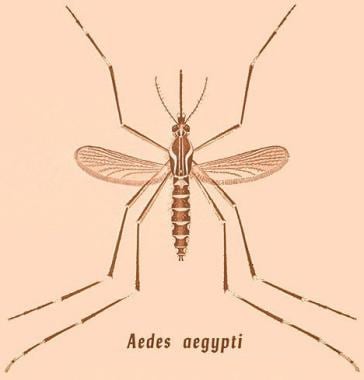 Drawing of Aedes aegypti mosquito. Courtesy of the Centers for Disease Control and Prevention (CDC).
A small percentage of persons who have previously been infected by one dengue serotype develop bleeding and endothelial leak upon infection with another dengue serotype. This syndrome is termed severe dengue (also known as dengue hemorrhagic fever and dengue shock syndrome). [2]
Dengue fever typically is a self-limited disease with a mortality rate of less than 1% when detected early and with access to proper medical care. When treated, severe dengue has a mortality rate of 2-5%, but, when left untreated, the mortality rate is as high as 20%.
See 7 Bug Bites You Need to Know This Summer, a Critical Images slideshow, for helpful images and information on various bug bites.
Signs and symptoms
On average, dengue becomes symptomatic after a 4- to 10-day incubation period (range, 3-14 days). Dengue symptoms usually last 2-7 days.
Many individuals with dengue may be asymptomatic. Many patients with dengue experience a prodrome of chills; rash, including erythematous mottling of the skin; and facial flushing, which may last 2-3 days. Children younger than 15 years who have dengue usually have a nonspecific febrile syndrome, which may be accompanied by a maculopapular rash. Dengue should be suspected in individuals who present with high fever (104°F/40°C), retro-orbital headache, muscle and joint pain, nausea, lymphadenopathy, vomiting, and rash and who have traveled within 2 weeks of symptom onset to an area where appropriate vectors are present and dengue transmission may be occurring.
Accompanying symptoms in patients with dengue may include any of the following:
-
Fever
-
Headache
-
Retro-orbital pain
-
Severe myalgias: Especially of the lower back, arms, and legs
-
Arthralgias: Usually of the knees and shoulders
-
Nausea and vomiting (diarrhea is rare)
-
Rash: A maculopapular or macular confluent rash over the face, thorax, and flexor surfaces, with islands of skin sparing
-
Weakness, malaise, and lethargy
-
Altered taste sensation
-
Anorexia
-
Sore throat
-
Mild hemorrhagic manifestations (eg, petechiae, bleeding gums, epistaxis, menorrhagia, hematuria)
-
Lymphadenopathy
Severe dengue (dengue hemorrhagic fever and dengue shock syndrome)
The initial phase of severe dengue is similar to that of dengue fever and other febrile viral illnesses. Shortly after the fever breaks (3-7 days after symptom onset or sometimes within 24 hours before), signs of plasma leakage appear, along with the development of hemorrhagic symptoms such as bleeding from sites of trauma, gastrointestinal bleeding, and hematuria. Patients may also present with severe abdominal pain, persistent vomiting that may contain blood, fatigue, and febrile seizures (in children).
The subsequent 24 hours frequently prove critical. If left untreated, hemorrhagic fever most likely progresses to shock. Common symptoms in impending shock include abdominal pain, vomiting, and restlessness. Patients also may have symptoms related to circulatory failure, such as pallor, tachypnea, tachycardia, dizziness/lightheadedness, and a decreased level of consciousness.
See Clinical Presentation for more detail.
Diagnosis
Laboratory criteria for the diagnosis of dengue include one or more of the following, which are used to detect the virus, viral nucleic acid, antibodies or antigens, or a combination thereof:
-
Demonstration of a fourfold or greater change in reciprocal immunoglobulin G (IgG) or IgM antibody titers to 1 or more dengue virus antigens in paired serum samples
-
Demonstration of dengue virus antigen in autopsy tissue via immunohistochemistry or immunofluorescence or in serum samples via enzyme immunoassay (MAC-ELISA, IgG ELISA, nonstructural protein 1 [NS1] ELISA, EIA)
-
Detection of viral genomic sequences in autopsy tissue, serum, or cerebral spinal fluid (CSF) samples via reverse-transcriptase polymerase chain reaction (RT-PCR) assay: RT-PCR provides earlier and more specific diagnosis.
-
Less frequently, isolation of the dengue virus from serum, plasma, leukocytes, or autopsy samples
During the early phase of the disease (first 4-5 days), virus can be detected in serum, plasma, circulating blood cells, and tissues. Virus isolation, nucleic acid detection, and antigen detection are more useful to diagnose infection. At the end of the acute phase of illness, serology becomes the method of choice.
The following laboratory tests should also be performed in the workup of patients with possible dengue:
-
Complete blood cell (CBC) count
-
Metabolic panel
-
Serum protein and albumin levels
-
Liver panel
-
Coagulation panel with or without disseminated intravascular coagulation (DIC) panel
Characteristic laboratory findings in dengue are as follows:
-
Thrombocytopenia (platelet count < 100 x 109/L)
-
Leukopenia
-
Mild to moderate elevation of aspartate aminotransferase and alanine aminotransferase values
In patients with severe dengue, the following may be present:
-
Increased hematocrit level secondary to plasma extravasation and/or third-space fluid loss
-
Hypoproteinemia
-
Prolonged prothrombin time
-
Prolonged activated partial thromboplastin time
-
Decreased fibrinogen
-
Increased amount of fibrin split products
Guaiac testing for occult blood in the stool should be performed on all patients in whom dengue virus infection is suspected. Urinalysis identifies hematuria.
Imaging studies include the following:
-
Chest radiography
-
Head computed tomography (CT) scanning without contrast: To detect intracranial bleeding or cerebral edema due to severe dengue
-
Ultrasonography: To detect fluid in the chest and abdominal cavities, pericardial effusion, and a thickened gallbladder wall in patients with severe dengue
See Workup for more detail.
Management
Oral rehydration therapy is recommended for patients with moderate dehydration caused by high fever and vomiting.
Patients who develop signs of severe dengue warrant closer observation. Admission for close volume status monitoring and intravenous fluid administration is indicated for patients who develop signs of dehydration, such as the following:
-
Tachycardia
-
Prolonged capillary refill time
-
Cool or mottled skin
-
Diminished pulse amplitude
-
Altered mental status
-
Decreased urine output
-
Rising hematocrit
-
Narrowed pulse pressure
-
Hypotension
Patients with internal or gastrointestinal bleeding may require transfusion, and patients with coagulopathy may require fresh frozen plasma.
See Treatment and Medication for more detail.
Background
Dengue is the most common and important arthropod-borne viral (arboviral) illness in humans. Globally, 2.5-3 billion individuals live in approximately 112 countries that experience dengue transmission. While the annual incidence is unclear owing to incomplete global reporting and misclassification of illness, approximately 3.2 million individuals were infected globally in 2015. Dengue is caused by infection with 1 of the 4 serotypes of dengue virus, which is a Flavivirus (a genus of single-stranded nonsegmented RNA viruses). Infection with one dengue serotype confers lifelong homotypic immunity to that serotype and a brief period (approximately 2 years) of partial heterotypic immunity to other serotypes, but an individual can eventually be infected by all 4 serotypes. [2, 3, 4] Several serotypes can be in circulation during an epidemic.
Dengue is transmitted by mosquitoes of the genus Aedes, which are widely distributed in subtropical and tropical areas of the world (see the image below). An individual with dengue is capable of transmitting the virus for 4-5 days (maximum, 12 days) to a capable vector. After an incubation period of 5-10 days, the infected mosquito can transmit virus for the rest of its life span (2 weeks to 1 month). Aedes albopictus is more cold tolerant than Aedes aegypti, so it can survive and transmit virus in the more temperate regions of the United States and Europe.
The global incidence of dengue has increased dramatically since the late 1900s, with an estimated 40-50% of the world’s population in 128 countries at risk. [6, 7, 8] Today, severe dengue largely affects Asian and Latin American countries, where it is a leading cause of hospitalization and death. The World Health Organization (WHO) ranked dengue as one of the top 10 threats to global health in 2019. [9]
Initial dengue infection may be asymptomatic (50-90%), [10] may result in a nonspecific febrile illness, or may produce the symptom complex of classic dengue fever (DF). Classic dengue fever is marked by rapid onset of high fever, headache, retro-orbital pain, diffuse body pain (both muscle and bone), weakness, vomiting, sore throat, altered taste sensation, and a centrifugal maculopapular rash, among other manifestations. The severity of the pain led to the term breakbone fever to describe dengue.
A small percentage of persons who have previously been infected by 1 dengue serotype develop bleeding and endothelial leak upon infection with another dengue serotype. This syndrome is termed severe dengue (reclassified in 2009 by the WHO, previously referred to as dengue hemorrhagic fever and dengue shock syndrome). [2, 4]
Severe dengue has also been termed dengue vasculopathy. Vascular leakage in these patients results in hemoconcentration and serous effusions and can lead to circulatory collapse. This, in conjunction with severe hemorrhagic complications, can lead to a shock syndrome, which poses a greater fatality risk than bleeding per se. [11]
Dengue virus transmission follows 2 general patterns: epidemic dengue and hyperendemic dengue. Epidemic dengue transmission occurs when dengue virus is introduced into a region as an isolated event that involves a single viral strain. If the number of vectors and susceptible pediatric and adult hosts is sufficient, explosive transmission can occur, with an infection incidence of 25-50%. Mosquito-control efforts, changes in weather, and herd immunity contribute to the control of these epidemics. Transmission appears to begin in urban centers and then spreads to the rest of the country. [12] This is the current pattern of transmission in parts of Africa and South America, areas of Asia where the virus has reemerged, and small island nations. Travelers to these areas are at increased risk of acquiring dengue during these periods of epidemic transmission.
Hyperendemic dengue transmission is characterized by the continuous circulation of multiple viral serotypes in an area where a large pool of susceptible hosts and a competent vector (with or without seasonal variation) are constantly present. This is the predominant pattern of global transmission. In areas of hyperendemic dengue, antibody prevalence increases with age, and most adults are immune. Hyperendemic transmission appears to be a major risk for dengue hemorrhagic fever. Travelers to these areas are more likely to be infected than are travelers to areas that experience only epidemic transmission. [13]
Because the signs and symptoms of dengue fever are nonspecific, attempting laboratory confirmation of dengue infection by serodiagnosis, reverse-transcriptase polymerase chain reaction (RT-PCR), or culture is important. Serodiagnosis is made on the basis of a rise in antibody titer in paired IgG or IgM specimens. Results vary depending on whether the infection is primary or secondary (see Presentation and Workup). Dengue is a reportable disease in the United States; known or suspected cases should be reported to public health authorities.
Dengue fever typically is a self-limited illness. Supportive care with analgesics, judicious fluid replacement, and bed rest usually are sufficient. Successful management of severe dengue requires intravascular volume replacement, with careful attention to fluid management and proactive treatment of hemorrhage. Admission to an intensive care unit is indicated for patients with severe dengue (see Treatment).
Historical background
The earliest known documentation of dengue fever–like illness was in the Chinese Encyclopedia of Symptoms during the Chin Dynasty (CE 265-420). The illness was called "the water poison" and was associated with flying insects near water.
Earliest recorded outbreaks
Outbreaks of febrile illnesses compatible with dengue fever have been recorded throughout history, with the first epidemic described in 1635 in the West Indies.
In 1779-1780, the first confirmed, reported outbreak of dengue fever occurred almost simultaneously in Asia, North America, and Africa. In 1789, the American physician Benjamin Rush published an account of a probable dengue fever epidemic that had occurred in Philadelphia in 1780. Rush coined the term breakbone fever to describe the intense symptoms reported by one of his patients.
A denguelike epidemic in East Africa in the early 1820s was called, in Swahili, ki denga pepo ("it is a sudden overtaking by a spirit"). The English version of this term, “Dandy fever,” was applied to an 1827-28 Caribbean outbreak, and in the Spanish Caribbean colonies, that term was altered to “dengue.”
Increased distribution after World War II
Probable outbreaks of dengue fever occurred sporadically every 10-30 years until after World War II. The socioeconomic disruptions caused by World War II resulted in increased worldwide spread of dengue viruses and capable vectors. The first epidemic of dengue hemorrhagic fever in the modern era was described in Manila in 1953. After that, outbreaks of dengue fever became more common.
A pattern developed in which dengue fever epidemics occurred with increasing frequency and were associated with occasional dengue hemorrhagic fever cases. Subsequently, dengue hemorrhagic fever epidemics occurred every few years. Eventually, dengue hemorrhagic fever epidemics occurred yearly, with major outbreaks occurring approximately every 3 years. This pattern has repeated itself as dengue fever has spread to new regions.
Although initial epidemics were located in urban areas, increased dengue spread has involved suburban and rural locales in Asia and Latin America. The only continents that do not experience dengue transmission are Europe and Antarctica. In the 1950s, 9 countries reported dengue outbreaks; currently, the geographic distribution includes more than 100 countries worldwide. Several of these countries had not previously reported dengue, and many had not reported dengue in 20 years.
Dengue transmission spread from Southeast Asia into surrounding subtropical and tropical Asian countries, southern China and southern Taiwan, the Indian subcontinent and Sri Lanka, and down the island nations of Malaysia, the Philippines, New Guinea, northeastern Australia, and several Pacific islands, including Tahiti, Palau, Tonga, and the Cook Islands. Hyperendemic transmission is reported in Vietnam, Thailand, Indonesia, Pakistan, India, Malaysia, and the Philippines. Dengue continues to extend its range.
In the Americas, dengue epidemics were rare post war because Aedes mosquitoes had been eradicated from most of the region through coordinated vector-control efforts. Systematic spraying was halted in the early 1970s because of environmental concerns. By the 1990s, A aegypti mosquitoes repopulated most of the countries in which they had been eliminated.
In 2014, increased cases of dengue were reported to the WHO in the Peoples Republic of China, Cook Island, Fiji, Malaysia, and Vanuatu, which experienced an outbreak of dengue serotype 3 (DENV-3) after a 10-year hiatus. In 2015, large outbreaks of dengue were reported in the Philippines (>169,000 cases), Malaysia (>111,000 suspected cases), and Brazil (>1.5 million cases). Delhi, India, experienced its worst outbreak since 2006.
DENV-1 and DENV-2
Serotype 1 dengue (DENV-1) was first reported in French Polynesia and Japan in 1943, followed by Hawaii. [14] DENV-1 was introduced into a largely susceptible population in Cuba in 1977. Serosurveys indicated that more than 44% of the population was infected, with only mild disease reported. The first dengue hemorrhagic fever epidemic in the Americas occurred in Cuba in 1981 and involved serotype 2 dengue (DENV-2), with hundreds of thousands of cases of dengue in both children and adults, 24,000 cases of dengue hemorrhagic fever, 10,000 cases of dengue shock syndrome, and 158 reported deaths.
In 1997, Asian genotype DENV-2 was reintroduced, and dengue shock syndrome and dengue hemorrhagic fever were seen only in adults who had previously been infected with DENV-1 in 1977. Disease and case-fatality rates were higher in those who had been infected with DENV-2 20 years after their initial DENV-1 infection than those who were infected 4 years apart.
Data from other countries supports the finding that the severity of secondary dengue infections appears to intensify with longer intervals between infections. [15, 16] Since then, dengue fever and dengue hemorrhagic fever cases have progressively increased.
United States
In 1986, the first clearly identified local transmission of dengue in the United States occurred in Texas. Carriers of the virus were believed to have crossed the border from Mexico; the local vector population was then infected. Since then, seasonal autochthonous infection has been reported in both Texas and Hawaii.
In 2001-2002, Hawaii experienced its first outbreak of dengue since World War II ended. The outbreak involved 2 variants of DENV-1 that were transmitted by A albopictus. Predominantly affecting young adults and adults, 122 cases of dengue fever spread slowly on Maui, Oahu, and Kauai. The epidemic was traced to viremic visitors from Tahiti, which was then experiencing a severe outbreak of the infection. In 2015, Hawaii reported more than 65,000 cases, with ongoing transmission reported in 2016.
Two competent vectors, A aegypti and A albopictus, are seasonally abundant in some areas of the southwestern and southeastern United States, including Texas, Arizona, New Mexico, Louisiana, Mississippi, Alabama, Georgia, and mid to south Florida. A aegypti also has been reported sporadically in portions of North Carolina, South Carolina, Tennessee, Arkansas, Maryland, and New Jersey. The range of A albopictus extends almost as far north as the Great Lakes.
As suggested by a reported case of a woman aged 63 years who died from complications of dengue acquired in New Mexico or Texas in 2012, the disease may not be adequately recognized in the United States as a source of potentially fatal acute febrile illness. The patient initially had been diagnosed with West Nile virus, but a postmortem bone marrow biopsy revealed the presence of dengue virus. [17, 18]
In addition, the patient’s records revealed that she met the clinical case definition for hemophagocytic lymphohistiocytosis, a hyperinflammatory syndrome that sometimes is associated with dengue and that, in this instance, was the cause of death.
Europe
Dengue fever did not naturally occur in the European Union and in continental Europe because these areas did not have an appropriate vector population to allow further spread of dengue from viremic individuals returning from other countries. However, dengue does occur in several overseas territories of European Union members. In recent decades, reports of dengue infections in long-term expatriates, aid workers, military personnel, immigrants, and travelers returning from the tropics and subtropics have been increasing. In 2010, local transmission was reported in France and Croatia. Another outbreak with more than 2000 cases occurred in Madeira in 2012.
Factors believed to be responsible for the spread of dengue include the following:
-
Explosive population growth
-
Unplanned urban overpopulation with inadequate public health systems
-
Poor control of standing water and vectors
-
Viral evolution
-
Increased international recreational, business, and military travel to endemic areas
All of these factors must be addressed to control the spread of dengue and other mosquito-borne infections. Unplanned urbanization is believed to have had the largest impact on disease amplification in individual countries, whereas travel is believed to have had the largest impact on global spread. [10, 12, 13, 16, 19]
Liu-Helmersson et al (2016) found that, over time, the duration and intensity of dengue transmission in Europe have increased. With increasing levels of greenhouse gas emissions, the authors predict that changes in intensity and duration will occur more rapidly. [20]
Travel surveillance
The GeoSentinel Network of Travel Medicine providers has demonstrated that dengue has become more frequently diagnosed than malaria in travelers returning from tropical areas other than Africa. Such sentinel travel surveillance can augment global and national public health surveillance. More recent studies have not supported an earlier suggestion that climate change also is directly responsible for increased transmission. [13, 15, 16]
Pathophysiology
Dengue fever is a mosquito-borne viral disease caused by 1 of 4 closely related but antigenically distinct serotypes of dengue virus, serotypes DENV-1 through DEN-4. [21, 1, 2, 3, 4] Infection with 1 dengue serotype confers lifelong homotypic immunity and a brief period of partial heterotypic immunity (2 years), but each individual eventually can be infected by all 4 serotypes. Several serotypes can be in circulation during an epidemic.
The Aedes mosquito
Dengue viruses are transmitted by the bite of an infected female Aedes (subgenus Stegomyia) mosquito. [22] Both males and females require nectar for energy. Females require a blood meal as a source of appropriate protein for egg development. Globally, Aedes aegypti is the predominant highly efficient mosquito vector for dengue infection, but the Asian tiger mosquito, Aedes albopictus, and other Aedes species can also transmit dengue with varying degrees of efficiency (see the images below).
Drawing of Aedes aegypti mosquito. Courtesy of the Centers for Disease Control and Prevention (CDC).
 Aedes albopictus. Courtesy of the Centers for Disease Control and Prevention (CDC).
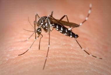
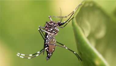 Aedes aegypti mosquito. Courtesy of Wikimedia Commons (https://commons.wikimedia.org/wiki/File:Aedes_aegypti.jpg; author Muhammad Mahdi Karim).
Aedes mosquito species have adapted well to human habitation, often breeding around dwellings in small amounts of stagnant water found in old tires or other small containers discarded by humans. Even a bottle cap filled with water can serve to incubate and hatch Aedes eggs. Eggs can survive periods of drying and will hatch when exposed to water. Humans are the preferred hosts.
Female Aedes mosquitoes are daytime feeders. They inflict an innocuous bite, usually on the back of the neck and the ankles, and are easily disturbed during a blood meal, causing them to move on to finish a meal on another individual, making them efficient vectors. Not uncommonly, entire families develop infection within a 24- to 36-hour period, presumably from the bites of a single infected mosquito.
Hosts for transmission
Humans serve as the primary reservoir for dengue. Certain nonhuman primates in Africa and Asia also serve as hosts but do not develop dengue hemorrhagic fever. Mosquitoes acquire the virus when they feed on a carrier of the virus. Persons with dengue viruses in their blood can transmit the viruses to the mosquito 1 day before the onset of the febrile period. The patient usually remains infectious for the subsequent 4-5 days (up to 12 days).
The mosquito can transmit dengue if it immediately bites another host. In addition, transmission occurs after 8-12 days of viral replication in the mosquito's salivary glands (extrinsic incubation period). The virus does not adversely affect the mosquito. The mosquito remains infected for the remainder of its life. The life span of A aegypti usually is 21 days but ranges from 15 to 65 days. Vertical transmission of dengue virus in mosquitoes has been documented. [23] The eggs of Aedes mosquitoes withstand long periods of desiccation, reportedly as long as 1 year, but are killed by temperatures of less than 10°C. Rare cases of vertical dengue transmission have been reported. In addition, rare reports of human-to-human transmission via needle-stick injuries have been published. [24] Dengue rarely has been described as transmitted by breast milk [25] or by sexual tranmission, [26, 27] though these are not common or significant modes of transmission.
Once inoculated into a human host, dengue has an incubation period of 3-14 days, (average 4-7 days) whereas viral replication takes place in target dendritic cells. Infection of target cells, primarily those of the reticuloendothelial system, such as dendritic cells, macrophages, hepatocytes, and endothelial cells, [28, 29, 30, 31] result in the production of immune mediators that serve to shape the quantity, type, and duration of cellular and humoral immune response to both the initial and subsequent virus infections. [28, 32, 33, 34, 35, 36, 37]
Dengue viral infections frequently are not apparent. In most cases, especially in children younger than 15 years, the patient is asymptomatic or has a mild undifferentiated febrile illness lasting 5-7 days. Classic dengue fever primarily occurs in nonimmune, nonindigenous adults and children and typically is self-limiting. Recovery usually is complete by 7-10 days. Severe dengue (dengue hemorrhagic fever/dengue shock syndrome) usually occur around the third to seventh day of illness during a second dengue infection in persons with preexisting actively or passively (maternally) acquired immunity to a heterologous dengue virus serotype.
Dengue fever
Dengue presents in a nonspecific manner similarly to that of many other viral and bacterial illnesses. Fever typically begins on the third day of illness and persists 5-7 days, abating with the cessation of viremia. Fever may reach 41C°. Occasionally, and more frequently in children, the fever abates for a day and recurs, a pattern that is termed a saddleback fever; however, this pattern more commonly is seen in dengue hemorrhagic fever.
Leukopenia, lymphopenia near the end of the febrile phase, and thrombocytopenia are common findings in dengue fever and are believed to be caused by direct destructive actions of the virus on bone marrow precursor cells. The resulting active viral replication and cellular destruction in the bone marrow are believed to cause the bone pain. Approximately one third of patients with dengue fever may have mild hemorrhagic symptoms, including petechiae, gingival bleeding, and a positive tourniquet test (>20 petechiae in an area of 2.5 X 2.5 cm). Dengue fever rarely is fatal.
Severe dengue (dengue hemorrhagic fever)
Severe dengue occurs less frequently than dengue fever but has a more dramatic clinical presentation. In most of Asia, where it first was described, severe dengue primarily is a disease of children. However, in the Americas, and more recently reported in Taiwan, severe dengue has an equal distribution in all ages.
Severe dengue typically begins with the initial manifestations of dengue fever. The acute febrile illness (temperatures ≤40°C), like that of dengue fever, lasts approximately 2-7 days. However, in persons with severe dengue, the fever reappears, giving a biphasic or saddleback fever curve.
Along with biphasic fever, patients with severe dengue have progressive thrombocytopenia, increasing hematocrit (20% absolute rise from baseline) and low albumin (signs of hemoconcentration preceding shock), more obvious hemorrhagic manifestations (>50% of patients have a positive tourniquet test), and progressive effusions (pleural or peritoneal). Lymphocytosis, often with atypical lymphocytes, commonly develops before defervescence or the onset of shock. Transaminase levels may be mildly elevated or present in the several thousands associated with hepatomegaly in those patients with acute hepatitis. Low fibrinogen and elevated fibrin split products are signs of disseminated intravascular coagulation. Severe metabolic acidosis and circulatory failure can occur.
The critical feature of severe dengue is plasma leakage. Plasma leakage is caused by increased capillary permeability and may manifest as hemoconcentration, as well as pleural effusion and ascites. Bleeding is caused by capillary fragility and thrombocytopenia and may manifest in various forms, ranging from petechial skin hemorrhages to life-threatening gastrointestinal bleeding.
Liver damage manifests as increases in levels of alanine aminotransferase and aspartate aminotransferase, low albumin levels, and deranged coagulation parameters (prothrombin time, partial thromboplastin time). [38, 39] In persons with fatal dengue hepatitis, infection was demonstrated in more than 90% of hepatocytes and Kupffer cells with minimal cytokine response (tumor necrosis factor [TNF]–alpha, interleukin [IL]–2). This is similar to that seen with fatal yellow fever and Ebola infections. [38]
As the term implies, severe dengue shock is essentially dengue hemorrhagic fever with progression into circulatory failure, with ensuing hypotension, narrow pulse pressure (< 20 mm Hg), and, ultimately, shock and death if left untreated. Death may occur 8-24 hours after onset of signs of circulatory failure. The most common clinical findings in impending shock include hypothermia, abdominal pain, vomiting, and restlessness.
Secondary infection
The immunopathology of severe dengue remains incompletely understood. Most patients who develop severe dengue have had prior infection with 1 or more dengue serotypes. When an individual is infected with another serotype (ie, secondary infection) and produces low levels of nonneutralizing antibodies, these antibodies, directed against 1 of 2 surface proteins (precursor membrane protein and envelope protein), when bound by macrophage and monocyte Fc receptors, have been proposed to fail to neutralize virus and instead form an antigen-antibody complex.
This results in increased viral entry into macrophages bearing IgG receptors, allowing unchecked viral replication with higher viral titers and increased cytokine production and complement activation, a phenomenon called antibody-dependent enhancement. [40, 41]
The affected macrophages release vasoactive mediators that increase vascular permeability, leading to vascular leakage, hypovolemia, and shock. This mechanism, along with individual host and viral genome variations, plays an active role in pathogenesis. Infants born to mothers who have had dengue, as maternally derived dengue neutralizing IgGs wane, also are thought to be at risk for enhanced disease. [40, 41]
Some researchers suggest that T-cell immunopathology may play a role, with increased T-cell activation and apoptosis. Increased concentrations of interferon have been recorded 1-2 days following fever onset during symptomatic secondary dengue infections. [42] The activation of cytokines, including TNF-alpha, TNF receptors, soluble CD8, and soluble IL-2 receptors, has been correlated with disease severity. [28]
Cuban studies have shown that stored serum sample analysis demonstrated progressive loss of cross-reactive neutralizing antibodies to DENV-2 as the interval since DENV-1 infection increased. [35] In addition, certain dengue strains, particularly those of DENV-2, have been proposed to be more virulent, in part because more epidemics of dengue hemorrhagic fever have been associated with DENV-2 than with the other serotypes.
DENV-2–activated platelets were phagocytized in large numbers when the platelet activation inhibitor prostacyclin was added. [43]
Several recent studies have investigated the causes of thrombocytopenia in dengue. Laboratory and human studies have suggested a direct correlation between activation and depletion of platelets, with a sharp drop occurring on day 4 of fever. A high number of dengue virus genome copies have been found in these activated platelets. Increased binding of complement C3 and IgG also have been found on the surface of these platelets. In addition to platelet activation, dengue infection has been found to activate the intrinsic pathway of apoptosis, with increased surface phosphatidylserine exposure, mitochondrial depletion, and activation of caspase 3 and 9. [44]
Etiology
Dengue infection is caused by dengue virus (DENV), which is a single-stranded RNA virus (approximately 11 kilobases long) with an icosahedral nucleocapsid and covered by a lipid envelope. The virus is in the family Flaviviridae, genus Flavivirus, and the type-specific virus is yellow fever.
The dengue virus has 4 related but antigenically distinct serotypes: DENV-1, DENV-2, DENV-3, and DENV-4. Genetic studies of sylvatic strains suggest that the 4 serotypes evolved from a common ancestor in primate populations approximately 1000 years ago and that all 4 separately emerged into a human urban transmission cycle 500 years ago in either Asia or Africa. [10, 45] Albert Sabin speciated these viruses in 1944. Each serotype is known to have several different genotypes. Viral genotype and serotype, and the sequence of infection with different serotypes, appear to affect disease severity.
Living in endemic areas of the tropics (or warm, moist climates such as the southern United States) where the vector mosquito thrives is an important risk factor for infection. [21, 46, 47, 48, 49] Poorly planned urbanization combined with explosive global population growth brings the mosquito and the human host into close proximity. Increased air travel easily transports infectious diseases between populations.
Epidemiology
United States statistics
In the United States, of the 100-200 reported cases per year, dengue principally occurs in travelers returning from endemic transmission areas. During 2006–2008, an average of 244 confirmed and probable travel-associated dengue cases were reported in the United States, according to the US Centers for Disease Control and Prevention (CDC). However, 2019 was an exception to these numbers, with over a thousand cases of dengue reported in US states. [50] The CDC reports that cases of dengue in returning US travelers have increased steadily, and dengue has become the leading cause of acute febrile illness in US travelers returning from the Caribbean, South America, and Asia. [51]
Dengue once was epidemic in the southeastern United States, and the potential exists for its reemergence. The principal mosquito vector for dengue, A aegypti, is found in the southern and southeastern United States, along with A albopictus, a less efficient vector species introduced in 1985. A aegypti breeds year-round in southern Florida.
Most dengue cases in US citizens occur in Puerto Rico, the US Virgin Islands, Guam, and Samoa. Puerto Rico has experienced several seasonal outbreaks since 2015, with more significant transmission occurring from August to November. The principal mosquito vector for dengue, A aegypti, is found in the southern and southeastern United States, along with A albopictus, a less efficient vector species introduced in 1985. The last dengue epidemic in Florida (in the Tampa and Miami areas) occurred in 1934-1935 and affected an estimated 15,000 people of the population of 135,000 in Miami. The last recorded epidemic in the southeastern United States occurred in Louisiana in 1945. Outbreaks of dengue also occurred in Laredo, Texas, in 1998. A aegypti breeds year-round in southern Florida. Dengue reemerged in Florida in 2009-2010, however, with 27 locally acquired cases in Key West. [51] The index case in this outbreak was diagnosed after returning home to New York from a visit to Key West. This illustrates the importance of awareness of dengue among physicians outside endemic areas. In 2013, 23 cases were reported in Martin County, Florida. During that year, 120 cases were imported into Florida from various countries. [52] Since January 2010, dengue has been a reportable disease in the United States. [51]
International statistics
The overall incidence of dengue, as well as the number of explosive outbreaks of dengue, has increased dramatically over the last few decades. Older data suggested an estimated 50-100 million cases of dengue fever and 500,000 cases of dengue hemorrhagic fever occur worldwide, with 22,000 deaths (mainly in children). [53, 54, 55] In 2015, official data from WHO member states reported more than 3.2 million cases, with 2.35 million cases in the Americas alone, including 10,200 cases of severe dengue and 1181 deaths. One study estimates that approximately 390 million dengue infections occur per year (95% CI; 284-528 million), with 96 million of these presenting clinically. [56] An estimated 2.5-3 billion people (approximately 40-50% of the world’s population) are estimated to be at risk for dengue infection. Some 128 countries worldwide are at risk for dengue infection, which includes 36 that once had been classified as dengue-free. [57] Globalization and expansion of the habitat for the Aedes aegypti mosquito both play a role in the changing epidemiology of dengue. The only continent that has not experienced dengue transmission is Antarctica.
According to the World Health Organization, dengue ranks as the most important mosquito-borne viral disease in the world. In the last 50 years, the incidence of dengue has increased 30-fold worldwide. [55] In the Americas alone, the incidence rose from 250,000 cases of dengue fever and 7,000 cases of dengue hemorrhagic fever in 1995 to more than 890,000 cases of dengue fever and 26,000 cases of dengue hemorrhagic fever in 2007. Dengue has a seasonal pattern, with the majority of cases in the Southern hemisphere occurring during the first half of the year and the majority of cases in the Northern hemisphere occuring during the second half of the year.
The world's largest known epidemic of dengue occurred in Cuba in 1981, with more than 116,000 persons hospitalized and as many as 11,000 cases reported in a single day. Current outbreaks can be monitored via the ProMed listserve by contacting owner-promed@promedmail.org.
Since 2000, at least 8 areas previously without dengue have reported outbreaks, including Nepal, Bhutan, Macau, Hong Kong, Taiwan, [58] Madagascar, the Galapagos, and Easter Island.
Southeast Asia
Dengue hemorrhagic fever is one of the leading causes of hospitalization and death in children in many Southeast Asian countries, with Indonesia reporting the majority of dengue hemorrhagic fever cases. Of interest and significance in prevention and control, 3 surveillance studies in Asia report an increasing age among infected patients and increasing mortality rate.
A 5-year prospective study in Thai children examined the relative economic burden of dengue infection in children on the local population. Most disability-adjusted life years (DALYs) lost to dengue resulted from long-duration illness in children who had not been hospitalized. The infecting serotype appeared to be a determining factor of DALYs lost, with DENV-2 and DENV-3 responsible for 30% and 29%, respectively. The mean cost of illness from dengue was significantly higher than that from other febrile illnesses. [59]
Since 1982 in Singapore, more than 50% of deaths have occurred in individuals older than 15 years. In Indonesia, young adults in Jakarta and provincial areas make up a larger percentage of infected patients. During the 2000 epidemic in Bangladesh, up to 82% of hospitalized patients were adults, and all deaths occurred in patients older than 5 years.
Africa
The epidemiology of dengue fever in Africa is more poorly characterized. Aedes aegypti is present in a large portion of the Middle East and sub-Saharan Africa. Dengue fever is present in 19 countries on the African continent. Of note, no major dengue hemorrhagic fever epidemics have occurred in Africa, despite the fact that all 4 dengue serotypes circulate on the continent. This may be explained by a genetic factor in these populations.
The Americas
Hyperendemic circulation of all 4 dengue serotypes is present in the northern countries of South America. Brazil, Colombia, and Venezuela report the most cases of dengue and dengue hemorrhagic fever, with low-level transmission occurring year-round but with most occurring during periods of epidemic transmission. Since the 1970s, outbreaks of dengue fever have increased in frequency and severity in the Caribbean.
Race-, sex-, and age-related demographics
The distribution of dengue is geographically determined. Dengue affects all races. Some African and Haitian data demonstrate a relative dearth of dengue hemorrhagic fever and dengue shock syndrome during dengue fever epidemics, suggesting that these populations may share a genetic advantage to the virus. This merits further study.
A study of dengue incidence in 6 different Asian countries reported a higher incidence in males than in females. The authors speculated that the difference may result from different gender roles and subsequent differences in exposure risks. [60]
Dengue affects people of all ages. However, children younger than 15 years typically present with only a nonspecific, self-limited, febrile illness. In endemic areas, a high prevalence of immunity in adults may limit outbreaks to children.
In Southeast Asia, where dengue is hyperendemic, dengue hemorrhagic fever usually affects children younger than 15 years. However, in the Americas, where dengue is becoming progressively hyperendemic, dengue hemorrhagic fever shows no age predilection.
Prognosis
Dengue fever typically is a self-limiting disease with a mortality rate of less than 1%. When treated, dengue hemorrhagic fever has a mortality rate of 2-5%. When left untreated, dengue hemorrhagic fever has a mortality rate as high as 50%. Survivors usually recover without sequelae and develop immunity to the infecting serotype.
The fatality rate associated with severe dengue varies by country, from 12-44%. In a 1997 Cuban epidemic, the fatality rate in patients who met criteria for severe dengue was approximately 6%. The mortality rate associated with dengue fever is less than 1%. Data from the 1997 Cuban epidemic suggest that, for every clinically apparent case of dengue fever, 13.9 cases of dengue infection went unrecognized because of absent or minimal symptoms.
A 2005 review from Singapore of 14,209 patients found that useful predictors of death included the following [61] :
-
Atypical presentations
-
Significant comorbid illness
-
Abnormal serum markers (including albumin and coagulation studies)
-
Secondary bacterial infections
Factors that affect disease severity include the following:
-
Patient age
-
Pregnancy
-
Nutritional status
-
Ethnicity
-
Sequence of infection with different dengue serotypes
-
Virus genotype
-
Quality and extent of available medical care
Complications and sequelae of dengue virus infections are rare but may include the following:
-
Cardiomyopathy
-
Seizures, encephalopathy, and viral encephalitis
-
Hepatic injury
-
Depression
-
Pneumonia
-
Iritis
-
Orchitis
-
Oophoritis
In 20-30% of dengue hemorrhagic fever cases, the patient develops shock, known as the dengue shock syndrome. Worldwide, children younger than 15 years constitute 90% of dengue hemorrhagic fever patients [53] ; however, in the Americas, dengue hemorrhagic fever occurs in both adults and children.
Although dengue is an extremely important arboviral illness globally, literature evaluating the economic impact is fairly sparse, with some conflicting findings. A recent expert panel assessment and 2 studies in the Americas recommended additional research to fill important information gaps, including disease outcomes and accurate statistics regarding disease burden, that could better inform future decision making regarding control and prevention. [62, 63, 64]
A 5-year prospective study in Thai children examined the relative economic burden of dengue infection in children on the local population. Most disability-adjusted life years (DALYs) lost to dengue resulted from long-term illness in children who had not been hospitalized. The infecting serotype appeared to be the major determinant of DALYs lost, with DEN-2 and DEN-3 responsible for 59%. The mean cost of illness from dengue was significantly higher than that from other febrile illnesses studied. [59]
A prospective study examined the direct and indirect costs of dengue infection in 1695 pediatric and adult patients in 8 countries. The average illness lasted 11.9 days for ambulatory patients and 11 days for hospitalized patients. Hospitalized students lost 5.6 days of school. Those at work lost 9.9 work days. Overall mean costs were more than double (1394 international dollars [I$]) for hospitalized cases. With an annual average of 594,000 cases the aggregate economic cost was estimated to be at least I$587 million, without factoring in underreporting of disease and dengue surveillance and vector control costs. This represents a significant global economic burden in low-income countries. [64]
Patient Education
Educate patients, especially those who have experienced prior dengue fever, to avoid mosquito bites, including the use of appropriate mosquito repellants and peridomestic vector control, when traveling to dengue-endemic areas. Current evidence suggests that those with a history of dengue fever are at highest risk for dengue hemorrhagic fever or dengue shock syndrome if they are infected with a different dengue strain.
Information for reducing risk of contracting dengue while traveling, as well as current information on dengue outbreaks, is available at the US Centers for Disease Control and Prevention Travel & Dengue Outbreaks Web page. Information on dengue and alerts on current outbreaks are also available through the World Health Organization Web site.
-
Drawing of Aedes aegypti mosquito. Courtesy of the Centers for Disease Control and Prevention (CDC).
-
Aedes albopictus. Courtesy of the Centers for Disease Control and Prevention (CDC).
-
Worldwide distribution of dengue in 2000. Courtesy of the Centers for Disease Control and Prevention (CDC).
-
Worldwide distribution of dengue in 2003. Courtesy of the Centers for Disease Control and Prevention (CDC).
-
Worldwide distribution of dengue in 2005. Courtesy of the Centers for Disease Control and Prevention (CDC).
-
Increasing rates of dengue infection by regions of the world. Courtesy of the World Health Organization (WHO).
-
Dengue transmission cycle. Courtesy of the Centers for Disease Control and Prevention (CDC).
-
Reinfestation by Aedes aegypti in the Americas after the 1970 (left) mosquito eradication program and most recent distribution as of 2002 (right). Courtesy of the Centers for Disease Control and Prevention (CDC).
-
A child with dengue hemorrhagic fever or dengue shock syndrome may present severely hypotensive with disseminated intravascular coagulation (DIC), as this severely ill pediatric ICU patient did. Crystalloid fluid resuscitation and standard DIC treatment are critical to the child's survival.
-
Delayed capillary refill may be the first sign of intravascular volume depletion. Hypotension usually is a late sign in children. This child's capillary refill at 6 seconds was delayed well beyond a normal duration of 2 seconds.
-
Signs of early coagulopathy may be as subtle as a guaiac test that is positive for occult blood in the stool. This test should be performed on all patients in whom dengue virus infection is suspected.
-
Aedes aegypti mosquito. Courtesy of Wikimedia Commons (https://commons.wikimedia.org/wiki/File:Aedes_aegypti.jpg; author Muhammad Mahdi Karim).
-
Global map of dengue risk. Frequent or continuous risk = frequent outbreaks or ongoing transmission. Sporadic or uncertain risk = variable and unpredictable risk, country-level data are unavailable. Courtesy of the Centers for Disease Control and Prevention (CDC) (https://www.cdc.gov/dengue/areaswithrisk/around-the-world.html).


