Practice Essentials
Chorioretinitis (CR) is an inflammatory process that involves the uveal tract of the eye. (See the image below.) In neonates, the inflammation is usually caused by congenital viral, bacterial, or protozoal infections. Medical care focuses on the establishment of specific therapies for treatable causes and on the stabilization of the patient with chorioretinitis to prevent further loss of vision, especially in immunocompromised infants and children.
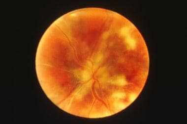 Chorioretinitis in a patients with acquired immunodeficiency syndrome (AIDS).
Signs and symptoms
If the inflammation is unilateral, the child may squint, favor the "good eye," or report blurred vision or an inability to see objects. Older children with chorioretinitis may present with photophobia and clumsiness with poor walking balance.
Ophthalmologic examination can reveal exudative "cotton balls" (ie, focal atrophic and pigmented scars of the retina). Vitreous inflammations can manifest as transient floating opacities.
See Presentation for more detail.
Diagnosis
Laboratory studies
Routine laboratory screening in chorioretinitis includes the following:
-
Complete blood cell count and platelet count
-
Liver function tests
-
Renal function tests
Imaging studies
A pediatric ocular imaging system (RetCam3) may be used in newborns, infants, and uncooperative children. Other imaging studies include the following:
-
Sonography, computed tomography (CT) scanning, and/or magnetic resonance imaging (MRI) of the central nervous system (CNS) or specific organs in patients in whom congenital infections are suspected or in an immunocompromised host
-
Chest radiography, CT scanning, or both in patients in whom tuberculosis or sarcoidosis is suspected
-
CT scanning of the abdomen for hepatic or splenic dissemination of disease
-
Radiography or bone scanning of the skeleton in patients in whom syphilis is suspected
-
Echocardiography in patients with congenital rubella
See Workup for more detail.
Treatment
Medical care in chorioretinitis focuses on the establishment of specific therapies for treatable etiologies and on the stabilization of the patient to prevent further loss of vision, especially in immunocompromised infants and children. Vitrectomy is usually not needed and is reserved for severe cases that are resistant to conservative medical treatment.
See Treatment and Medication for more detail.
Background
Inflammation associated with chorioretinitis is usually caused by congenital viral, bacterial, or protozoal infections in neonates. Congenital toxoplasma and cytomegalovirus (CMV) infection are the most common etiologies in this age group. Fungal infections are commonly identified, and emergent pathogens such as West Nile virus and lymphocytic choriomeningitis virus (LCMV) have been described. [1, 2] In rare instances, chorioretinitis is part of a systemic noninfectious process.
Chorioretinitis associated with congenital viral infections like CMV tends to be stable or improve in infancy, whereas chorioretinitis associated with asymptomatic congenital toxoplasmosis (CTP) progresses for years after birth and is more likely to be clinically significant at an older age.
Although CMV is the most common congenital infection in the developed world, affecting approximately 1% of all infants born in the United States, only 10% of all infants born in the United States with congenital CMV infection have symptomatic disease at birth, including chorioretinitis. [3]
Congenital disseminated infections such as CMV and toxoplasmosis may also manifest with extraocular findings such as intrauterine growth retardation, microcephaly, microphthalmia, cataract, uveitis, hearing defect, osteomyelitis, hepatosplenomegaly, lymphadenopathy, dermal erythropoiesis, carditis, and congenital heart disease.
Beyond the neonatal period, chorioretinitis can be diagnosed in diverse clinical conditions and can reflect newly acquired diseases or reactivation. CTP is the most common cause of infectious chorioretinitis in immunocompetent children. [4] Chorioretinitis can also result from a dissemination of parasitic infections like Toxocara or Baylisascaris (the raccoon roundworm) in immunocompetent patients. [5] In severely immunodeficient patients, including those with acquired immunodeficiency syndrome (AIDS), chorioretinitis may be associated with Epstein-Barr virus (EBV), CMV, varicella-zoster virus, various fungi (eg, Candida, Aspergillus, Fusarium, dimorphic fungi), and Toxoplasma. [6] See the image below.
Chorioretinitis in a patients with acquired immunodeficiency syndrome (AIDS).
In addition, with increasing air travel and globalization, several emerging infectious diseases have been recognized as causing ocular disease, including retinitis, chorioretinitis, retinal vasculitis, and optic nerve involvement. These include rickettsiosis, Rift Valley fever, dengue fever, and Chikungunya virus. [7]
Pathophysiology
Chorioretinitis affects the uveal tract, which consists of the iris, ciliary body, and choroid. Inflammatory conditions are generally classified according to the predominant compartment of involvement (eg, anterior and posterior uveitis). Inflammation of the posterior uveal tract of the eye is generally termed choroiditis; because the retina is invariably involved, the terms chorioretinitis or retinochoroiditis are generally used. [8]
The extent of ocular involvement depends on the organism. Bilateral focal or extensive exudative chorioretinitis or panuveitis may be seen in patients with Toxoplasma gondii infection. A single large choroidal lesion with extensive inflammation or endophthalmitis is usually observed in patients with Toxocara canis, whereas interstitial keratitis or iritis is most common in patients with Treponema pallidum. Strabismus and optic atrophy may accompany chorioretinitis caused by CMV. The central retinal lesions of CMV cannot be clinically distinguished from those of toxoplasmosis. However, unlike congenital toxoplasma infection, the retinitis caused by CMV does not progress. [8, 9]
Vessel trauma caused by other organisms, such as Toxocara or Baylisascaris larvae, may be associated with severe inflammatory responses.
Etiology
Congenital infection
In immunocompetent children, chorioretinitis is usually associated with congenital infection; acquired infection is a less likely cause. T gondii and CMV are the leading causes of congenital infections associated with chorioretinitis.
Viral etiologies include vertical or perinatal infections, including HSV, rubella, varicella, Epstein-Barr virus (EBV), lymphocytic choriomeningitis virus (LCMV) and, possibly, flavivirus. With the recent increase in the incidence of congenital infection after being at a nadir since 1991, syphilis should be considered in an infant born with chorioretinitis whose mother has untreated or inadequately treated syphilis, particularly if she also has human immunodeficiency virus infection (HIV). [10, 11] Distinguishing these infections from perinatal transmission of other viral illnesses, including HSV, hepatitis B, and HIV is important. The risk of intrauterine infection is highest in infants of women with primary infection and is much less with recurrent infections.
Acquired chorioretinitis may occur in immunocompetent children. Some children who ingest embryonated T canis or Baylisascaris procyonis eggs may develop visceral larva migrans or ocular larva migrans. Another acquired infection that may lead to chorioretinitis is B henselae. [12] More than 90% of patients with catscratch disease have a history of recent contact with a cat, often a kitten, and 50-87% of these patients have been scratched.
Immunocompromised children
Chorioretinitis may be associated with systemic infection due to a vast array of pathogens. Any of the infections discussed above may be seen; however, the presentation in an immunocompromised individual may be atypical. Other infections may include congenital or acquired Lyme disease, Yersinia enterocolitica, and Mycobacterium tuberculosis (MTB). [13, 14]
Invasive fungal infections may result from Candida, Cryptococcus species, and histoplasmosis. [15] A species of blackfly (Simulium species) can transmit onchocerciasis (in tropical Africa, Yemen, Saudi Arabia, and parts of Latin America). [16]
Noninfectious disease
Systemic noninfectious disease, such as sarcoidosis, collagen vascular disease, chronic granulomatous disease (CGD), Behcet disease, and juvenile rheumatoid arthritis may cause chorioretinitis. [17]
Other possible noninfectious processes include chronic infantile, neurological, cutaneous, and articular syndrome (CINCA) syndrome, also known as neonatal onset multisystemic inflammatory disease. [18]
Epidemiology
United States data
Chorioretinitis due to CTP occurs much less frequently in the United States than in Europe. Rates of seroprevalence vary and depend on the population studied. An estimated 400-4,000 cases of CTP occur in the United States each year. [19] Rates of seroprevalence are much higher in certain European countries (eg, France, Denmark, Germany) where active surveillance systems are in place to detect symptomatic and asymptomatic cases. [20, 21] The risk of retinochoroiditis rises from 10% in infancy to approximately one third by age 12 years in children whose infection was identified by screening. By school age, 20% of infected children with CTP have one or more retinochoroidal lesion. [22] More than 90% of children have normal vision in their best eye; severe bilateral impairment is rare.
One of the most commonly acquired childhood eyesight impairments in the United States is due to T canis, probably because of the high prevalence of young pet dogs. The incidence is higher in people living in the south-central and southeastern parts of the country. Annually, more than 700 people infected with Toxocara experience permanent partial loss of vision. [23]
Age-related demographics
Chorioretinitis due to congenital infections or occasionally other causes is usually evident at birth; progression and prognosis depends on the etiology. Acquired chorioretinitis occurs at any age, depending on the underlying illness.
Prognosis
Except when caused by congenital toxoplasmosis (CTP), prognosis for individuals with chorioretinitis depends on the originating process but tends to be self-limited. Chorioretinitis due to CTP is progressive, and the outcome is not usually predictable. Late-onset retinal lesions can occur many years after birth, but the overall ocular prognosis of congenital toxoplasmosis is satisfactory when the infection is identified early and treatment is instituted appropriately. [24]
In patients with symptomatic congenital CMV infection, chorioretinitis was found to be a risk factor associated with the development of cortical visual impairment. [25] Thus, the researchers recommend yearly ophthalmologic examinations for such patients.
Morbidity/mortality
If left untreated or if the condition does not respond to treatment, severe chorioretinitis can result in partial or total loss of vision in the affected eye. Morbidity is due to concurrent damage to major organ systems, especially damage to the brain (eg, developmental delays, seizures). Mortality due to chorioretinitis depends on the nature and progression of the underlying illness.
Complications
A delay in the initiation of specific therapies for treatable etiologies of the patient with chorioretinitis results in further loss of vision especially in immunocompromised infants and children.
Patient Education
Aim educational efforts at reducing the incidence of primary toxoplasmosis in pregnant women. Screen pregnant women for the presence of toxoplasmosis immunoglobin (Ig)G and educate these individuals to avoid consuming undercooked meat and handling a cat litter box.
In the summer and fall seasons, public health measures can be used to reduce the risk of mosquito-borne viral encephalitis or tick-borne Lyme disease. During peak season for mosquitos and ticks, educate pregnant women to avoid insect bites (eg, cover up, apply insecticide to clothing items) and carry out limited larvicidal spraying to control mosquito infestation.
-
Chorioretinitis in a patients with acquired immunodeficiency syndrome (AIDS).


