Background
Worldwide, shock is a leading cause of morbidity and mortality in the pediatric population. Shock is defined as a state of acute energy failure due to inadequate glucose substrate delivery, oxygen delivery, or mitochondrial failure at the cellular level. The clinical state of shock is diagnosed on the basis of vital signs, physical examination, and laboratory data, although its recognition in the pediatric patient can be difficult.
Delay in recognizing and quickly treating a state of shock results in anaerobic metabolism, tissue acidosis, and a progression from a compensated reversible state to an irreversible state of cellular and organ damage. Morbidity from shock may be widespread and can include central nervous system (CNS) failure, respiratory failure (ie, from muscle fatigue or acute respiratory distress syndrome [ARDS]), renal failure, hepatic dysfunction, gastrointestinal ischemia, disseminated intravascular coagulation (DIC), metabolic derangements, and ultimately death. [1, 2, 3] (See Pathophysiology and Etiology.)
This article reviews the common physiologic foundations of shock that underpin all patients with this condition as well as defines the different pathophysiologic classifications of shock and their etiologies. The defining clinical findings of shock are described, and current diagnostic and therapeutic strategies are presented to help guide the most effective and appropriate treatment for resuscitating the child in shock. (See Pathophysiology, Presentation, Workup, Treatment, and Medication.)
Pathophysiology
Shock and shock-states are ultimately due to circulatory failure to deliver adequate substrate and remove toxins at the tissue and cellular levels.
In the nonstressed physiologic state, adequate oxygen and glucose are delivered intracellularly to mitochondria that generate 36 adenosine triphosphate (ATP) molecules per glucose molecule via aerobic metabolism and the Krebs cycle. In the stressed state in children, the ability to compensate via gluconeogenesis and glycogenolysis is limited due to small hepatic and skeletal muscle masses. Thus, glycolysis and secondary fat metabolism become the primary sources of energy substrate.
Cellular metabolism then becomes much less efficient as pyruvate is converted to lactate instead of acetyl-CoA. This shift in metabolic pathways generates only two ATP molecules per molecule of glucose and results in the accumulation of lactic acid. Ultimately, cell membrane ion pump dysfunction occurs, acidosis progresses, intracellular edema develops, intracellular contents leak into the extracellular spaces, and cell death ensues.
Key physiologic parameters that affect metabolic homeostasis include tissue blood flow, the balance between oxygen delivery and demand, and the oxygen content. Although the sufficiency of tissue oxygenation cannot be directly measured and varies by time and tissue-type, the relationship of oxygenation delivery and consumption is key to understanding the pathophysiology of shock. It is defined as follows:
VO2 = DO2 x O2 ER
Where VO2 represents oxygen consumption, DO2 represents total arterial flow of oxygen, and O2 ER is the oxygen extraction ratio (%).
In the normal state, oxygen demand is independent of delivery, as DO2 is greater than VO2. When demand increases, physiologic compensation occurs to match the need, such as through an increased heart rate and stroke volume. In shock, as DO2 declines, O2 ER increases to maintain adequate tissue oxygenation. At a critical point (DO2 crit), however, O2 ER can no longer compensate, and VO2 becomes dependent on DO2. Beyond this threshold, oxygen debt develops and blood lactate levels rise. Thus, further understanding the components of DO2 is also fundamental to the pathophysiology and treatment of shock.
Oxygen delivery
DO2 depends on the amount of blood pumped per minute, or cardiac output (CO), and the arterial oxygen content of that blood (CaO2). Thus, DO2 may be defined by the following equation:
DO2 = CaO2 × CO
The CaO2 depends on how much oxygen-carrying capacity is available, which is primarily a function of the hemoglobin (Hb) level and the arterial oxygen saturation (SaO2). A small, but typically insignificant, amount of oxygen is directly dissolved in the blood rather than bound to Hb. Therefore, CaO2 may be defined by the following formula:
CaO2 = (Hb × SaO2 × 1.34) + (0.003 X PaO2)
CO depends on the amount of blood pumped with each heartbeat, known as stroke volume (SV), and the heart rate (HR). SV depends on the ventricular end-diastolic filling volume (commonly referred to as ventricular preload), the state of myocardial contractility, and the afterload (systemic vascular resistance [SVR]) on the heart. Each of these variables affect CO and can be impaired in clinical shock states. Thus, the following relationships are observed [1] :
CO = HR × SV
SV ∝ preload, contractility, and afterload
See the image below for the factors that determine cardiac function and oxygen delivery to tissues.
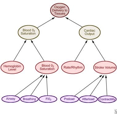 Determinants of cardiac function and oxygen delivery to tissues. FiO2 = fraction of inspired oxygen. Adapted from Strange GR. APLS: The Pediatric Emergency Medicine Course. 3rd ed. Elk Grove Village, Ill: American Academy of Pediatrics; 1998:34.
The recognition and treatment of pediatric shock is dependent on an understanding of these physiologic principles and definitions. Once understood, the different clinical presentations and causes of shock, as well as their most appropriate treatment strategies, are more easily appreciated.
Etiology
Several etiologic classifications of shock are recognized. The major categories are as follows:
-
Hypovolemic
-
Cardiogenic
-
Distributive
-
Obstructive
In each of these categories of shock, one or more of the physiologic principles that govern oxygen delivery or consumption is disturbed.
Hypovolemia leads to decreased cardiac filling, lower end-diastolic volume, and decreased stroke volume in accordance with the Frank-Starling curve and, therefore, results in decreased cardiac output. Hypovolemia due to hemorrhage additionally decreases oxygen-carrying capacity through the direct loss of available hemoglobin.
Cardiogenic shock resulting from congenital heart disease or cardiomyopathies develops from primary pump failure and inadequate cardiac output.
Distributive shock from sepsis, anaphylaxis, or high-level spinal cord injury results in peripheral vasodilation and decreased systemic vascular resistance (SVR), with venous pooling and inadequate arterial tissue perfusion to meet demand metabolic demands.
Obstructive causes of shock such as pulmonary embolism, pneumothorax, and cardiac tamponade impede either pulmonary outflow, systemic outflow, or both, thereby directly decreasing cardiac output.
Hypovolemic shock
Hypovolemic shock results from an absolute deficiency of intravascular blood volume. It is a leading cause of pediatric mortality in the United States and worldwide, although the specific causative agents may be different globally. Causes of hypovolemic shock include the following:
-
Intravascular volume loss (eg, from gastroenteritis, burns, diabetes insipidus, heat stroke)
-
Hemorrhage (eg, from trauma, surgery, gastrointestinal bleeding) (see the image below)
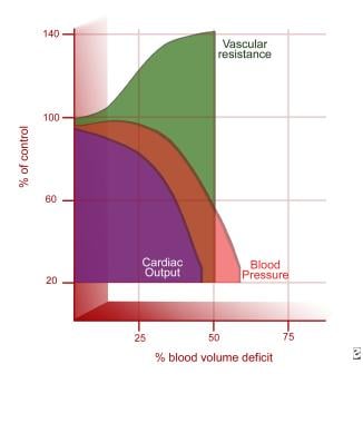 Hemodynamic response to shock hemorrhage model (based on normal data). Adapted from Adapted from Schwaitzberg SD, Bergman KS, Harris BH. A Pediatric Trauma Model of Continuous Hemorrhage. J Pediatr Surg. Jul 1988;23(7):605-9.
-
Interstitial loss (eg, from burns, sepsis, nephrotic syndrome, intestinal obstruction, ascites)

Gastroenteritis
Children with gastroenteritis may lose 10-20% of their circulating volume within 1-2 hours. [2] Rehydration is often impeded by concurrent vomiting, and clinical deterioration may be rapid. Common infectious causes of gastroenteritis include bacteria such as Salmonella, Shigella,Campylobacter species (spp), Escherichia coli, and Vibrio cholerae as well as viruses such as rotaviruses, adenoviruses, noroviruses, and enteroviruses. Worldwide, amebiasis and cholera are also important causes.
Hemorrhage
In the United States, the leading cause of death in children younger than 1 year is unintentional injury. [4] A major component of traumatic death is hemorrhage. In the pediatric patient, primary sites of hemorrhage include intracranial, intrathoracic, intra-abdominal, pelvic, and external. In the pediatric patient in shock without a clear etiology and absent history, occult hemorrhage secondary to nonaccidental trauma should be considered. [5]
Third spacing
Other causes of hypovolemia include capillary leak and tissue third spacing, which results in leakage of fluid out of the intravascular space into the interstitial tissues. Etiologies include burns, sepsis, and other causes of systemic inflammatory response syndrome (SIRS) (eg, pancreatitis, anaphylaxis). Patients with such etiologies may appear edematous and overloaded with total-body fluid; however, they may be significantly intravascularly depleted, with inadequate preload, and in significant shock. Through understanding of the physiologic disturbance affecting the intravascular volume and preload, it becomes clear that such patients need additional fluid administration despite their overall edematous appearance in order to improve total arterial flow of oxygen (DO2) and to prevent or correct a state of shock.
Distributive shock
In certain clinical states such as distributive shock, normal peripheral vascular tone becomes inappropriately relaxed. Vasodilation results in increased venous capacitance, causing relative hypovolemia even if the patient has not actually had any net fluid loss. As a result, the common physiologic disturbance that affects DO2 in distributive shock is a decrease in preload caused as a result of massive vasodilation and inadequate effective intravascular volume.
Common causes of distributive shock include anaphylaxis, neurologic injury (eg, head injury, spinal shock), sepsis, and drug-related causes. [6] Causes of anaphylaxis include the following:
-
Medications (eg, antibiotics, vaccines, other drugs)
-
Blood products
-
Envenomation
-
Foods
-
Latex
Anaphylaxis results in mast cell degranulation with resultant histamine release and vasodilation. Neurologic injury can interrupt sympathetic input to vasomotor neurons, resulting in vasodilation. Spinal shock may result from cervical cord injuries above T1, which interrupt the sympathetic chain, allowing for unopposed parasympathetic stimulation. Such patients may present with the clinical picture of hemodynamic instability and hypotension accompanied by bradycardia, because they lose sympathetic vascular tone (resulting in vasodilation) while being unable to mount an appropriate sympathetic-mediated tachycardic response. Medications may also cause vasodilation.
Finally, sepsis causes the release of many vasoactive mediators that may produce profound vasodilation, resulting in distributive shock.
Sepsis
Sepsis may be defined as a dysregulated, systemic inflammatory state that is triggered by the presence of probable or documented infection. [7, 8] Disturbances of virtually every variable in the DO2 equation may result from the presence of infectious agents such as endotoxin or gram-positive bacterial cell wall components. Systemic molecular cascade activation leads to the release of inflammatory mediators and cytokines (eg, tumor necrosis factor–alpha [TNF-alpha]), interleukins (such as IL-1, IL-2, IL-6), products of the coagulation cascade, bradykinins, and complement activation.
Nitric oxide synthase induction results in production of the potent direct vasodilator nitric oxide, leading to inappropriate and often massive regional and systemic vasodilation. This distributive effect reduces effective preload and impairs cardiac output (CO) and DO2. Circulating toxins and inflammatory mediators can also directly depress myocardial function and reduce cardiac contractility, adding a cardiogenic component to impaired CO. Sepsis may also disrupt capillary integrity, resulting in intravascular fluid leak into tissue third spaces, causing hypovolemia. Overactivation of the clotting cascade can result in disseminated intravascular coagulation (DIC)—DIC can directly obstruct critical tissue capillary beds, resulting in microvascular obstructive shock as well as hemorrhage.
Cardiogenic shock
Impairment of cardiac contractility defines cardiogenic shock. A decreased contractile state results in decreased stroke volume (SV) and CO and, therefore, in decreased DO2. Causes of cardiogenic shock include the following:
-
Arrrhythmias
-
Cardiomyopathies/carditis: Hypoxic/ischemic, infectious, metabolic, connective tissue diseases, neuromuscular disease, toxic reaction, idiopathic
-
Congenital heart disease
-
Trauma
-
Iatrogenic (ie, postoperative low cardiac output syndrome)
Obstructive shock
Obstructive shock occurs when either pulmonary or systemic blood flow is impaired as a result of either congenital or acquired obstruction, leading to CO impairment and shock. Causes include acute cardiac tamponade, tension pneumothorax, massive pulmonary embolism, and other forms of pulmonary or systemic circulation obstruction such as acute or acquired pulmonary hypertension or hypertrophic cardiomyopathy. Additional causes in the neonatal period include coarctation of the aorta, interrupted aortic arch, and severe aortic valvular stenosis.
In addition to medical management for obstructive shock, treatment often depends on prompt recognition and relief of the physical obstruction, such as through pericardiocentesis for tamponade or tube thoracostomy for pneumothorax. Neonates may require maintenance of the patency of the ductus arteriosus in order to bypass the obstruction until more definitive surgery can be performed.
Epidemiology
Pediatric practitioners treating acutely ill children, from neonates to young adults, are faced with different degrees and causes of shock on a regular basis, making shock in infants and children one of the most common and, often, life-threatening conditions encountered.
The frequencies of the different etiologies of shock vary around the world by country and age.
Worldwide in 2015, 2.6 million neonates younger than 1 month died. In the same year, among children aged 1-59 months, 5.8 million children died. The leading causes of death in children were preterm birth, neonatal encephalopathy, lower respiratory infections, diarrheal illness, congenital anomalies, malaria and sepsis. Although these etiologies may result in death via multiple mechanisms, they suggest that sepsis from communicable diseases and hypovolemia due to infectious gastroenteritis remain major causes of shock in developing countries. [9]
In developed countries such as the United States, pediatric shock accounts for 37% of transfers of care from community hospitals to tertiary medical centers. [10] These children have a higher mortality rate compared with patients not in shock (11.4% vs 2.6%, respectively) regardless of their trauma status. Additionally, early use of the recommended Pediatric Advanced Life Support (PALS) guidelines is associated with decreased mortality (8.69% vs 15.01%, respectively) and decreased functional morbidity (1.24% vs 4.23%, respectively). [10]
Of pediatric patients who present to the emergency department in shock, sepsis is the leading cause (57%), followed by hypovolemic shock (24%), distributive shock (14%), and cardiogenic shock (5%). [11] Between 2004 and 2012, the overall incidence of sepsis/septic shock appears to have increased from 3.7% to 4.4%, although mortality has declined 10.9% over the same period. [12]
-
Chest radiograph in a patient with cardiomegaly, which may accompany cardiogenic shock.
-
Determinants of cardiac function and oxygen delivery to tissues. FiO2 = fraction of inspired oxygen. Adapted from Strange GR. APLS: The Pediatric Emergency Medicine Course. 3rd ed. Elk Grove Village, Ill: American Academy of Pediatrics; 1998:34.
-
Hemodynamic response to shock hemorrhage model (based on normal data). Adapted from Adapted from Schwaitzberg SD, Bergman KS, Harris BH. A Pediatric Trauma Model of Continuous Hemorrhage. J Pediatr Surg. Jul 1988;23(7):605-9.
-
Pediatric shock management algorithm. ACTH = adrenocorticotropic hormone; CI = cardiac index; ECMO = extracorporeal membrane oxygenation; MAP-CVP = mean arterial pressure-central venous pressure; PALS = Pediatric Advanced Life Support; PDE = phosphodiesterase; PICU = pediatric intensive care unit; SVC O2 = superior vena cava oxygen saturation.
Tables

What would you like to print?
- Overview
- Presentation
- Workup
- Approach Considerations
- Comprehensive Metabolic Panel
- Blood Gas Analysis
- Complete Blood Count and Coagulation Studies
- Fluid Culture
- Chest Radiography
- Point-of-Care Ultrasound
- Mixed Venous Oxygen Saturation
- Central Venous Pressure
- Cardiac Output Monitoring
- Near-Infrared Spectroscopy
- B-Type Natriuretic Peptide
- Sepsis Biomarker Risk Model
- Show All
- Treatment
- Medication
- Media Gallery
- References