Lynch T, Kilgar J, Al Shibli A. Pediatric Abdominal Trauma. Curr Pediatr Rev. 2018. 14 (1):59-63. [QxMD MEDLINE Link].
Chaudhari PP, Rodean J, Spurrier RG, Hall M, Marin JR, Ramgopal S, et al. Epidemiology and management of abdominal injuries in children. Acad Emerg Med. 2022 Aug. 29 (8):944-953. [QxMD MEDLINE Link]. [Full Text].
Schmidt B, Schimpl G, Höllwarth ME. Blunt liver trauma in children. Pediatr Surg Int. 2004 Dec. 20 (11-12):846-50. [QxMD MEDLINE Link].
Stylianos S. Compliance with evidence-based guidelines in children with isolated spleen or liver injury: a prospective study. J Pediatr Surg. 2002 Mar. 37 (3):453-6. [QxMD MEDLINE Link].
Stylianos S. Evidence-based guidelines for resource utilization in children with isolated spleen or liver injury. The APSA Trauma Committee. J Pediatr Surg. 2000 Feb. 35 (2):164-7; discussion 167-9. [QxMD MEDLINE Link].
Clark P, Letts M. Trauma to the thoracic and lumbar spine in the adolescent. Can J Surg. 2001 Oct. 44 (5):337-45. [QxMD MEDLINE Link].
Hazen BJ, Keane OA, Vandewalle RJ, Grady Z, Wetzel M, Chern JJ, et al. Difference in Presentation and Concomitant Intra-Abdominal Injury with Chance Fracture in Pediatric and Adult Populations. Am Surg. 2023 Jun. 89 (6):2486-2491. [QxMD MEDLINE Link].
Cooper A, Barlow B, DiScala C, String D. Mortality and truncal injury: the pediatric perspective. J Pediatr Surg. 1994 Jan. 29 (1):33-8. [QxMD MEDLINE Link].
Nance ML, Sing RF, Branas CC, Schwab CW. Shotgun wounds in children. Not just accidents. Arch Surg. 1997 Jan. 132 (1):58-61; discussion 62. [QxMD MEDLINE Link].
Hobbs CJ. Abdominal injury due to child abuse. Lancet. 2005 Jul 16-22. 366 (9481):187-8. [QxMD MEDLINE Link].
Advanced Trauma Life Support. American College of Surgeons. Available at https://www.facs.org/quality-programs/trauma/atls. 2024; Accessed: April 4, 2024.
Stylianos S, Egorova N, Guice KS, Arons RR, Oldham KT. Variation in treatment of pediatric spleen injury at trauma centers versus nontrauma centers: a call for dissemination of American Pediatric Surgical Association benchmarks and guidelines. J Am Coll Surg. 2006 Feb. 202 (2):247-51. [QxMD MEDLINE Link].
Tataria M, Nance ML, Holmes JH 4th, Miller CC 3rd, Mattix KD, Brown RL, et al. Pediatric blunt abdominal injury: age is irrelevant and delayed operation is not detrimental. J Trauma. 2007 Sep. 63 (3):608-14. [QxMD MEDLINE Link].
Rothrock SG, Green SM, Morgan R. Abdominal trauma in infants and children: prompt identification and early management of serious and life-threatening injuries. Part I: injury patterns and initial assessment. Pediatr Emerg Care. 2000 Apr. 16 (2):106-15. [QxMD MEDLINE Link].
Rothrock SG, Green SM, Morgan R. Abdominal trauma in infants and children: prompt identification and early management of serious and life-threatening injuries. Part II: Specific injuries and ED management. Pediatr Emerg Care. 2000 Jun. 16 (3):189-95. [QxMD MEDLINE Link].
Stafford PW, Blinman TA, Nance ML. Practical points in evaluation and resuscitation of the injured child. Surg Clin North Am. 2002 Apr. 82 (2):273-301. [QxMD MEDLINE Link].
Stafford PW, Nance ML. Managing pediatric solid organ injury. J Am Coll Surg. 2002 Mar. 194 (3):394-5. [QxMD MEDLINE Link].
Wright MS. Update on pediatric trauma care. Curr Opin Pediatr. 1995 Jun. 7 (3):292-6. [QxMD MEDLINE Link].
Taylor GA, Kaufman RA, Sivit CJ. Active hemorrhage in children after thoracoabdominal trauma: clinical and CT features. AJR Am J Roentgenol. 1994 Feb. 162 (2):401-4. [QxMD MEDLINE Link].
Mizzi A, Shabani A, Watt A. The role of follow-up imaging in paediatric blunt abdominal trauma. Clin Radiol. 2002 Oct. 57 (10):908-12. [QxMD MEDLINE Link].
Kuas C, Acar N, Ozakin E, Karakilic E, Arda MS, Bastug BT, et al. The diagnostic value of laboratory tests in detecting solid organ injuries in pediatric patients with blunt abdominal trauma. Am J Emerg Med. 2022 Jul. 57:133-137. [QxMD MEDLINE Link].
Lim-Dunham JE, Narra J, Benya EC, Donaldson JS. Aspiration after administration of oral contrast material in children undergoing abdominal CT for trauma. AJR Am J Roentgenol. 1997 Oct. 169 (4):1015-8. [QxMD MEDLINE Link].
Bixby SD, Callahan MJ, Taylor GA. Imaging in pediatric blunt abdominal trauma. Semin Roentgenol. 2008 Jan. 43 (1):72-82. [QxMD MEDLINE Link].
Strouse PJ, Close BJ, Marshall KW, Cywes R. CT of bowel and mesenteric trauma in children. Radiographics. 1999 Sep-Oct. 19 (5):1237-50. [QxMD MEDLINE Link].
Shankar KR, Lloyd DA, Kitteringham L, Carty HM. Oral contrast with computed tomography in the evaluation of blunt abdominal trauma in children. Br J Surg. 1999 Aug. 86 (8):1073-7. [QxMD MEDLINE Link].
Tsang BD, Panacek EA, Brant WE, Wisner DH. Effect of oral contrast administration for abdominal computed tomography in the evaluation of acute blunt trauma. Ann Emerg Med. 1997 Jul. 30 (1):7-13. [QxMD MEDLINE Link].
Fang JF, Chen RJ, Wong YC, Lin BC, Hsu YB, Kao JL, et al. Pooling of contrast material on computed tomography mandates aggressive management of blunt hepatic injury. Am J Surg. 1998 Oct. 176 (4):315-9. [QxMD MEDLINE Link].
Acker SN, Stewart CL, Roosevelt GE, Partrick DA, Moore EE, Bensard DD. When is it safe to forgo abdominal CT in blunt-injured children?. Surgery. 2015 Aug. 158 (2):408-12. [QxMD MEDLINE Link].
Nellensteijn DR, Greuter MJ, El Moumni M, Hulscher JB. The Use of CT Scan in Hemodynamically Stable Children with Blunt Abdominal Trauma: Look before You Leap. Eur J Pediatr Surg. 2016 Aug. 26 (4):332-5. [QxMD MEDLINE Link].
Streck CJ, Vogel AM, Zhang J, Huang EY, Santore MT, Tsao K, et al. Identifying Children at Very Low Risk for Blunt Intra-Abdominal Injury in Whom CT of the Abdomen Can Be Avoided Safely. J Am Coll Surg. 2017 Apr. 224 (4):449-458.e3. [QxMD MEDLINE Link].
Yoshii H, Sato M, Yamamoto S, Motegi M, Okusawa S, Kitano M, et al. Usefulness and limitations of ultrasonography in the initial evaluation of blunt abdominal trauma. J Trauma. 1998 Jul. 45 (1):45-50; discussion 50-1. [QxMD MEDLINE Link].
Coley BD, Mutabagani KH, Martin LC, Zumberge N, Cooney DR, Caniano DA, et al. Focused abdominal sonography for trauma (FAST) in children with blunt abdominal trauma. J Trauma. 2000 May. 48 (5):902-6. [QxMD MEDLINE Link].
McGahan JP, Richards J, Gillen M. The focused abdominal sonography for trauma scan: pearls and pitfalls. J Ultrasound Med. 2002 Jul. 21 (7):789-800. [QxMD MEDLINE Link].
Kennedy C, Kempf J. FAST exams in pediatric abdominal trauma (abstr 435). Acad Emerg Med. 2002. 9 (5):519. [Full Text].
McGahan PJ, Richards JR, Bair AE, Rose JS. Ultrasound detection of blunt urological trauma: a 6-year study. Injury. 2005 Jun. 36 (6):762-70. [QxMD MEDLINE Link].
Baka AG, Delgado CA, Simon HK. Current use and perceived utility of ultrasound for evaluation of pediatric compared with adult trauma patients. Pediatr Emerg Care. 2002 Jun. 18 (3):163-7. [QxMD MEDLINE Link].
Ben-Ishay O, Daoud M, Peled Z, Brauner E, Bahouth H, Kluger Y. Focused abdominal sonography for trauma in the clinical evaluation of children with blunt abdominal trauma. World J Emerg Surg. 2015. 10:27. [QxMD MEDLINE Link].
Holmes JF, Kelley KM, Wootton-Gorges SL, Utter GH, Abramson LP, Rose JS, et al. Effect of Abdominal Ultrasound on Clinical Care, Outcomes, and Resource Use Among Children With Blunt Torso Trauma: A Randomized Clinical Trial. JAMA. 2017 Jun 13. 317 (22):2290-2296. [QxMD MEDLINE Link].
Calder BW, Vogel AM, Zhang J, Mauldin PD, Huang EY, Savoie KB, et al. Focused assessment with sonography for trauma in children after blunt abdominal trauma: A multi-institutional analysis. J Trauma Acute Care Surg. 2017 Aug. 83 (2):218-224. [QxMD MEDLINE Link].
Kornblith AE, Addo N, Plasencia M, Shaahinfar A, Lin-Martore M, Sabbineni N, et al. Development of a Consensus-Based Definition of Focused Assessment With Sonography for Trauma in Children. JAMA Netw Open. 2022 Mar 1. 5 (3):e222922. [QxMD MEDLINE Link]. [Full Text].
Firnberg M, Addo N, Lin-Martore M, Shaahinfar A, Kornblith A. Evaluation of Focused Assessment With Sonography for Trauma Completeness of Children in the Clinical Setting. J Ultrasound Med. 2024 Jan 28. [QxMD MEDLINE Link].
Dantes G, Kolousek A, Doshi N, Dutreuil V, Sciarretta JD, Sola R Jr, et al. Utilization of Angiography in Pediatric Blunt Abdominal Injury at Adult versus Pediatric Trauma Centers. J Surg Res. 2024 Jan. 293:561-569. [QxMD MEDLINE Link].
Krausz MM, Abbou B, Hershko DD, Mahajna A, Duek DS, Bishara B, et al. Laparoscopic diagnostic peritoneal lavage (L-DPL): A method for evaluation of penetrating abdominal stab wounds. World J Emerg Surg. 2006 Mar 24. 1:3. [QxMD MEDLINE Link]. [Full Text].
Willmann JK, Roos JE, Platz A, Pfammatter T, Hilfiker PR, Marincek B, et al. Multidetector CT: detection of active hemorrhage in patients with blunt abdominal trauma. AJR Am J Roentgenol. 2002 Aug. 179 (2):437-44. [QxMD MEDLINE Link].
Gaines BA, Rutkoski JD. The role of laparoscopy in pediatric trauma. Semin Pediatr Surg. 2010 Nov. 19 (4):300-3. [QxMD MEDLINE Link].
Park YC, Jo YG, Ki YJ, Kang WS, Kim J. Efficacy and Safety of Laparoscopy for Mild and Moderate Pediatric Abdominal Trauma: A Systematic Review and Meta-Analysis. J Clin Med. 2022 Mar 31. 11 (7):[QxMD MEDLINE Link]. [Full Text].
Marwan A, Harmon CM, Georgeson KE, Smith GF, Muensterer OJ. Use of laparoscopy in the management of pediatric abdominal trauma. J Trauma. 2010 Oct. 69 (4):761-4. [QxMD MEDLINE Link].
Ameh EA, Nmadu PT. Gastrointestinal injuries from blunt abdominal trauma in children. East Afr Med J. 2004 Apr. 81 (4):194-7. [QxMD MEDLINE Link].
DiScala C, Sege R, Li G, Reece RM. Child abuse and unintentional injuries: a 10-year retrospective. Arch Pediatr Adolesc Med. 2000 Jan. 154 (1):16-22. [QxMD MEDLINE Link].
Mezhir JJ, Glynn L, Liu DC, Statter MB. Handlebar injuries in children: should we raise the bar of suspicion?. Am Surg. 2007 Aug. 73 (8):807-10. [QxMD MEDLINE Link].
Nayci A, Stavlo PL, Zarroug AE, Zietlow SP, Moir CR, Rodeberg DA. Snowmobile injuries in children and adolescents. Mayo Clin Proc. 2006 Jan. 81 (1):39-44. [QxMD MEDLINE Link].
Shapiro MB, Nance ML, Schiller HJ, Hoff WS, Kauder DR, Schwab CW. Nonoperative management of solid abdominal organ injuries from blunt trauma: impact of neurologic impairment. Am Surg. 2001 Aug. 67 (8):793-6. [QxMD MEDLINE Link].
Reppucci ML, Stevens J, Cooper E, Meier M, Phillips R, Shahi N, et al. Discreet Values of Shock Index Pediatric Age-Adjusted (SIPA) to Predict Intervention in Children With Blunt Organ Injuries. J Surg Res. 2022 Jun 15. 279:17-24. [QxMD MEDLINE Link].
Gilley M, Beno S. Damage control resuscitation in pediatric trauma. Curr Opin Pediatr. 2018 Jun. 30 (3):338-343. [QxMD MEDLINE Link].
Wegner Araya A. [Damage control resuscitation in pediatric severe trauma]. Rev Chil Pediatr. 2018 Feb. 89 (1):118-127. [QxMD MEDLINE Link].
Eichelberger MR, Randolph JG. Thoracic trauma in children. Surg Clin North Am. 1981 Oct. 61 (5):1181-97. [QxMD MEDLINE Link].
Kaya SO, Karabulut N, Yuncu G, Sevinc S, Kiroğlu Y. Sinus cut-off sign: a helpful sign in the CT diagnosis of diaphragmatic rupture associated with pleural effusion. Eur J Radiol. 2006 Aug. 59 (2):253-6. [QxMD MEDLINE Link].
Abadir J, Emil S, Nguyen N. Abdominal foregut perforations in children: a 10-year experience. J Pediatr Surg. 2005 Dec. 40 (12):1903-7. [QxMD MEDLINE Link].
Polites SF, Habermann EB, Glasgow AE, Zielinski MD. Damage control laparotomy for abdominal trauma in children. Pediatr Surg Int. 2017 May. 33 (5):587-592. [QxMD MEDLINE Link].
Villalobos MA, Hazelton JP, Choron RL, Capano-Wehrle L, Hunter K, Gaughan JP, et al. Caring for critically injured children: An analysis of 56 pediatric damage control laparotomies. J Trauma Acute Care Surg. 2017 May. 82 (5):901-909. [QxMD MEDLINE Link].
Feliz A, Shultz B, McKenna C, Gaines BA. Diagnostic and therapeutic laparoscopy in pediatric abdominal trauma. J Pediatr Surg. 2006 Jan. 41 (1):72-7. [QxMD MEDLINE Link].
Simon RJ, Rabin J, Kuhls D. Impact of increased use of laparoscopy on negative laparotomy rates after penetrating trauma. J Trauma. 2002 Aug. 53 (2):297-302; discussion 302. [QxMD MEDLINE Link].
Eichelberger MR, Mangubat EA, Sacco WJ, Bowman LM, Lowenstein AD. Outcome analysis of blunt injury in children. J Trauma. 1988 Aug. 28 (8):1109-17. [QxMD MEDLINE Link].
Eichelberger MR, Mangubat EA, Sacco WS, Bowman LM, Lowenstein AD. Comparative outcomes of children and adults suffering blunt trauma. J Trauma. 1988 Apr. 28 (4):430-4. [QxMD MEDLINE Link].
Jensen AR, Hughes WB, Grewal H. Secondary abdominal compartment syndrome in children with burns and trauma: a potentially lethal complication. J Burn Care Res. 2006 Mar-Apr. 27 (2):242-6. [QxMD MEDLINE Link].
Keller MS. Blunt injury to solid abdominal organs. Semin Pediatr Surg. 2004 May. 13 (2):106-11. [QxMD MEDLINE Link].
Keller MS, Stafford PW, Vane DW. Conservative management of pancreatic trauma in children. J Trauma. 1997 Jun. 42 (6):1097-100. [QxMD MEDLINE Link].
Navarro O, Babyn PS, Pearl RH. The value of routine follow-up imaging in pediatric blunt liver trauma. Pediatr Radiol. 2000 Aug. 30 (8):546-50. [QxMD MEDLINE Link].
Sharpe RP, Nance ML, Stafford PW. Nonoperative management of blunt extrahepatic biliary duct transection in the pediatric patient: case report and review of the literature. J Pediatr Surg. 2002 Nov. 37 (11):1612-6. [QxMD MEDLINE Link].
Minarik L, Slim M, Rachlin S, Brudnicki A. Diagnostic imaging in the follow-up of nonoperative management of splenic trauma in children. Pediatr Surg Int. 2002 Sep. 18 (5-6):429-31. [QxMD MEDLINE Link].
Nance ML, Mahboubi S, Wickstrom M, Prendergast F, Stafford PW. Pattern of abdominal free fluid following isolated blunt spleen or liver injury in the pediatric patient. J Trauma. 2002 Jan. 52 (1):85-7. [QxMD MEDLINE Link].
Davis DH, Localio AR, Stafford PW, Helfaer MA, Durbin DR. Trends in operative management of pediatric splenic injury in a regional trauma system. Pediatrics. 2005 Jan. 115 (1):89-94. [QxMD MEDLINE Link].
Wegner S, Colletti JE, Van Wie D. Pediatric blunt abdominal trauma. Pediatr Clin North Am. 2006 Apr. 53 (2):243-56. [QxMD MEDLINE Link].
Dervan LA, King MA, Cuschieri J, Rivara FP, Weiss NS. Pediatric solid organ injury operative interventions and outcomes at Harborview Medical Center, before and after introduction of a solid organ injury pathway for pediatrics. J Trauma Acute Care Surg. 2015 Aug. 79 (2):215-20. [QxMD MEDLINE Link].
Oosterveld MJ, Van Der Kuip M, De Meer K, De Greef HJ, Gemke RJ. Energy expenditure and balance following pediatric intensive care unit admission: a longitudinal study of critically ill children. Pediatr Crit Care Med. 2006 Mar. 7 (2):147-53. [QxMD MEDLINE Link].
[Guideline] Williams RF, Grewal H, Jamshidi R, Naik-Mathuria B, Price M, Russell RT, et al. Updated APSA Guidelines for the Management of Blunt Liver and Spleen Injuries. J Pediatr Surg. 2023 Aug. 58 (8):1411-1418. [QxMD MEDLINE Link].
[Guideline] Coccolini F, Coimbra R, Ordonez C, Kluger Y, Vega F, Moore EE, et al. Liver trauma: WSES 2020 guidelines. World J Emerg Surg. 2020 Mar 30. 15 (1):24. [QxMD MEDLINE Link]. [Full Text].
Levy JB, Baskin LS, Ewalt DH, Zderic SA, Bellah R, Snyder HM 3rd, et al. Nonoperative management of blunt pediatric major renal trauma. Urology. 1993 Oct. 42 (4):418-24. [QxMD MEDLINE Link].
Gaines BA, Rutkoski JD. The role of laparoscopy in pediatric trauma. Semin Pediatr Surg. 2010 Nov. 19 (4):300-3. [QxMD MEDLINE Link].
Marwan A, Harmon CM, Georgeson KE, Smith GF, Muensterer OJ. Use of laparoscopy in the management of pediatric abdominal trauma. J Trauma. 2010 Oct. 69 (4):761-4. [QxMD MEDLINE Link].
Evans PT, Phelps HM, Zhao S, Van Arendonk KJ, Greeno AL, Collins KF, et al. Therapeutic laparoscopy for pediatric abdominal trauma. J Pediatr Surg. 2020 Jul. 55 (7):1211-1218. [QxMD MEDLINE Link].
Philpott JM, Nance ML, Carr MC, Canning DA, Stafford PW. Ureteral stenting in the management of urinoma after severe blunt renal trauma in children. J Pediatr Surg. 2003 Jul. 38 (7):1096-8. [QxMD MEDLINE Link].
Davidson RN, Wall RA. Prevention and management of infections in patients without a spleen. Clin Microbiol Infect. 2001 Dec. 7 (12):657-60. [QxMD MEDLINE Link].
Gandhi RR, Keller MS, Schwab CW, Stafford PW. Pediatric splenic injury: pathway to play?. J Pediatr Surg. 1999 Jan. 34 (1):55-8; discussion 58-9. [QxMD MEDLINE Link].
Haller JA Jr, Papa P, Drugas G, Colombani P. Nonoperative management of solid organ injuries in children. Is it safe?. Ann Surg. 1994 Jun. 219 (6):625-8; discussion 628-31. [QxMD MEDLINE Link]. [Full Text].
Holmes JH 4th, Wiebe DJ, Tataria M, Mattix KD, Mooney DP, Scaife ER, et al. The failure of nonoperative management in pediatric solid organ injury: a multi-institutional experience. J Trauma. 2005 Dec. 59 (6):1309-13. [QxMD MEDLINE Link].
Nance ML, Peden GW, Shapiro MB, Kauder DR, Rotondo MF, Schwab CW. Solid viscus injury predicts major hollow viscus injury in blunt abdominal trauma. J Trauma. 1997 Oct. 43 (4):618-22; discussion 622-3. [QxMD MEDLINE Link].
Jones VS, Soundappan SV, Cohen RC, Pitkin J, La Hei ER, Martin HC, et al. Posttraumatic small bowel obstruction in children. J Pediatr Surg. 2007 Aug. 42 (8):1386-8. [QxMD MEDLINE Link].
Nance ML, Keller MS, Stafford PW. Predicting hollow visceral injury in the pediatric blunt trauma patient with solid visceral injury. J Pediatr Surg. 2000 Sep. 35 (9):1300-3. [QxMD MEDLINE Link].
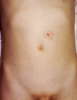
Durbin DR, Arbogast KB, Moll EK. Seat belt syndrome in children: a case report and review of the literature. Pediatr Emerg Care. 2001 Dec. 17 (6):474-7. [QxMD MEDLINE Link].
Santschi M, Echavé V, Laflamme S, McFadden N, Cyr C. Seat-belt injuries in children involved in motor vehicle crashes. Can J Surg. 2005 Oct. 48 (5):373-6. [QxMD MEDLINE Link].
Lopez PP, Benjamin R, Cockburn M, Amortegui JD, Schulman CI, Soffer D, et al. Recent trends in the management of combined pancreatoduodenal injuries. Am Surg. 2005 Oct. 71 (10):847-52. [QxMD MEDLINE Link].
Shilyansky J, Pearl RH, Kreller M, Sena LM, Babyn PS. Diagnosis and management of duodenal injuries in children. J Pediatr Surg. 1997 Jun. 32 (6):880-6. [QxMD MEDLINE Link].
Voss M, Bass DH. Traumatic duodenal haematoma in children. Injury. 1994 May. 25 (4):227-30. [QxMD MEDLINE Link].
Touloukian RJ. Protocol for the nonoperative treatment of obstructing intramural duodenal hematoma during childhood. Am J Surg. 1983 Mar. 145 (3):330-4. [QxMD MEDLINE Link].
Moremen JR, Nakayama DK, Ashley DW, Astin M, Nolan TL. Traumatic disruption of the abdominal wall: lap-belt injuries in children. J Pediatr Surg. 2013 Apr. 48 (4):e21-4. [QxMD MEDLINE Link].