Practice Essentials
Influenza is one of the most significant causes of acute upper respiratory tract infections worldwide. Influenza viruses are highly contagious and can cause seasonal epidemics, manifesting as an acute febrile illness with variable degrees of severity, ranging from mild fatigue to respiratory failure and death. See the image below.
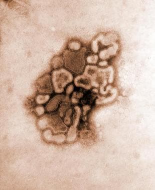 Swine influenza virus. Colorized transmission electron micrograph (37,800X) of the A/New Jersey/76 (Hsw1N1) virus under plate magnification. Image taken during the virus' first developmental passage through a chicken egg. Courtesy of the CDC/Dr. E. Palmer; R.E. Bates.
Signs and symptoms
Signs and symptoms of pediatric seasonal influenza include the following:
-
High fever
-
Chills
-
Myalgia
-
Headache
-
Fatigue
-
Sore throat/pharyngitis
-
Nasal congestion
-
Rhinitis
-
Nonproductive cough
-
Cervical lymphadenopathy
-
Conjunctivitis
Conjunctivitis, rhinitis, and gastrointestinal symptoms are more common in infants and young children than in adults.
Avian influenza
Symptoms of avian influenza include the following:
-
Fever: More than 90% of the time
-
Cough
-
Sore throat
-
Respiratory distress
-
Myalgia
-
Conjunctivitis
-
Vomiting
-
Diarrhea
-
Pleurisy
-
Abdominal pain
-
Rhinorrhea
-
Lymphadenitis
-
Nasal and gingival bleeding
-
Signs and/or symptoms of encephalitis: Occasionally noted on presentation
See Clinical Presentation for more detailed information on the signs and symptoms of pediatric influenza.
Diagnosis
The diagnosis and evaluation of influenza A or B can involve the following:
-
Viral culture of nasopharyngeal and/or throat samples
-
Hemagglutination inhibition techniques
-
Immunofluorescence assay
-
Enzyme-linked immunosorbent assay (ELISA)
-
Rapid influenza diagnostic tests (RIDTs) [1]
Avian influenza
Tests for avian influenza include the following:
-
Blood culture
-
Complete blood count with differential
-
Electrolyte level measurement
-
Liver enzyme assay
-
Blood urea nitrogen and creatinine level measurement
-
RIDTs
Histologic findings may include pulmonary changes with alveolar damage similar to seasonal influenza.
See Clinical Presentation and Workup for more detailed information on the diagnosis of pediatric influenza.
Management
Supportive care for pediatric influenza can include the following:
-
Acetaminophen for fever
-
Cough suppressants and expectorants
-
Steam inhalation
-
Oral or intravenous fluids if dehydration occurs
-
Antiviral therapy for selected patients [2]
Avian influenza
World Health Organization (WHO) recommendations for the management of avian influenza are as follows [3] :
-
Patients with confirmed or suspected H5N1 infection should be treated with oseltamivir as soon as possible
-
Zanamivir may be considered as an alternative if the patient is capable of using an inhaler
-
If neuraminidase inhibitors are available, amantadine and rimantadine should not be used as first-line therapies, because of potential resistance
-
If neuraminidase inhibitors are available and if the virus is susceptible, a combination of neuraminidase inhibitors and M2 inhibitors can be used in confirmed cases of H5N1 infection
-
If neuraminidase inhibitors are not available, amantadine can be used as a first-line therapy, provided the virus is susceptible
-
If neuraminidase inhibitors are not available, rimantadine can be used if the virus is known to be susceptible, because it has fewer side effects than amantadine
-
For prophylaxis in high-risk and moderate-risk exposures, give oseltamivir for 7-10 days from the day of exposure
-
Prophylaxis is not recommended for low-risk groups
See Treatment and Medication for more detailed information on the management of pediatric influenza.
Background
Influenza is the one of the most significant acute upper respiratory tract infections. Influenza viruses cause a broad array of respiratory illnesses responsible for significant morbidity and mortality in children. Influenza viruses cause epidemic disease (influenza virus types A and B) and sporadic disease (type C) in humans. Seasonal human influenza causes about 36,000 deaths and 226,000 hospitalizations in the United States annually. [4, 5]
The word influenza may have been derived from the Latin word influo, which means "to flow in," indicating airborne transmission, or from the Italian word influence, which indicates influence of weather or an astrological influence. [6]
In addition to humans, influenza also infects a variety of animal species. Some of these influenza strains are species specific, but new strains of influenza may spread from other animal species to humans (see Pathophysiology). The term avian influenza used in this context refers to zoonotic human infection with an influenza strain that primarily affects birds.
Swine influenza refers to infections from strains derived from pigs. For more information on the 2009 influenza pandemic, a recombinant influenza consisting of a mix of swine, avian, and human gene segments, see the article H1N1 Influenza (Swine Flu).
Typical symptoms of influenza begin 2-3 days after exposure to the virus. Influenza produces an acute febrile respiratory illness with cough, headache, and myalgia for 3-4 days, with symptoms that may persist for as long as 2 weeks (see Clinical). Accurately diagnosing influenza A or B infection based solely on clinical criteria is difficult because of the overlapping symptoms caused by the various viruses associated with upper respiratory tract infection (see Differentials).
As with other diseases, prevention of influenza is the most effective strategy. Each year in the United States, a vaccine that contains antigens from the strains most likely to cause infection during the winter flu season is produced. The vaccine provides good protection against immunized strains, becoming effective 10-14 days after administration. Antiviral agents are also available that can prevent some cases of influenza; when given soon after the development of influenza, they can reduce the duration and severity of illness. (See Treatment.)
For discussion of influenza in adults, see Influenza.
Pathophysiology
Influenza viruses are negative-sense, single-strand RNA viruses that belong to the family Orthomyxoviridae. [7] Human influenza viruses are divided into 3 major types: A, B, and C.
Influenza type A viruses cause disease in humans and many animal species. Waterfowl (eg, ducks, geese) are the natural reservoir for type A. In addition, in freshwater lakes, influenza A virus can stay alive for 4 days at 22o C and for more than a month at 0o C.
Influenza type B viruses primarily cause disease in humans, particularly children. Infections with influenza type C viruses are rare.
Influenza viral RNA has 8 genetic elements. The RNA has a lipid envelope with 2 major antigenic components on its surface, hemagglutinin (H) and neuraminidase (N). These components enable the replication and subsequent release of the virus, leading to its spread. Influenza type A viruses also have ionic channel proteins, termed M2 proteins. [7]
The H antigen is the major virulence determinant because these antigens help viral attachment to the cell. H proteins are divided into 16 types, whereas N proteins are divided into 9 types. N act on the sialic acid component of the cell, which enables viral detachment. [8, 9]
Different subtypes of influenza viruses are identified based on the combinations of these antigenic structures, with 144 combinations possible. For example, influenza A subtype H3N2 expresses hemagglutinin 3 and neuraminidase 2. Influenza A subtype H5N1, or avian influenza, has been found in chickens, ducks, and migratory fowl throughout Asia and is now spreading west through Europe and North Africa. It is highly virulent in humans but is poorly transmissible between humans.
Currently, influenza A/H1N1, A/H3N2, and influenza type B viruses are the circulating influenza strains that cause seasonal human infections. The influenza vaccine components for the current year are intended to provide protection from these strains. [10] In contrast with the typical course of disease caused by seasonal influenza, a higher viral load and prolonged viral replication are observed in infections caused by avian influenza virus A/H5N1. [11, 12]
Current research is focusing on broadly neutralizing antibodies against influenza viruses for potential treatments and vaccines. One group recently reported isolating and characterizing human monoclonal antibody CR8020 with broad neutralizing activity against most group 2 viruses, including H3N2 and H7N7, [13] and another group isolated a neutralizing monoclonal antibody that recognized the hemagglutinin glycoprotein of all 16 subtypes and neutralized both group 1 and group 2 influenza A viruses. [14]
Antigenic drift and shift
Influenza virus type A is readily prone to mutations, which can lead to minor antigenic changes (antigenic drift) or major antigenic changes (antigenic shift). These changes can result in viral strains capable of causing either an epidemic or a pandemic, because overall immunity in the population is lower or nonexistent. [15]
With antigenic drift, the surface hemagglutinin or neuraminidase proteins are slightly altered by accumulated point mutations and nucleotide substitutions, insertions, or deletions. Because the antigens are only slightly altered, an infected person’s immune system still recognizes the virus to some extent, and the infection is usually less severe.
Antigenic shift refers to a major change in the viral RNA caused by gene reassortment. This results in replacement of the surface hemagglutinin or neuraminidase proteins, and these new proteins may be unrecognizable by an infected person’s immune system. The newly reasserted viral strain may cause a pandemic in an immunologically naïve population.
Because avian influenza viruses attach to receptors specific to birds, whereas human influenza viruses attach to receptors specific to humans, transmission of influenza from birds to humans is rare. However, an intermediate host that has receptors used by both viruses (eg, pigs) can become co-infected with both avian and human influenza viruses; this permits reassortment that may produce major changes in the genetic component of surface proteins. The resulting viral strain may be more capable of transmission to humans.
Infection
Influenza is an acute infection of the respiratory tract that involves the nose, throat, and, sometimes, the lungs. Following respiratory transmission, the virus attaches to and penetrates respiratory epithelial cells in the trachea and bronchi. Viral replication occurs, which results in the destruction of the host cell. Viremia does not occur. The virus is shed in respiratory secretions for 5-10 days.
Influenza occurs as sporadic illness, epidemics, or pandemics. Epidemic disease occurs annually, especially in the winter months. Global pandemics occur in part because of the high degree of transmissibility and the emergence of an influenza virus with a major antigenic shift (major antigenic variations on the hemagglutinin surface protein) in a nonimmune population.
The most recent pandemics included the 1889 pandemic, the 1918-1919 pandemic (influenza virus subtype H1), the 1957 pandemic (subtype H2N2), the 1968-1969 pandemic (Hong Kong subtype H3N2), and, to a lesser extent, the mild pediatric pandemic in 1977 (subtype H1N1). Approximately 21 million persons died worldwide in the 1918-1919 influenza pandemic, with 549,000 deaths in the United States.
Geographic eponyms for these pandemics (eg, Spanish flu for the 1918-1919 pandemic, Russian flu for the 1977 pandemic) typically reflect areas strongly affected, rather than sites of origin. [16]
Avian influenza
An enormous release of cytokines is probably responsible for the severity of avian influenza in humans, resulting in rapid progression. [17] The major entry site seems to be conjunctiva and oropharynx.
Hyperinduction of cytokine storm with elevated interferon in serum samples is observed. [10, 18] Increased cytokines and chemokines with high levels of tumor necrosis factor alpha, interleukin 6, and interferon gamma lead to tissue destruction. [10] Pulmonary and intestinal involvement with isolation of viral messenger RNA (mRNA) in these tissues is reported.
Prolonged viral RNA detection in the respiratory tracts results in systemic dissemination in fatal cases of avian influenza. [10] Lung changes include diffuse alveolar damage, interstitial pneumonia, bronchiolitis, and hemorrhages. The type II pneumocytes showed reactive hyperplasia without cytopathic changes. Predominant macrophage infiltrates in the lung are reported.
Superimposed opportunistic infection by fungus and aspergillosis has been reported. Histiocytic hyperplasias have been noted in the spleen, bone marrow, and lymph nodes. Hemophagocytic syndrome with multisystem involvement is also reported. [19] Minimal fatty changes in liver with portal infiltrates with lymphocytes were reported. Edema and focal areas of necrosis in the brain have been reported. The intestines, heart, kidneys, and other organs are unremarkable.
Autopsy studies of cases of avian influenza A/H5N1 infection in pregnancy reveal that, unlike human influenza, A/H5N1 causes viremia and disseminates across the placenta. It infects cytotrophoblasts and macrophages similar to cytomegalovirus. The avian influenza A/H5N1 virus genomic sequence has been identified in fetal lung tissue without severe lung injury compared with severe adult lung injury. This could be due to the poor immune status and low cytokine storm. [20]
The images below show the avian influenza A/H5N1 virus as viewed on electron micrographs.
 Colorized transmission electron micrograph shows avian influenza A/H5N1 viruses (gold) grown in Madin-Darby canine kidney (MDCK) cells (green). Image courtesy of Centers for Disease Control and Prevention.
 Transmission electron micrograph (original magnification X 150,000) shows ultrastructural details of an avian influenza A/H5N1 virion, a subtype of avian influenza A. Note the stippled appearance of the roughened surface of the proteinaceous coat encasing the virion. Image courtesy of Centers for Disease Control and Prevention.
Etiology
Influenza is an acute infection caused by any of 3 types of influenza viruses (A, B, C). Types A and B cause epidemic disease, and type C causes sporadic disease. Type A is the most common.
Influenza is highly contagious. The virus is spread when an individual inhales contaminated airborne droplets (following coughing or sneezing by an infected person) or comes in direct contact with an infected person's secretions (eg, kissing, sharing of handkerchiefs and other items, sharing of objects such as spoons and forks). Viruses may also be transmitted via touching of smooth surfaces, such as doorknobs, handles, and telephones.
Certain populations are at higher risk for influenza. Risk factors include the following:
-
Advanced age
-
Chronic respiratory disease
-
Chronic cardiac disease
-
Chronic renal failure
-
Diabetes mellitus
-
Immunosuppression
-
Residence in residential care homes and long-stay facilities
Avian influenza
The main reservoir for influenza A viruses are migratory birds, especially waterfowl. Birds with low-pathogenic influenza A virus infection can be asymptomatic and can be a source of infection for domestic poultry. The influenza A viruses are shed in oral secretions and feces.
Domesticated birds are infected via secretions and excretions and via contaminated utensils. Highly pathogenic (HP) avian influenza A viruses can kill domesticated birds in 48 hours. HP avian influenza H5N1 viruses have crossed the species barrier in Asia, resulting in several human cases. [21] This poses a potential pandemic threat.
Most H5N1 cases have been in eastern Asia; some cases have been reported in Eastern Europe and North Africa. Infection has been associated with exposure to infected poultry and poultry products. No cases of infection with the H5N1 avian influenza virus have been reported in North America.
Epidemiology
In tropical areas, influenza occurs throughout the year. In temperate areas, the influenza season typically starts in early fall, peaks in mid-February, and ends in the late spring of the following year. Approximately 250,000-500,000 cases of seasonal influenza occur each year in the United States, with about 200,000 hospitalizations.
By convention, mortality statistics for influenza are combined with those for pneumonia. For the population as a whole, influenza and pneumonia result in approximately 20,000 deaths each year in the United States. There were 162 influenza-associated pediatric deaths in the 2022-2023 season. In the 2021-2022 season, there were 47 influenza-associated pediatric deaths. [22]
H1N1 influenza statistics
The 2009-2010 influenza season was affected by the H1N1 (“swine flu”) global pandemic, the first wave of which hit the United States in the spring of 2009, followed by a second, larger wave in the fall and winter; activity peaked in October and then declined quickly to below baseline levels by January, but small numbers of cases were reported through the spring and summer of 2010. [23, 24, 25] For more information, see the article H1N1 Influenza (Swine Flu).
The H1N1 pandemic in 2009 in Argentina was associated with pediatric death rates that were 10 times the rates for seasonal influenza in previous years; the overall rate of death was 1.1 per 100,000 children, compared with 0.1 per 100,000 children for seasonal influenza in 2007. [26] In the United States, from August 30, 2009 to March 27, 2010, at least 81%% of deaths associated with influenza were due to H1N1 disease. [27]
An analysis of data from a statewide public health surveillance in California, including 345 children (< 18 y), noted that more than 25% of children hospitalized for 2009 novel influenza A (H1N1) required intensive care (27%) and/or died (3%). [28] This study documents the severe nature of H1N1 infection in children.
Avian influenza statistics
A potential influenza pandemic could have a major impact economically and in health care. In the United States, a medium-level pandemic could lead to approximately 89,000-207,000 deaths, 314,000-734,000 hospitalizations, 20-47 million people with infection, and probably 18-42 million outpatient visits. [29]
Previous pandemics have occurred in waves, with new waves occurring in intervals of 3-7 months. Approximately 15-35% of the US population may be involved by an influenza pandemic, and the economic loss could be $71.3-166.5 billion. [29]
In 1997, the first report of human cases infected with HP avian influenza A/H5N1 documented 6 deaths among 18 hospitalized patients in Hong Kong. [30] In 1999, HP avian influenza A/H9N2 infection was reported in 2 girls with fever and rhinorrhea; they recovered and no human-to-human transmission was noted. [31]
Between January-March of 2004, 34 human cases of HP avian influenza A/H5N1 virus infections were confirmed from Vietnam and Thailand. Significant respiratory illness that required hospitalization resulted in a 68% fatality rate. In Vietnam, 15 deaths were reported among 22 cases; in Thailand, 8 deaths were reported among 12 cases. [31] Children and young adults were predominantly involved, and all human cases were associated with outbreaks of HP avian influenza A/H5N1 among domestic poultry in the back yard and in the farms. More than 100 million domestic poultry have been culled. [32]
Migratory birds are presumed to be responsible for the recent spread of HP avian influenza virus A/H5N1 from Southeast Asia, where the poultry outbreak has been reported, to other regions across the globe. The HP avian influenza virus A/H5N1 was confirmed in ducks, chicken, turkey, and geese in Russia and Kazakhstan in July 2005, resulting in an implementation of restrictions on importation of birds from these areas. [15] In January 2006, 2 cases of HP avian influenza virus A/H5N1 infection were reported in humans in Turkey.
By August 2007, a total of 12 cases and 4 deaths were reported. [33] Iraq, the seventh country to have reported human cases, has reported 3 HP avian influenza A/H5N1–related human cases. Laboratory confirmation is performed by the US Naval Medical Research Unit, located in Cairo, Egypt. [34] In Egypt, from 2006-2007, 38 human cases were reported, with 15 deaths due to severe respiratory illness. [33] Although poultry deaths are noted, HP avian influenza A/H5N1 viruses have not been isolated from poultry in these areas.
Globally, as of March 2011, 532 cases of avian influenza A/H5N1 had been reported worldwide, with 315 deaths. [35]
During the 2003-2004 influenza season, one case of avian influenza virus A/H7N3 was reported in New York; the patient had respiratory symptoms and required hospitalization but recovered, although the source of infection was unknown. [31] In March 2004, 2 poultry workers were involved in the culling of poultry during an avian influenza outbreak in British Columbia, Canada; one worker reportedly had conjunctivitis and headache, whereas the other had nasal drainage. Both workers recovered without hospitalization. [31]
The avian influenza A/H5N1 outbreak noted in Manipur, India was unique and was unrelated to the 2006 virus. [36]
Sexual and racial differences in incidence
No difference based on sex has been identified. No difference based on race has been identified. Race appears to be a factor in avian influenza only to the extent that geographic differences in the rate of avian influenza A/H5N1 among birds and the degree of bird-to-human contact are significant.
Age-related differences in incidence
The infection rate of influenza viruses is high in all age groups. The infection rate and the frequency of isolation of influenza viruses are highest in young children. The infection rate in healthy children is 10-30% annually.
Prognosis
Generally, the prognosis for recovery from seasonal influenza is excellent, although full return to normal levels of activity and freedom from cough usually requires weeks rather than days. However, a Centers for Disease Control and Prevention (CDC) review of data on 794 pediatric influenza-related deaths that occurred between October 2004 and September 2012 revealed that 341 children (43%) had no high-risk medical conditions such as neurologic disorders, asthma, or diseases of the heart, kidney, liver, or immune system. [37, 38] Median age of death was 7 years. Otherwise healthy children were more likely to die before hospital admission than children with other high-risk conditions (relative risk, 1.9; 95% confidence interval [CI], 1.6 - 2.4) and were 1.6 times more likely to die within 3 days of the onset of symptoms (95% CI, 1.3 - 2.0). [37, 38]
In contrast, the prognosis for patients with avian influenza is poor. Through August 2007, overall mortality from avian influenza A/H5N1 was 60% [34] ; the case-fatality rate in children younger than 15 years was approximately 89%. [17] The average duration from onset of illness to death varied from 1-3 weeks. [34] Poor prognostic features include a high viral load, leukopenia, lymphopenia, longer duration of illness prior to treatment, and old age. [18]
Children with neurological and neurodevelopmental conditions are at increased risk for severe outcomes from influenza, including death. This was evident in April 2011 during an outbreak of influenza at an Ohio residential facility for 130 such children. Ten were hospitalized and 7 died. A high index of suspicion and low threshold to empirically test for influenza and initiate empiric antiviral therapy during influenza season should be considered if there is worsening respiratory status among these patients. [39]
Patient Education
Education regarding covering the mouth when coughing and strict handwashing is helpful. Household contacts of patients should be instructed to monitor fever, cough, and respiratory distress symptoms. These household contacts should be given prophylactic antiviral therapy with oseltamivir. If they are symptomatic, they must be tested for the virus. Avoiding close contact with the patient is recommended.
For patient education information, see Colds, Flu in Adults, and Flu in Children.
For information on avian influenza, follow the travel advisories recommended by the CDC and the World Health Organization. For example, people traveling to certain countries should avoid poultry farms and markets or other places where live poultry are present.
-
Swine influenza virus. Colorized transmission electron micrograph (37,800X) of the A/New Jersey/76 (Hsw1N1) virus under plate magnification. Image taken during the virus' first developmental passage through a chicken egg. Courtesy of the CDC/Dr. E. Palmer; R.E. Bates.
-
Colorized transmission electron micrograph shows avian influenza A/H5N1 viruses (gold) grown in Madin-Darby canine kidney (MDCK) cells (green). Image courtesy of Centers for Disease Control and Prevention.
-
Transmission electron micrograph (original magnification X 150,000) shows ultrastructural details of an avian influenza A/H5N1 virion, a subtype of avian influenza A. Note the stippled appearance of the roughened surface of the proteinaceous coat encasing the virion. Image courtesy of Centers for Disease Control and Prevention.