Continually Updated Clinical Reference
 
 
  All Sources     eMedicine     Medscape     Drug Reference     MEDLINE
 
eMedicine - Stomas of the Small and Large Intestine : Article by

Quick Find
Authors & Editors
Introduction
Indications
Relevant Anatomy
Contraindications
Workup
Treatment
Complications
Outcome and Prognosis
Future and Controversies
Multimedia
References


Patient Education
Click here for patient education.


Author: Robert K Minkes, MD, PhD, Staff Pediatric Surgeon, Houston Pediatric Surgeons, Texas Children's Hospital

Robert K Minkes is a member of the following medical societies: Alpha Omega Alpha, American College of Surgeons, American Medical Association, American Pediatric Surgical Association, and Phi Beta Kappa

Coauthor(s): Sean E McLean, MD, Consulting Staff, General Surgery, Washington University, Barnes-Jewish Hospital, St Louis Children's Hospital; Mark V Mazziotti, MD, Assistant Professor of Pediatric Surgery, Department of Surgery, Baylor College of Medicine, Texas Children's Hospital; Jacob C Langer, MD, Professor, Department of Surgery, University of Toronto Faculty of Medicine

Editors: Kurt D Newman, MD, Vice Chairman, Department of Pediatric Surgery, Children's National Medical Center; Professor, Departments of Surgery and Pediatrics, George Washington University School of Medicine; Mary L Windle, PharmD, Adjunct Assistant Professor, University of Nebraska Medical Center College of Pharmacy, Pharmacy Editor, eMedicine.com, Inc; Andre Hebra, MD, Clinical Associate Professor, Department of Surgery, University of South Florida School of Medicine; Director, Minimally Invasive Pediatric Surgery Program, Chief of Surgery, All Children's Hospital; H Biemann Othersen Jr, MD, Professor of Surgery and Pediatrics, Emeritus Head, Division of Pediatric Surgery, Medical University of South Carolina; Harsh Grewal, MD, FACS, FAAP, Professor of Surgery and Pediatrics, Temple University School of Medicine; Chief, Section of Pediatric Surgery, Temple University Children's Medical Center

Author and Editor Disclosure

Synonyms and related keywords: stomas of the small and large intestine, ostomy, ostomies, enterostomy, enterostomies, jejunostomy, jejunostomies, ileostomy, ileostomies, colostomy, colostomies, mucus fistula, mucus fistulae, GI stomas, gastrointestinal stomas, mucous fistula, mucous fistulae, intestinal stomas, gastrointestinal access, gastrointestinal decompression, gastrointestinal diversion, gastrointestinal evacuation, GI access, GI decompression, GI diversion, GI evacuation

The use and management of gastrointestinal stomas in children have evolved since the early success with colostomy formation in the 1800s. Improved surgical techniques, better understanding of the physiologic and psychological consequences of intestinal stomas, and advances in stoma care have contributed to more rational use of ostomies by pediatric surgeons and a wider acceptance in the medical and lay communities. Nevertheless, treating a child with multiple abdominal stomas can be intimidating and challenging (see Image 1), especially when the anatomy is not clear and the fluid and electrolyte abnormalities are difficult to control.
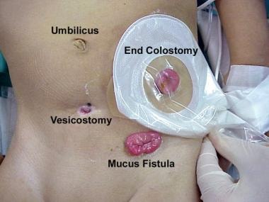
Several differences between adult and pediatric ostomies exist. Most stomas in adults are formed in the distal ileum or colon for the treatment of inflammatory bowel disease, malignant conditions, and trauma; more proximal stomas are only rarely created. In contrast, stomas in infants and children may be required anywhere along the gastrointestinal tract because of the wide variety of congenital and acquired conditions that require stoma formation. The effect of a stoma on physical and emotional development and on growth is an additional consideration in children.

While great advances have been made with regard to stoma formation and management, both early and late complications are common. Fortunately, most pediatric stomas are temporary, and many of the complications associated with intestinal stomas are eliminated when the stoma is closed. Understanding enterostomal construction and physiology is essential for providing these children with optimal care.

History of the Procedure

Colostomies were used in the late 1800s to treat intestinal obstruction. Some of the earliest survivors were children with an imperforate anus. Intestinal stomas were considered a drastic procedure and were avoided because of the high incidence of complications and mortality. With improvements in surgical technique and practice, the need for stomas increased as children with formerly fatal conditions survived.

Problem

Pediatric ostomies include any surgically created opening between a hollow organ and the skin connected either directly (stoma) or with the use of a tube. In infants and children, stomas are used for a variety of purposes, including access, decompression, diversion, and evacuation. As a rule, most ostomies are for temporary use and are typically reversible in children. Certain medical conditions may dictate the need for a permanent stoma.

Frequency

The frequency of intestinal stomas is difficult to determine in the pediatric population.

Etiology

Many diseases may require a stoma or placement of a tube within the bowel. Small bowel stomas are used for patients with intestinal perforation or ischemia in whom an anastomosis is considered unsafe. A proximal ileostomy is often used to protect the distal anastomosis after restorative proctocolectomy for familial polyposis or ulcerative colitis. Similarly, colostomy is often used both before and after a pull-through procedure for imperforate anus or Hirschsprung disease, although many surgeons are now performing primary pull-through procedures without colostomy for both of these conditions. Tube cecostomy or Malone appendicocecostomy have been used for antegrade bowel irrigation in children with intractable constipation and a variety of medical conditions.

Children with severe perineal burns or trauma (see Image 2) often require a temporary colostomy to allow the injury to heal.
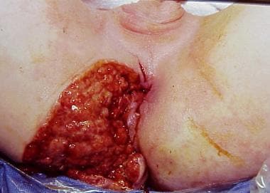
Patients with the following conditions may require a stoma:

Pathophysiology

The pathophysiologic features depend on the specific disease process.

Clinical

The clinical presentation depends on the specific diagnosis and age of the patient.


Stomas are used in situations in which diversion of, decompression of, or access to the bowel lumen is needed.


The clinical scenario and relevant anatomy may affect the technique used for stoma creation and the location of the stoma.

Technique

Several types of intestinal stomas exist (see Image 3). The clinical scenario often dictates the segment of intestine selected, the type of stoma created, and its external location. In children, most stomas are created as an end or loop ostomy; however, be familiar with the many potential variations in their construction. Roux-en-Y construction can also be performed for tube stomas such as feeding jejunostomy.

For an end stoma (see Images 3-6), the bowel is divided, and the proximal end is brought through the abdominal wall. The distal nonfunctioning limb can be brought out through the same abdominal wall opening as the end stoma (ie, double-barrel stoma), it can be brought out through a separate incision (ie, mucous fistula), or it can be closed and left in the peritoneal cavity (ie, Hartmann procedure). When the distal segment is left inside the abdomen, many surgeons fasten it to the abdominal wall adjacent to the end ostomy or tag it with a nonabsorbable suture to facilitate identification when the stoma is reversed. A loop stoma is created by maturing a segment of bowel over a rod or tube without completely dividing the bowel.

Loop stomas provide excellent decompression and have the advantage of simple closure without the need for a separate laparotomy in most cases. However, loop stomas are not completely diverting because proximal contents can spill over into the distal limb. Therefore, they should be used with caution in patients in whom stool in the distal bowel may be problematic. A decompressing stoma, or blowhole, is created in patients in unstable condition by opening the antimesenteric border of bowel without mobilizing the entire loop of bowel.

Stomas can also be formed in association with an anastomosis for proximal or distal venting or irrigation (ie, Bishop-Koop and Santulli stomas [see Image 3 and Image 7]). These stomas were initially designed for the treatment of infants with meconium ileus, but they have been adapted for many other purposes. In children with necrotizing enterocolitis, multiple intestinal atresia, or midgut volvulus in which multiple bowel anastomoses may be unsafe and in whom preservation of intestinal length is desired, one or more discontinuous segments of bowel may be externalized. The operating surgeon should clearly describe the anatomic configuration in the surgical notes and provide an illustration in the patient's chart.

In general, a stoma is easier to manage when it is not flush with the skin. Everting the bowel prior to suturing the edge to the skin (ie, Brooke technique) produces a spigot conformation that holds a stoma appliance and prevents serositis (see Image 3, inset in part A). Eversion is not always possible in neonates (in whom the blood supply may be tenuous) and in situations in which the bowel is markedly edematous. In these cases, the bowel is left to protrude through the skin without eversion, and the stoma automatically matures as the mucosa rapidly grows over the exposed serosal surface.

Stoma location

Intestinal stomas can be exteriorized on the neck, chest, or abdomen. The abdomen is by far the most common site for intestinal stomas. Enterostomies can be brought through the abdominal wall in the laparotomy incision or through a separate site. Theoretical disadvantages of bringing a stoma through a large laparotomy incision include the risk of wound infection, dehiscence, and evisceration. Nevertheless stomas are often incorporated into the incision, especially when the only goal of surgery is to create a stoma. Whenever clinically feasible, a primary stoma site, as well as alternative sites, should be selected and marked before surgery. The ideal location for an abdominal stoma in older children and adolescents is similar to that in adults. The stoma is distant from the incision, through the midportion of the rectus muscle away from skin folds (eg, groin, flank), bony prominences (eg, rib cage, iliac spine), and umbilicus (see Images 8-9).

Stoma location in infants and neonates follows these same principles whenever possible; however, the small size of the abdominal wall in infants and the short mesentery of the bowel chosen for the stoma often limit the options. For temporary stomas in infants, the bowel can be brought out directly through or adjacent to the umbilicus (see Images 10-11). This site is easier for appliance placement and results in a cosmetically superior scar when the stoma is ultimately closed.


No specific contraindications for the use of intestinal stomas exist, other than those for surgery in general.


Lab Studies

  • Laboratory investigations depend on the indication for a stoma.

Imaging Studies

  • Imaging studies may be performed as part of the evaluation of the specific disease process and may include the following:
    • Plain radiography
    • Contrast enema examination (particularly before closure of stomas)
    • CT scanning
    • Ultrasonography
  • Prior to reversal of most ostomies in infants, consider performing a distal contrast study to evaluate the unused portion of the bowel. This is particularly important in babies treated for neonatal necrotizing enterocolitis with a diverting ostomy. Such patients may have an unrecognized distal intestinal stricture. Repair of the stricture is essential prior to, or during, the procedure for reversal of the ostomy.

Diagnostic Procedures

  • Intestinal biopsy: In selected cases in which a colostomy or ileostomy was performed for intestinal obstruction of unclear etiology, a mucosal biopsy might be helpful to rule out the possibility of Hirschsprung disease.


Medical therapy

Stomas are created for the treatment of surgical diseases when no medical alternatives exist.

Surgical therapy

The operation performed depends on the specific disease being treated.

Injuries to the rectum and sphincter are difficult to repair in the initial setting. Minor injuries may be primarily repaired. For more extensive injuries, exploratory laparotomy should be performed to rule out intra-abdominal injuries, and a diverting colostomy should be created. Debridement of the perineum should be avoided in the initial setting. The anal sphincter can be reconstructed later.

Preoperative details

Stomas are created in both elective and emergency settings. A physician, enterostomal therapist, or nurse specialist should counsel children undergoing elective colostomy as well as their families. This preparation reduces their anxiety and makes postoperative management easier. When safe to do so, the bowel is prepared mechanically and with antibiotics; however, this preparation should be avoided when an obstruction is present.

The potential stoma sites should be marked preoperatively. In older children, the site should be marked with the patient in both the sitting and supine positions to ensure that the stoma appliance fits securely. The stoma site should be selected to avoid fat folds, scars, and bony prominences. A useful selection technique is drawing a vertical line through the umbilicus to the pubis and a transverse line through the inferior margin of the umbilicus. A disk the size of a stoma faceplate can then be used to determine the location. When an adequate site for all positions is found, the site should be marked with ink. After the induction of general anesthesia in the operating room, a fine needle can be used to scratch the site prior to preparation of the abdominal skin.

Intraoperative details

Construction of a stoma requires the bowel to be mobile enough to be brought through the abdominal wall. Tension on the mesentery should be avoided. Ideally, stomas are brought out through separate skin incisions, but the clinical situation ultimately dictates the site used. In older children and adolescents, stomas should be brought through the rectus abdominus muscle to prevent parastomal hernias. A disk of skin is removed, and the fascia is incised longitudinally. The stoma is matured with absorbable suture after the abdominal incision is closed. In neonates and infants, the stomas may be covered with Vaseline gauze until bowel function returns. In older children, a 1-piece stoma appliance that can be cut to just fit around the stoma should be applied in the operating room.

Postoperative details

Return of bowel function varies and depends on the clinical setting. Ileostomies and colostomies usually begin to function in 4-5 days. In the first few days, clear or serosanguinous fluid may appear. This finding should not be mistaken for an indication of bowel activity. An enterostomal therapist or nurse specialist should be involved early in the care of the newly formed stoma and in teaching the patient and family long-term care of the stoma.

Follow-up

Small bowel stomas (ie, jejunostomy, ileostomy) and proximal colostomies have liquid output. Volumes may be large, and the fluid may irritate the skin. Use of an appropriately fitting stoma bag is essential. A variety of stoma appliances and products are available. Stoma appliances should be able to remain in place for several days. Improperly fitted appliances leak and lead to more complications.

Local irritation, skin excoriation, and yeast infections can be treated with appropriate topical medication and skin care. Mild stoma bleeding is not uncommon and usually stops spontaneously. Children with high ostomy output require close follow-up for dehydration and electrolyte imbalances.


Stoma-related complications

Many potential stoma-related complications exist.

Skin irritation and infection are the most common complications with pediatric stomas. Excoriation from stoma effluent, candidal infection (see Image 13), and dermatitis are frequent; improper location or construction of the stoma and poor stoma care are often responsible. Local wound care and patient or caretaker education often corrects the problem. Occasionally, surgical revision is needed. Wound infection, wound separation, dehiscence, and postoperative sepsis may also occur after formation of a stoma, particularly if the stoma has been brought out through the wound (see Image 14).

Stoma prolapse also is common in children. Prolapse can occur in end or loop stomas. Both proximal and distal bowel segments can protrude many centimeters (see Image 15), although this complication may be more common in the distal limb. A variety of surgical techniques have been used, with mixed success, to reduce the incidence of prolapse. Once prolapse occurs, it often becomes a chronic problem that can be difficult to correct. In most cases, prolapse is an unsightly nuisance, but it is well tolerated by the child. Surgical revision is indicated for the rare instances of ischemia, obstruction, ulceration, or chronic bleeding. Stoma reversal is the optimal treatment.

Stoma retraction results in a stoma that is flush with the skin, and difficulty in controlling the effluent may result in significant skin breakdown. Retraction of a loop colostomy results in a blowhole configuration that allows proximal contents to spill into the distal segment. Revision may be required if distal diversion is necessary. A retracted end stoma often re-protrudes spontaneously. In most cases in which the stoma is temporary, only short-term measures are needed until the stoma is reversed. Permanent stomas that have retracted may require surgical revision.

Intestinal obstruction also is common. Stoma strictures can occur at the skin and/or fascial levels. Partial obstruction can result in hyperperistalsis and hypersecretion; massive fluid losses through the stoma may result in dehydration. If a stoma stricture is suspected, the size of the opening can be determined by carefully passing metal sounds through the stoma. Attempts at dilating the stoma are usually unsuccessful and may cause intestinal perforation. Passage of a soft catheter proximal to the stricture can provide temporary decompression. Most significant stoma strictures require surgical revision; a local procedure with minimal morbidity is often possible. Parastomal hernias usually require surgical intervention.

Other causes of obstruction include luminal plugging caused by ingested food, adhesive intestinal obstruction, internal hernia, and volvulus. Obstruction is usually obvious, and the diagnosis is based on the patient's history and findings at physical examination and on plain radiography. In all patients with a bowel obstruction, a nasogastric tube should be placed for decompression and the patient should receive intravenous hydration. A study with water-soluble contrast material administered through the stoma provides diagnostic information, and in most cases of luminal obstruction, it is therapeutic. Most adhesive obstructions improve with nasogastric decompression and bowel rest. Prompt surgical exploration is required in patients with suspected ischemic or gangrenous bowel, clinical deterioration, or obstruction that does not rapidly resolve with nonsurgical therapy.

Psychological issues can be significant for the child and the family. These effects can be particularly important in adolescents, who are dealing with body image and sexuality. A team approach to providing preoperative counseling, postoperative care, and rehabilitation is crucial to the well-being of the patient. An enterostomal therapist or nurse specialist is essential. The age of the patient and an understanding of physical and psychological changes that children with stomas experience must be carefully considered.

In summary, potential stoma-related problems include the following:

  • Skin irritation - Chemical, mechanical, allergic
  • Intestinal obstruction - Adhesion, volvulus, internal hernia
  • Wound-related complications - Infection, separation, dehiscence
  • Infections
  • Prolapse
  • Retraction
  • Stricture
  • Fistula
  • Ulceration
  • Leakage
  • Bleeding
  • Parastomal hernia
  • Fluid and electrolyte imbalances
  • Psychological trauma

Fluid and electrolyte problems

Physiologic abnormalities related to loss of fluid and electrolytes are common in young patients with stomas, particularly when the stoma is in the proximal gastrointestinal tract. Fluid and electrolyte losses from any stoma can be significant, and replacement is usually required. The electrolyte composition of enteral fluids is listed in the following Table.

Electrolyte Composition of Enteral Fluids

FluidNa+
mEq/L
Cl-
mEq/L
K+
mEq/L
HCO3-
mEq/L
H+
mEq/L
Saliva30-6015-402015-50N/A
Gastric60-10090-14010-20N/A30-100
Duodenal14080550N/A
Bile1401005-1040-50N/A
Pancreatic140755-1590N/A
Jejunal1001005-1010-20N/A
Ileal1301101030N/A
Colonic60403020N/A
Diarrhea1303090N/AN/A

Duodenal stomas are rare, but duodenal fistulas may occur after trauma or surgery. Duodenal fistulas typically have high output and, therefore, are difficult to control. Proximal jejunal stomas also tend to have high-volume output, with fluid and electrolyte losses similar to those of duodenal fistulas, except for a slightly lower sodium loss from the jejunum. Pancreatic and biliary fluids also empty into the duodenum, and secretion of these fluids may substantially increase if feeding into the stomach is initiated.

Often, the proximal bowel can adapt to the fluid and electrolyte losses of a distal small bowel stoma. After a period of adaptation, the absorptive capacity of the small bowel proximal to the ileostomy increases, and the bowel can reduce ileostomy electrolyte losses by as much as two thirds of its initial output. Adaptation of water absorption is a much slower process. Ileostomy output should average 10-15 mL/kg/d. A doubling of usual stoma output should be considered abnormal. Normal ileostomy output results in the loss of 2-3 times the normal amount of salt and water, and children with these stomas are susceptible to dehydration. In infants, sodium and bicarbonate losses may exceed renal conservation mechanisms. Infants are prone to dehydration, and they may not gain appropriate amounts of weight unless salt and bicarbonate supplementation is provided.

Patients with long-standing ileostomies often have hypomagnesemia and decreased absorption of vitamin B-12 and folic acid. Patients with ileostomies also have a higher incidence of renal calculi and gallstones than that of the general population. Also, they may have iron deficiency and fat malabsorption.

Intestinal contents become progressively more solid as they pass through the colon to the rectum. The contents of the ileum and cecum are liquid and erosive to the skin. The contents of transverse colon stomas are typically semiliquid and less erosive, while those of the descending colon tend to be solid and nonreactive to the skin. These generalizations are true when the proximal bowel has sufficient length to allow absorption of nutrients and fluid. In children with short bowel syndrome, fluid and electrolyte losses can be massive, even with a distal colostomy.


The outcome of patients with intestinal stomas depends on the underlying condition. Fortunately, most stomas in infants and children are reversible. Reestablishing bowel continuity depends on factors such as the underlying disease, the general medical condition of the child, and the presence of stoma-related complications. Understanding the anatomy prior to stoma closure is crucial. In most instances, a preoperative distal contrast-enhanced study should be performed.

In general, the prognosis for patients with intestinal stomas is good. The exception is in patients with stomas and short gut syndrome. In such cases, reversal of the stoma should be attempted as soon as possible in order to maximize the absorptive capacity of the intestines. However, in many cases of short gut syndrome, ostomy reversal is not possible because of other associated comorbid conditions. The most common cause of short gut syndrome in North America is necrotizing enterocolitis.


In selected cases, performing ostomies using minimally invasive surgical technique (laparoscopic surgery) is possible.


Media file 1:  Multiple stomas in the abdomen of a 3-year-old child who underwent surgery as an infant for high imperforate anus. A divided colostomy was performed to divert the stool. The other end was brought out through the skin (mucous fistula) to allow evacuation of mucus and gas. A vesicostomy was performed because of a neurogenic bladder.
Click to see larger pictureClick to see detailView Full Size Image
Media type:  Photo

Media file 2:  Severe injury to the perineum and anal sphincter (caused by a lawn mower) in a 9-year-old boy. A diverting colostomy was performed. Several weeks later, a skin graft was placed over the defect. After reconstruction of the anal sphincter, the colostomy was reversed.
Click to see larger pictureClick to see detailView Full Size Image
Media type:  Photo

Media file 3:  Diagrams illustrate pediatric stomas. A) End stoma (inset shows everting maturation), B) double barrel stoma - End stoma and mucous fistula are divided and brought through the same incision (inset shows closed mucus fistula sutured to abdominal wall), C) end stoma with mucous fistula brought out separate incision, D) end ileostomy with closed mucous fistula left inside abdominal cavity, E) loop stoma, F) decompressing blowhole stoma, G) Bishop-Koop end-to-side anastomosis with distal vent for irrigation, H) Santulli side-to-end anastomosis with proximal stoma to promote decompression.
Click to see larger pictureClick to see detailView Full Size Image
Media type:  Image

Media file 4:  Divided end ileostomy and ileal mucous fistula. The ileostomy is brought out through a separate skin incision.
Click to see larger pictureClick to see detailView Full Size Image
Media type:  Image

Media file 5:  End ileostomy with closed distal limb (also known as Hartman pouch).
Click to see larger pictureClick to see detailView Full Size Image
Media type:  Image

Media file 6:  End ileostomy with closed distal intestine after multiple resections. The segment without function is left in situ, and a stoma is used for decompression.
Click to see larger pictureClick to see detailView Full Size Image
Media type:  Image

Media file 7:  End-to-side anastomosis created in an infant with a complicated meconium ileus, with distal stoma used for irrigation (also known as the Bishop-Koop ileostomy).
Click to see larger pictureClick to see detailView Full Size Image
Media type:  Photo

Media file 8:  Potential sites for neonate and infant stomas.
Click to see larger pictureClick to see detailView Full Size Image
Media type:  Image

Media file 9:  Potential sites for stomas in an older child or adolescent. Ideally, a stoma is brought through the rectus muscle in a position that allows placement of a stoma appliance.
Click to see larger pictureClick to see detailView Full Size Image
Media type:  Image

Media file 10:  Stoma brought directly through the umbilicus.
Click to see larger pictureClick to see detailView Full Size Image
Media type:  Photo

Media file 11:  Stoma matured through a separate inferior umbilical incision.
Click to see larger pictureClick to see detailView Full Size Image
Media type:  Photo

Media file 12:  Appearance of violaceous rash commonly due to Candida albicans. String coming out of the distal stoma was passed through a distal stricture and out of the anus.
Click to see larger pictureClick to see detailView Full Size Image
Media type:  Photo

Media file 13:  Skin irritation, stoma retraction, and wound infection after placement of a stoma through a laparotomy incision.
Click to see larger pictureClick to see detailView Full Size Image
Media type:  Photo

Media file 14:  Prolapse of loop colostomy. Both proximal and distal limbs have prolapsed. This may or may not cause intestinal obstruction.
Click to see larger pictureClick to see detailView Full Size Image
Media type:  Photo

Media file 15:  Wound infection and postoperative sepsis may also occur after formation of a stoma. Note the redness of the skin at the laparotomy incision site and evidence of soft tissue abdominal wall infection.
Click to see larger pictureClick to see detailView Full Size Image
Media type:  Photo

Media file 16:  An 8-month-old infant with a stoma.
Click to see larger pictureClick to see detailView Full Size Image
Media type:  Photo


  • Albanese CT, Jennings RW, Lopoo JB. One-stage correction of high imperforate anus in the male neonate. J Pediatr Surg. May 1999;34(5):834-6. [Medline].
  • Beiler HA, Zachariou Z, Daum R. Impalement and anorectal injuries in childhood: a retrospective study of 12 cases. J Pediatr Surg. Aug 1998;33(8):1287-91. [Medline].
  • Bishop HC, Koop CE. Management of meconium ileus: resection, Roux-en-Y anastomosis and ileostomy irrigation with pancreatic enzymes. Ann Surg. 1957;145:410.
  • Bower TR, Pringle KC, Soper RT. Sodium deficit causing decreased weight gain and metabolic acidosis in infants with ileostomy. J Pediatr Surg. Jun 1988;23(6):567-72. [Medline].
  • Brown H, Randle J. Living with a stoma: a review of the literature. J Clin Nurs. Jan 2005;14(1):74-81. [Medline].
  • Cameron GS, Lau GY. The umbilicus as a site for temporary colostomy in infants. J Pediatr Surg. Aug 1982;17(4):362-4. [Medline].
  • Curry JI, Osborne A, Malone PS. The MACE procedure: experience in the United Kingdom. J Pediatr Surg. Feb 1999;34(2):338-40. [Medline].
  • Fonkalsrud EW, Dunn JC, Kawaguchi AI. Simplified technique for antegrade continence enemas for fecal retention and incontinence. J Am Coll Surg. Oct 1998;187(4):457-60. [Medline].
  • Fonkalsrud EW. Long-term results after colectomy and ileoanal pull-through procedure in children. Arch Surg. Aug 1996;131(8):881-5; discussion 885-6. [Medline].
  • Griffiths DM, Malone PS. The Malone antegrade continence enema. J Pediatr Surg. Jan 1995;30(1):68-71. [Medline].
  • Haberlik A, Hollwarth ME, Windhager U. Problems of ileostomy in necrotizing enterocolitis. Acta Paediatr Suppl. 1994;396:74-6. [Medline].
  • Harberg FJ, Senekjian EK, Pokorny WJ. Treatment of uncomplicated meconium ileus via T-tube ileostomy. J Pediatr Surg. Feb 1981;16(1):61-3. [Medline].
  • Haut ER, Nance ML, Keller MS. Management of penetrating colon and rectal injuries in the pediatric patient. Dis Colon Rectum. Sep 2004;47(9):1526-32. [Medline].
  • Hollinworth H, Howlett S, Tallett J. Professional holistic care of the person with a stoma: online learning. Br J Nurs. Nov 25-Dec 8 2004;13(21):1268-75. [Medline].
  • Janik JS, Ein SH, Filler RM. An assessment of the surgical treatment of adhesive small bowel obstruction in infants and children. J Pediatr Surg. Jun 1981;16(3):225-35. [Medline].
  • Kock NG, Darle N, Hulten L. Ileostomy. Curr Probl Surg. Aug 1977;14(8):1-52. [Medline].
  • Kootstra G, Kamann HL, Okken A. The Bishop-Koop anastomosis-a find in pediatric surgery. Neth J Surg. 1980;32(3):92-6. [Medline].
  • Langer JC, Fitzgerald PG, Winthrop AL. One-stage versus two-stage Soave pull-through for Hirschsprung''s disease in the first year of life. J Pediatr Surg. Jan 1996;31(1):33-6; discussion 36-7. [Medline].
  • Langer JC, Minkes RK, Mazziotti MV. Transanal one-stage Soave procedure for infants with Hirschsprung''s disease. J Pediatr Surg. Jan 1999;34(1):148-51; discussion 152. [Medline].
  • Malone PS, Ransley PG, Kiely EM. Preliminary report: the antegrade continence enema. Lancet. Nov 17 1990;336(8725):1217-8. [Medline].
  • Millar AJ, Lakhoo K, Rode H. Bowel stomas in infants and children. A 5-year audit of 203 patients. S Afr J Surg. Sep 1993;31(3):110-3. [Medline].
  • Mollitt DL, Malangoni MA, Ballantine TV. Colostomy complications in children. An analysis of 146 cases. Arch Surg. Apr 1980;115(4):455-8. [Medline].
  • Mousa HM, van den Berg MM, Caniano DA. Cecostomy in children with defecation disorders. Dig Dis Sci. Jan 2006;51(1):154-60. [Medline].
  • Musemeche CA, Kosloske AM, Ricketts RR. Enterostomy in necrotizing enterocolitis: an analysis of techniques and timing of closure. J Pediatr Surg. Jun 1987;22(6):479-83. [Medline].
  • Nour S, Beck J, Stringer MD. Colostomy complications in infants and children. Ann R Coll Surg Engl. Nov 1996;78(6):526-30. [Medline].
  • Pearl RK, Prasad ML, Orsay CP. Early local complications from intestinal stomas. Arch Surg. Oct 1985;120(10):1145-7. [Medline].
  • Quarmby CJ, Millar AJ, Rode H. The use of diverting colostomies in paediatric peri-anal burns. Burns. Nov 1999;25(7):645-50. [Medline].
  • Ratliff CR, Scarano KA, Donovan AM. Descriptive study of peristomal complications. J Wound Ostomy Continence Nurs. Jan-Feb 2005;32(1):33-7. [Medline].
  • Rothstein FC, Halpin TC Jr, Kliegman RJ. Importance of early ileostomy closure to prevent chronic salt and water losses after necrotizing enterocolitis. Pediatrics. Aug 1982;70(2):249-53. [Medline].
  • Santulli TV, Blanc WA. Congenital atresia of the intestine: pathogenesis and treatment. Ann Surg. 1961;154:939.
  • Schwarz KB, Ternberg JL, Bell MJ. Sodium needs of infants and children with ileostomy. J Pediatr. Apr 1983;102(4):509-13. [Medline].
  • Sherkin-Langer F. If This Is A Test, Have I Passed Yet?. Toronto, Canada: Macmillan. 1994.
  • Steinau G, Ruhl KM, Hörnchen H. Enterostomy complications in infancy and childhood. Langenbecks Arch Surg. Aug 2001;386(5):346-9. [Medline].
  • Woodward MN, Foley P, Cusick EL. Colostomy for treatment of functional constipation in children: a preliminary report. J Pediatr Gastroenterol Nutr. Jan 2004;38(1):75-8. [Medline].
  • Yoshida NR, Webber EM, Gillis DA. Roux-en-Y jejunostomy in the pediatric population. J Pediatr Surg. Jun 1996;31(6):791-3. [Medline].
  • Yu SC, Petty JK, Bensard DD. Laparoscopic-assisted percutaneous endoscopic gastrostomy in children and adolescents. JSLS. Jul-Sep 2005;9(3):302-4. [Medline].
  • al-Salem AH, Grant C, Khawaja S. Colostomy complications in infants and children. Int Surg. Jul-Sep 1992;77(3):164-6. [Medline].
  • el Hefny A, el Beshlawy A, Nour S. Ketotifen in the treatment of infants and young children with wheezy bronchitis and bronchial asthma. J Int Med Res. 1986;14(5):267-73. [Medline].

Stomas of the Small and Large Intestine excerpt

Article Last Updated: Oct 25, 2006