Practice Essentials
Necrotizing enterocolitis (NEC), which typically occurs in the second to third week of life in premature, formula-fed infants, is characterized by variable damage to the intestinal tract, ranging from mucosal injury to full-thickness necrosis and perforation (see the image below). NEC affects close to 10% of infants who weigh less than 1500 g, with mortality rates of 50% or more depending on severity, but may also occur in term and near-term babies.
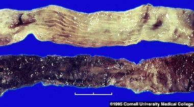 Normal (top) versus necrotic section of bowel. Photo courtesy of the Department of Pathology, Cornell University Medical College.
Signs and symptoms
In premature infants, onset of NEC is typically during the first several weeks after birth, with the age of onset inversely related to gestational age at birth. In term infants, the reported median age of onset is 1-3 days, but onset may occur as late as age 1 month.
Initial symptoms may be subtle and can include 1 or more of the following:
-
Vomiting
-
Diarrhea
-
Delayed gastric emptying
-
Abdominal distention, abdominal tenderness, or both
-
Ileus/decreased bowel sounds
-
Abdominal wall erythema (advanced stages)
-
Hematochezia
Systemic signs are nonspecific and can include any combination of the following:
-
Apnea
-
Lethargy
-
Decreased peripheral perfusion
-
Shock (in advanced stages)
-
Cardiovascular collapse
-
Bleeding diathesis (consumption coagulopathy)
Physical findings in patients with NEC can be primarily GI, primarily systemic, indolent, fulminant, or any combination of these. Gastrointestinal signs can include any or all of the following:
-
Increased abdominal girth
-
Visible intestinal loops
-
Obvious abdominal distention and decreased bowel sounds
-
Change in stool pattern
-
Hematochezia
-
Palpable abdominal mass
-
Erythema of the abdominal wall
Systemic signs can include any of the following:
-
Respiratory failure
-
Decreased peripheral perfusion
-
Circulatory collapse
See Clinical Presentation for more detail.
Diagnosis
Obtain radiographic studies if any concern about NEC is present. Pursue laboratory studies, especially if the abdominal study findings are worrisome or the baby is manifesting any systemic signs. A CBC with manual differential is usually repeated at least every 6 hours if the patient's clinical status continues to deteriorate. Relevant findings may include the following:
-
WBC – Moderate to profound neutropenia (absolute neutrophil count [ANC] < 1500/μL) strongly suggests established sepsis
-
Hematocrit and hemoglobin – Blood loss from hematochezia and/or a developing consumptive coagulopathy can manifest as an acute decrease in hematocrit; an elevated hemoglobin level and hematocrit may mark hemoconcentration due to notable accumulation of extravascular fluid
-
Platelet count – Thrombocytopenia may be present
Other laboratory findings
-
Blood culture is usually negative
-
Hyponatremia – An acute decrease in serum sodium (< 130 mEq/dL) is alarming
-
Low serum bicarbonate (< 20) may be seen in babies with poor tissue perfusion, sepsis, and bowel necrosis
-
Reducing substances may be identified in the stool of formula-fed infants
-
A breath hydrogen test may be positive
-
Arterial blood gas levels may indicate the infant's need for respiratory support and can provide information on the acid-base status
Abdominal radiography
-
The mainstay of diagnostic imaging
-
An AP and a left lateral decubitus view are essential for initial evaluation
-
Should be performed serially at 6-hour or greater intervals, depending on presentation acuity and clinical course, to assess disease progression
-
Characteristic findings on AP views include an abnormal gas pattern, dilated loops, and thickened bowel walls
-
A fixed and dilated loop that persists over several examinations is especially worrisome
-
Scarce or absent intestinal gas is more worrisome than diffuse distention that changes over time
Other radiographic findings include the following:
-
Pneumatosis intestinalis – Pathognomic of NEC
-
Abdominal free air – Ominous; patients usually require emergency surgical intervention
-
Portal gas – A poor prognostic sign
-
Distended loops of small bowel – Common but nonspecific
-
Intraperitoneal free fluid
Abdominal ultrasonography
-
Available at bedside
-
Noninvasive
-
Can identify areas of loculation and/or abscess consistent with a walled-off perforation
-
Excellent for identifying and quantifying ascites
-
Limited availability at some medical centers
-
Requires extensive training to discern subtle ultrasonographic appearance of some pathologies
-
Abdominal air can interfere with assessing intra-abdominal structures
See Workup for more detail.
Management
The initial course of treatment consists of the following:
-
Stop enteral feedings
-
Perform nasogastric decompression
-
Initiate broad-spectrum antibiotics (eg, ampicillin, gentamicin, and clindamycin or metronidazole)
Bell stages IA and IB – suspected disease
-
NPO diet and antibiotics for 3 days
-
IV fluids, including total parenteral nutrition (TPN)
Bell stages IIA and IIB – definite disease
Support for respiratory and cardiovascular failure, including fluid resuscitation
-
NPO diet and antibiotics for 14 days
-
Consider surgical consultation
-
After stabilization, provide TPN while the infant is NPO
Bell stage IIIA – advanced disease
-
NPO for 14 days
-
Fluid resuscitation
-
Inotropic support
-
Ventilator support
-
Obtain surgical consultation
-
Provide TPN during the period of NPO
-
Surgical intervention
Surgery
The principal indication for operative intervention in NEC is perforated or necrotic intestine, which is most compellingly predicted by pneumoperitoneum. Other indications include the following:
-
Erythema in the abdominal wall
-
Gas in the portal vein
-
Positive paracentesis
-
Clinical deterioration
See Treatment and Medication for more detail.
Background
Necrotizing enterocolitis (NEC) is the most common gastrointestinal (GI) medical/surgical emergency occurring in neonates. An acute inflammatory disease with a multifactorial and controversial etiology, the condition is characterized by variable damage to the intestinal tract ranging from mucosal injury to full-thickness necrosis and perforation (see the image below). (See Etiology.)
Normal (top) versus necrotic section of bowel. Photo courtesy of the Department of Pathology, Cornell University Medical College.
Necrotizing enterocolitis represents a significant clinical problem and affects close to 10% of infants who weigh less than 1500 g, with mortality rates of 50% or more depending on severity. Although it is more common in premature infants, it can also be observed in term and near-term babies. (See Epidemiology and Prognosis.)
NEC most commonly affects the terminal ileum and the proximal ascending colon. However, varying degrees of NEC can affect any segment of the small intestine or colon. The entire bowel may be involved and may be irreversibly damaged.
Numerous, vague reports in 19th-century literature report described infants who died from peritonitis in the first few weeks of life. The first half of the 20th century brought more reports of peritonitis with ileal perforation due to what was called infectious enteritis. In 1953, Scmid and Quaiser called this condition newborn NEC. [1] The first clear report of NEC did not appear until 1964, when Berdon from the New York Babies Hospital described the clinical and radiographic findings of 21 infants with the disease. [2]
As neonatal intensive care has progressed and as premature newborns have come to survive long enough for the disease to develop, the incidence of NEC in neonatal intensive care units (NICUs) has increased. NEC remains one of the most challenging diseases confronted by pediatric surgeons. It likely represents a spectrum of diseases with variable causes and manifestations, and surgical care must therefore be individualized. (See Etiology, Epidemiology, and Prognosis.)
NEC typically occurs in the second to third week of life in the infant who is premature and has been formula fed. Although various clinical and radiographic signs and symptoms are used to make the diagnosis, the classic clinical triad consists of abdominal distension, bloody stools, and pneumatosis intestinalis. Occasionally, signs and symptoms include temperature instability, lethargy, or other nonspecific findings of sepsis. (See Presentation and Workup.)
Disease characteristics
Necrotizing enterocolitis affects the gastrointestinal tract and, in severe cases, can cause profound impairment of multiple organ systems. Initial symptoms may be subtle and can include 1 or more of the following (See Presentation.):
-
Feeding intolerance
-
Delayed gastric emptying
-
Abdominal distention, abdominal tenderness, or both
-
Ileus/decreased bowel sounds
-
Abdominal wall erythema (advanced stages)
-
Hematochezia
Systemic signs are nonspecific and can include any combination of the following:
-
Apnea
-
Lethargy
-
Decreased peripheral perfusion
-
Shock (in advanced stages)
-
Cardiovascular collapse
-
Bleeding diathesis (consumption coagulopathy)
Nonspecific laboratory abnormalities can include the following (See Workup.):
-
Thrombocytopenia
-
Leukopenia or leukocytosis with left shift
-
Neutropenia
-
Prolonged prothrombin time (PT) and activated partial thromboplastin time (aPTT), decreasing fibrinogen, rising fibrin split products (in cases of consumption coagulopathy)
Etiology
Although the exact etiology of necrotizing enterocolitis (NEC) remains unknown, research suggests that it is multifactorial; ischemia and/or reperfusion injury, exacerbated by activation of proinflammatory intracellular cascades, may play a significant role. Cases that cluster in epidemics suggest an infectious etiology. Gram-positive and gram-negative bacteria, fungi, and viruses have all been isolated from affected infants; however, many infants have negative culture findings.
Furthermore, the same organisms isolated in stool cultures from affected babies have also been isolated from healthy babies. Extensive experimental work in animal models suggests that translocation of intestinal flora across an intestinal mucosal barrier rendered vulnerable by the interplay of intestinal ischemia, immunologic immaturity, and immunological dysfunction may play a role in the etiology of the disease, spreading it and triggering systemic involvement. Such a mechanism could account for the apparent protection breast-fed infants have against fulminant NEC.
Animal model research studies have shed light on the pathogenesis of this disease. Regardless of the triggering mechanisms, the resultant outcome is significant inflammation of the intestinal tissues, the release of inflammatory mediators (eg, leukotrienes, tumor necrosis factor [TNF], platelet-activating factor [PAF]) and intraluminal bile acids, and downregulation of cellular growth factors, all of which lead to variable degrees of intestinal damage.
Abnormal intestinal flora
In healthy individuals, the intestinal milieu is characterized by a predominance of bifidobacteria. Such colonization is enhanced by the presence of oligofructose, a component of human milk, in the intestinal lumen. Infants who receive formula feedings without oligofructose as a constituent have been noted to have a predominance of clostridial organisms.
Although infectious organisms have long been thought to play a key role in the development of NEC, specifics regarding this role continue to be elusive. Whether bacterial infection has a primary inciting role in NEC or whether an initial intestinal mucosal injury allows secondary bacterial invasion is unclear. Is it a straight-forward "infection" with a pathogenic organism that starts the disease cascade, or is it more complex? Positive blood cultures are found in 30% of patients; the most commonly identified organisms are Escherichia coli and Klebsiella pneumoniae. Proteus mirabilis, Staphylococcus aureus, S epidermidis, Enterococcus species, Clostridium perfringens, and Pseudomonas aeruginosa have also been identified.
However, more recent research is focusing not on individual species but rather the role of the premature gut microbiome as a risk factor. Unlike term infants, the premature gut becomes colonized with a limited number of bacterial species, the majority of which are gram-negative organisms in the Gammaproteobacteria class. [3, 4] Termed "inappropriate colonization," or "dysbiosis," this class of bacteria has been shown in animal models to produce short-chain fatty acids and other bioactive substances, affecting epithelial cell health and integrity. How these mediators impact the immature gut continues to be explored. [5] This line of inquiry has been further supported by observational studies that have shown breastfed infants (not breast-milk fed) and those advanced more quickly to full enteral feeds were less likely to develop NEC than their counterparts. [5, 6]
E coli, Klebsiella species, Enterobacter cloacae, P aeruginosa, Salmonella species, S epidermidis, C perfringens, C difficile, and C butyricum commonly grow in stool cultures. Klebsiella species, E coli, S epidermidis, and yeast are most commonly identified on peritoneal cultures. Fungal infection is believed to be an opportunistic infection in the presence of an altered host intestinal defense system.
New opportunistic and pathologic bacteria are being identified and speciated, especially as the protective role of probiotics continues to be elucidated. [4] Infant formulas contaminated with organisms such as Cronobacter species (previously called Enterobacter sakazakii) [7] further complicate the picture as to whether the formula or the bacteria are implicated in the disease or, conversely, whether the breast milk or the bacteria are protective.
The observation of an epidemic or cluster of cases in a short period in one nursery after sporadic cases supports the key role of infectious organisms in NEC. Nursery personnel are known to experience acute gastrointestinal (GI) illnesses in association with these outbreaks, and the institution of infection control measures has accordingly reduced the rates of NEC.
Rat pups colonized with Staphylococcus aureus and Escherichia coli demonstrated increased incidence and severity of necrotizing enterocolitis compared with those whose intestines were populated with various bacterial species. [8] Toll-like receptor signaling of intestinal mucosal transmembrane proteins is accomplished by binding of specific bacterial ligands that mediate the inflammatory response; the character of the intestinal bacterial milieu is thought to play a role in the up-regulation or down-regulation of intestinal inflammation via toll-receptor signaling.
Many preterm infants receive frequent exposure to broad-spectrum antibacterial agents, further altering the intra-intestinal bacterial environment. Advances in genome sequencing of the gut microbiome of healthy as well as affected premature infants, along with the roles of host molecular and immune factors, continue to raise more questions than answers about the multifactorial etiology of this devastating condition. [3, 5]
That NEC is related to gut colonization of pathogenic flora is supported by findings showing administration of physiologic flora decreases the incidence of NEC. The protective effect of breast milk is thought, in part, to be related to delivery of Lactobacillus species that somehow repair the intestinal milieu. Work by Blackwood, et al suggests that the mechanism is more elegant: Both in vitro and in vivo models showed improved intestinal integrity in rat pups fed L rhamnosus and L plantarum probiotics. [9] By increasing capillary tight junctions, tested animals experienced less intestinal injury compared to control animals when challenged with a known cellular toxin. [9]
Intestinal ischemia
Epidemiologically, some have noted that infants exposed to intrauterine environments marked by compromised placental blood flow (ie, maternal hypertension, preeclampsia, cocaine exposure) have an increased incidence of NEC. Similarly, infants with postnatally diminished systemic blood flow, as is found in patients with patent ductus arteriosus or congenital heart disease (both considered risk factors for NEC), also have an increased incidence. Infants with patent ductus arteriosus are at particularly high risk for developing NEC if pharmacologic closure is attempted.
A retrospective analysis compared outcomes of NEC in patients with congenital heart disease with outcomes of NEC in patients without congenital heart disease; the study demonstrated improved outcomes in patients with heart disease. This somewhat counterintuitive finding further emphasizes the multifactorial pathophysiology underlying NEC. [10]
Animal models of induced intestinal ischemia have identified its significant role in the development of NEC. Pathologically, ischemia induces a local inflammatory response that results in activation of a proinflammatory cascade with mediators such as PAF, TNF-a, complement, prostaglandins, and leukotrienes such as C4 and interleukin 18 (IL-18). This potential role of perhaps even antenatal inflammation in the eventual clinical occurrence of NEC is further supported by a recent systematic review of the evidence showing a strong correlation between antenatal clinical and/or histologic chorioamnionitis and subsequent NEC. [11]
Alterations in hepatobiliary cell junction integrity result in leakage of these proinflammatory substances and bile acids into the intestinal lumen, increasing intestinal injury. Cellular protective mechanisms such as epidermal growth factor (EGF), transforming growth factor β1 (TGF-β1), and erythropoietin are down-regulated, further compromising the infant's ability to mount a protective response. Subsequent norepinephrine release and vasoconstriction result in splanchnic ischemia, followed by reperfusion injury.
Intestinal necrosis results in breach of the mucosal barrier, allowing for bacterial translocation and migration of bacterial endotoxin into the damaged tissue. The endotoxin then interacts synergistically with PAF and a multitude of other proinflammatory molecules to amplify the inflammatory response.
Activated leukocytes and intestinal epithelial xanthine oxidase may then produce reactive oxygen species, leading to further tissue injury and cell death. Experimental administration of PAF inhibitors in animal models has not been shown to mitigate intestinal mucosal injury. Many other modulators of the inflammatory response are being studied both in vivo in animal models and in vitro in an attempt to mitigate or prevent the morbidity and mortality caused by fulminant necrotizing enterocolitis.
Intestinal mucosal immaturity
NEC is principally a disease of premature infants. Although approximately 5-25% of infants with NEC are born full term, studies have found a markedly decreased risk of NEC with increasing gestational age. This finding suggests that maturation of the GI system plays an important role in the development of NEC.
The premature neonate has numerous physical and immunologic impairments that compromise intestinal integrity. Gastric acid and pepsin production are decreased during the first month of life. Pancreatic exocrine insufficiency is associated with low levels of enterokinase, the enzyme that converts trypsinogen to trypsin, which allows hydrolysis of intestinal toxins. Mucus secretion from immature goblet cells is decreased. Gut motility is impaired, and peristaltic activity is poorly coordinated. Finally, secretory immunoglobulin A (IgA) is deficient in the intestinal tract of premature infants not fed breast milk.
In the preterm infant, mucosal cellular immaturity and the absence of mature antioxidative mechanisms may render the mucosal barrier more susceptible to injury. Intestinal regulatory T-cell aggregates are a first-line defense against luminal pathogens and may be induced by collections of small lymphoid aggregates, which are absent or deficient in the premature infant.
Experimental and epidemiologic studies have noted that feeding with human milk has a protective effect; however, donor human milk that has been pasteurized is not as protective. Human milk contains secretory immunoglobulin A (IgA), which binds to the intestinal luminal cells and prohibits bacterial transmural translocation. Other constituents of human milk, such as IL-10, EGF, TGF-β1, and erythropoietin, may also play a major role in mediating the inflammatory response. Oligofructose encourages replication of bifidobacteria and inhibits colonization with lactose-fermenting organisms.
Human milk has been found to contain PAF acetylhydrolase, which metabolizes PAF; preterm human milk has higher PAF acetylhydrolase activity (as much as 5 times greater in one study [12] ) than milk collected from women who delivered at term.
It has long been suspected that the initiation of early enteral feedings is associated with NEC, sparked in part by the observation that unfed infants rarely develop NEC. Some series have reported decreased rates of NEC when feeding volumes are reduced, however, a more recent multicenter, matched case-control study (EPIFLORE: Epidemiologic Study of Flora) comprising data from 64 neonatal intensive care units, reported the opposite. [5] Earlier, in a prospective randomized trial, Book et al found a significant increase in the development of NEC among preterm infants fed a hyperosmolar elemental formula compared with those fed a milk formula. [13] The complex relationship between feeding and NEC is further confounded by the recognition that NEC is much more likely to occur in infants who receive packed red blood cell (RBC) transfusion, especially in infants who are enterally feeding. [14] Up to one third of all NEC cases in very low birth weight infants may occur within 24-48 hours after RBC transfusion, with the highest risk potentially in infants transfused with the most severe anemia. [15]
Innate genetic predisposition
Twin studies have suggested susceptibility to NEC may be affected by a genetic component. [16] Given the frequent subtle and nonspecific nature of presenting symptoms, identification of a biomarker for infants at higher risk of developing necrotizing enterocolitis could have significant impact on morbidity and mortality rates.
Animal models have focused on single-nucleotide polymorphisms (SNPs) that negatively affect innate immune responses to bacterial antigens. One such SNP, discovered in the gene that encodes carbamoyl-phosphate synthetase I (the rate-limiting enzyme for the production of arginine), has been reportedly associated with an increased risk of NEC. [17]
A more recent study of 184 neonates reported an association between the functional SNP IL-6 (rs1800795) and the development of NEC (6-fold increase) as well as an increased severity of NEC (7-fold increase in stage III disease) in white neonates. [18] There was also an association between TRIM21 (rs660) (increased incidence) and TGFβ-1 (rs2241712) (decreased incidence) with NEC- related perforation. [18]
Infants with distinct genotypes of various cytokines have also been associated with higher frequencies of NEC. Given the interplay of inherent, infectious, ischemic, inflammatory, iatrogenic, and environmental factors, alterations in expression of proinflammatory and/or anti-inflammatory mediators may play a role in neonatal susceptibility to the disease. [19, 20]
Medications
Numerous medications have been implicated as a risk factor in NEC. Xanthine derivatives, such as theophylline and aminophylline, slow gut motility and produce oxygen free radicals during their metabolism to uric acid. Indomethacin, used to treat patent ductus arteriosus, may cause splanchnic vasoconstriction leading to impaired intestinal integrity. Vitamin E, used to treat retinopathy of prematurity, is known to impair leukocyte function and has been associated with NEC. Inhibitors of gastric acid secretion alter the pH of the intestinal milieu, which subsequently affects the intestinal flora. Several recent studies, including meta-analysis, have identified a higher incidence of NEC in infants exposed to gastric antacids. [21]
The results from a multicenter, prospective, observational study suggest that ranitidine treatment in very low birth weight infants is associated with an increased risk of infections, a 6.6-fold higher risk of NEC, and a significantly higher mortality rate. [22]
Many of the most premature infants are exposed in utero to magnesium sulfate (MgSO4) administered to the mother for a variety of obstetric indications. Data exist suggesting a neuroprotective effect of MgSO4 on the extremely premature infant has further increased its use, fueling concerns of an increased risk of NEC in these infants. However, in a retrospective (2011-2014) cohort of over 4,000 extremely premature infants, no difference in odds ratios were seen for the risk of NEC in MgSO4-treated infants (n = 2,055) compared to those without such exposure (n = 2,300). [23]
Epidemiology
Although some studies indicate a higher frequency of NEC in black babies than in white babies, other studies show no difference based on race. Most studies indicate that male and female babies are equally affected.
Occurrence in the United States
The frequency of necrotizing enterocolitis (NEC) varies among nurseries, without correlation with season or geographic location. Outbreaks of NEC seem to follow an epidemic pattern within nurseries, suggesting an infectious etiology, although a specific causative organism has not been isolated.
Population studies conducted in the United States over the past 25 years indicate a relatively stable incidence, ranging from 0.3-2.4 cases per 1000 live births. The disease classically presents among the smallest preterm infants. Although it is reported among term infants with perinatal asphyxia or congenital heart disease, differences in severity and outcome suggest presentation in this population may represent a distinct pathophysiologic entity. [10]
International occurrence
Population-based studies from other countries suggest a frequency similar to the United States. However, nations with a lower rate of premature births than that in the United States generally have a lower rate of NEC as well. For example, a large study of NICUs in Japan identified a 0.3% incidence of NEC, which is significantly lower than that in similar patient populations in the United States. [24]
An epidemiologic review of the disease in infants born at less than 32 weeks' gestation who survived past 5 days of life in Canada reported an incidence of 6.4%. [25]
Age-related demographics
NEC is more prevalent in premature infants, with incidence inversely related to birth weight and gestational age. Although specific numbers range from 4% to more than 50%, infants who weigh less than 1000 g at birth have the highest attack rates. This rate dramatically drops to 3.8 per 1000 live births for infants who weigh 1501-2500 g at birth. Similarly, rates profoundly decrease for infants born after 35-36 weeks' postconceptional age.
The average age of onset in premature infants seems to be related to postconceptional age, with babies born earlier developing NEC at a later chronologic age. The average age of onset has been reported to be 20.2 days for babies born at less than 30 weeks' estimated gestational age (EGA), 13.8 days for babies born at 31-33 weeks' EGA, and 5.4 days for babies born after 34 weeks' gestation.
Term infants develop necrotizing enterocolitis much earlier, with the average age of onset within the first week of life or, sometimes, within the first 1-2 days of life. Observational studies have suggested the etiology of the disease in term and near-term infants may be different than that postulated in the premature infant and could include entities such as cow's milk protein–induced enterocolitis and glucose-6-phosphate dehydrogenase deficiency.
Prognosis
With improved supportive intensive care, including ventilatory management, anesthetic techniques, and total parenteral nutrition, the survival of infants with necrotizing enterocolitis (NEC) has steadily improved since the late 20th century. The improved prognosis is most notable in critically ill neonates younger than 28 weeks' gestational age who weigh less than 1000 g. However, these neonates are still at significantly increased risk for pan involvement and are more likely than other premature infants to require surgery.
The mortality rate in NEC ranges from 10% to more than 50% in infants who weigh less than 1500 g, depending on the severity of disease, compared with a mortality rate of 0-20% in babies who weigh more than 2500 g. Extremely premature infants (1000 g) are still particularly vulnerable, with reported mortality rates of 40-100%. One study comparing mortality rates for term versus preterm infants reported rates of 4.7% for term infants and 11.9% for premature babies. [26]
The improvement in treatment efficacy in infants with NEC is underscored by the fact that if patients with pan involvement are excluded, the survival in surgically treated infants with NEC is 95%. However, comparison between reported series is difficult because of wide variations in patient populations, extent of disease, coexisting conditions, and severity categorization between centers.
Of those patients who survive, 50% develop a long-term complication. The 2 most common complications are intestinal stricture and short-gut syndrome.
Intestinal stricture
This complication, the incidence of which is 25-33%, can develop in infants with or without a preceding perforation. Intestinal stricture occurs when an area of intestinal ischemia heals with resultant fibrosis and scar formation that impinges on the diameter of the lumen. The most common site of stricture is the left colon, followed by the terminal ileum.
Intestinal stricture is most common in infants treated nonoperatively, because infants treated operatively commonly undergo contrast enema before closure of the ostomy, and any area of stricture is resected when the ostomy is closed.
Intestinal stricture should be suspected in any infant who receives nonoperative treatment for NEC and who fails to thrive and/or has bloody stools or bowel obstruction. Symptoms of feeding intolerance and bowel obstruction typically occur 2-3 weeks after recovery from the initial event.
Short-gut syndrome
Short-gut syndrome is the most serious postoperative complication in NEC, occurring in as many as 23% of patients after intestinal resection. This is a malabsorption syndrome resulting from the removal of excessive or critical portions of small bowel necessary for absorption of essential nutrients from the intestinal lumen.
Symptoms are most profound in babies who either have lost most of their small bowel or have lost a smaller portion that includes the ileocecal valve. Loss of small bowel can result in malabsorption of nutrients, as well as of fluids and electrolytes.
The neonatal gut grows and adapts over time, but long-term studies suggest that this growth may take as long as 2 years to occur. During that time, maintenance of an anabolic and complete nutritional state is essential for the growth and development of the baby. This is achieved by parenteral provision of adequate vitamins, minerals, and calories; appropriate management of gastric acid hypersecretion; and monitoring for bacterial overgrowth. The addition of appropriate enteral feedings during this time is important for gut nourishment and remodeling.
Babies who can never successfully feed enterally and/or who develop life-threatening hyperalimentation liver disease may be candidates for organ transplantation. Centers specializing in neonatal and infant small bowel and liver transplantation may consider referrals on a case-by-case basis.
Cholestatic liver disease
Cholestatic liver disease is a multifactorial condition caused by prolonged fasting and total parenteral nutrition. It is characterized by hepatomegaly and elevated aminotransferase and direct bilirubin levels. The treatment is initiation of enteral feedings as early as possible to stimulate bile flow.
Recurrent NEC
Recurrent NEC is an uncommon complication that can occur after either operative or nonoperative management of NEC. It is seen in only 4-6% of patients with NEC. Recurrent NEC has not been associated with the method of managing the initial episode, the timing of enteral feedings, or the site of initial disease.
Neurodevelopmental disorders
Infants who survive NEC are at increased risk for neurodevelopmental disorders. [27] As many as 50% of infants who survive NEC have some abnormality in intelligence and motor skills. However, the incidence of non-gastrointestinal sequelae in matched cohorts with and without NEC are similar, implying that neurodevelopmental problems may be a function of underlying prematurity rather than of NEC itself.
Additional complications
A multicenter, retrospective study in Switzerland reported a 29% rate of catheter-related sepsis in patients with Bell stage II kept on a diet of nothing by mouth (NPO) for longer than 5 days. [28] Prolonged hyperalimentation and the absence of enteral nutrition can cause cholestasis, direct hyperbilirubinemia, and other metabolic complications.
-
Normal (top) versus necrotic section of bowel. Photo courtesy of the Department of Pathology, Cornell University Medical College.
-
Pneumatosis intestinalis. Photo courtesy of Loren G Yamamoto, MD, MPH, Kapiolani Medical Center for Women & Children, University of Hawaii, with permission.
-
Pneumatosis intestinalis. Photo courtesy of Loren G Yamamoto, MD, MPH, Kapiolani Medical Center for Women & Children, University of Hawaii, with permission.
-
Pneumatosis intestinalis. Photo courtesy of Loren G Yamamoto, MD, MPH, Kapiolani Medical Center for Women & Children, University of Hawaii, with permission.
-
Pneumatosis intestinalis. Photo courtesy of Loren G Yamamoto, MD, MPH, Kapiolani Medical Center for Women & Children, University of Hawaii, with permission.
-
Pneumoperitoneum. Photo courtesy of the Department of Pathology, Cornell University Medical College.
-
Resected portion of necrotic bowel. Photo courtesy of the Department of Pathology, Cornell University Medical College.
-
Micrograph of mucosal section showing transmural necrosis. Photo courtesy of the Department of Pathology, Cornell University Medical College.
-
Histologic section of mucosal wall demonstrating pneumatosis. Photo courtesy of the Department of Pathology, Cornell University Medical College.
-
Histologic section of bowel mucosa showing regeneration of normal cellular architecture. Photo courtesy of the Department of Pathology, Cornell University Medical College.
-
Extensive pneumatosis intestinalis.
-
Necrotizing enterocolitis totalis. Pneumatosis intestinalis and multiple areas of perforation were seen.
-
Pneumatosis intestinalis.


