Practice Essentials
Budd-Chiari syndrome is an uncommon condition induced by thrombotic or nonthrombotic obstruction of the hepatic venous outflow and is characterized by hepatomegaly, ascites, and abdominal pain. [49] See the image below.
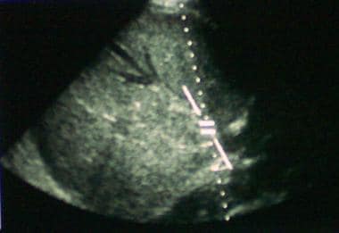 Sonogram showing hepatic vein thrombus, with new vessels forming. The arrow is pointing to the thrombus.
The prognosis is poor in patients with Budd-Chiari syndrome who remain untreated, with death resulting from progressive liver failure in 3 months to 3 years from the time of the diagnosis. [1] Following portosystemic shunting, however, the 5-year survival rate for patients with the syndrome is 38-87%. The actuarial 5-year survival rate following liver transplantation is 70%. [2, 3, 4]
Signs and symptoms
Physical examination may reveal the following:
-
Jaundice
-
Ascites
-
Hepatomegaly
-
Splenomegaly
-
Ankle edema
-
Stasis ulcerations
-
Prominence of collateral veins
The clinical variants of Budd-Chiari syndrome have been described as follows [5, 6, 7] :
-
Acute and subacute forms: Characterized by rapid development of abdominal pain, ascites (which can cause abdominal distention), hepatomegaly, jaundice, and renal failure.
-
Chronic form: Most common presentation; patients present with progressive ascites; jaundice is absent; approximately 50% of patients also have renal impairment
-
Fulminant form: Uncommon presentation; fulminant or subfulminant hepatic failure is present, along with ascites, tender hepatomegaly, jaundice, and renal failure.
See Clinical Presentation for more detail.
Diagnosis
Laboratory studies
Examination of the ascitic fluid provides useful clues to the diagnosis of Budd-Chiari syndrome, including the following:
-
Patients usually have high protein concentrations (>2 g/dL); this may not be present in persons with the acute form of the disease
-
The white blood cell (WBC) count is usually less than 500/µL
-
The serum ascites–albumin gradient is usually less than 1.1 (except in the acute forms of Budd-Chiari syndrome)
Imaging studies
-
Ultrasonography
-
Computed tomography (CT) scanning
-
Magnetic resonance imaging (MRI)
-
Venography
Biopsy
Pathologic findings in liver biopsy are (1) high-grade venous congestion and centrilobular liver cell atrophy, and, possibly, (2) thrombi within the terminal hepatic venules.
See Workup for more detail.
Management
Pharmacologic therapy
-
Anticoagulants
-
Thrombolytics
-
Diuretics
Procedures and surgery
-
Balloon angioplasty
-
Localized thrombolysis
-
Placement of a stent or transjugular intrahepatic portacaval shunt (TIPS)
-
Variceal treatment
-
Paracentesis
-
Portal decompression
-
Percutaneous transhepatic balloon angioplasty (PTBA)
-
Liver transplantation
See Treatment and Medication for more detail.
Background
Budd-Chiari syndrome is an uncommon condition induced by thrombotic or nonthrombotic obstruction of the hepatic venous outflow and is characterized by hepatomegaly, ascites, and abdominal pain. [49] It most often occurs in patients with an underlying thrombotic diathesis, including in those who are pregnant or who have a tumor, a chronic inflammatory disease, a clotting disorder, an infection, or a myeloproliferative disorder, such as polycythemia vera or paroxysmal nocturnal hemoglobinuria. (See Etiology and Presentation.)
Obstruction of large- or small-caliber veins leads to hepatic congestion as blood flows into, but not out of, the liver. Microvascular ischemia due to congestion causes hepatocellular injury. Portal hypertension and liver insufficiency result. [5, 6, 49] (See Pathophysiology.)
Budd-Chiari syndrome should be considered separate from veno-occlusive disease (VOD), also known as sinusoidal obstruction syndrome, which is characterized by toxin-induced, nonthrombotic obstruction of prehepatic veins (see the images below). (See Presentation and Workup.)
Sonogram showing hepatic vein thrombus, with new vessels forming. The arrow is pointing to the thrombus.
Pathophysiology
Occlusion of a single hepatic vein is usually silent. Overt Budd-Chiari syndrome generally requires the occlusion of at least 2 hepatic veins. [8] Venous congestion of the liver causes hepatomegaly, which can stretch the liver capsule and be very painful. Enlargement of the caudate lobe is common because blood is shunted through it directly into the inferior vena cava (IVC).
Hepatic function can be affected to a degree, depending on the amount of stasis and the resultant hypoxia. Increased sinusoidal pressure can itself cause hepatocellular necrosis. [49] The literature also suggests that upregulation of specific genes in chronic Budd-Chiari syndrome contributes to liver destruction through the stimulation of extracellular matrix proliferation, which contributes to liver fibrosis.
The most prominent genes involved include matrix metalloproteinase 7 and superior cervical ganglion 10 (SCG10), which are increased in expression, and thrombospondin-1, which is decreased. [9] Overexpression of the proliferating cell nuclear antigen gene, the c -MYC oncogene, and the tumor protein p53 gene may also be etiologic factors for Budd-Chiari syndrome. [10]
Etiology
Most patients with Budd-Chiari syndrome have an underlying thrombotic diathesis, although in approximately one third of patients, the condition is idiopathic. Thus, it may be a primary venous problem or an intra-/extrahepatic space-occupying lesion compressing/invading the venous outflow. [49] Causes of Budd-Chiari syndrome include the following:
-
Hematologic disorders
-
Inherited or acquired thrombotic diathesis [49]
-
Pregnancy and postpartum [11]
-
Oral contraceptives [49]
-
Chronic infections
-
Chronic inflammatory diseases
-
Tumors
-
Congenital membranous obstruction
-
Hepatic venous stenosis
-
Hypoplasia of the suprahepatic veins
-
Postsurgical obstruction
-
Posttraumatic obstruction
-
Malnutrition [49]
-
Total parenteral nutrition (TPN): Budd-Chiari syndrome has been reported as a complication of TPN via an IVC catheter in a neonate
Hematologic disorders
These include the following:
-
Polycythemia rubra vera
-
Paroxysmal nocturnal hemoglobinuria
-
Unspecified myeloproliferative disorder
-
Antiphospholipid antibody syndrome
-
Essential thrombocytosis
Inherited thrombotic diathesis
Coagulopathies include the following:
-
Protein C deficiency
-
Protein S deficiency
-
Antithrombin III deficiency
-
Factor V Leiden deficiency
Chronic infections
These include the following:
-
Hydatid cysts
-
Aspergillosis
-
Amebic abscess
Chronic inflammatory diseases
These include the following:
-
Inflammatory bowel disease
-
Mixed connective-tissue disease
Tumors
These include the following:
-
Hepatocellular carcinoma (HCC) [12]
-
Renal cell carcinoma
-
Leiomyosarcoma
-
Adrenal carcinoma
-
Right atrial myxoma
Congenital membranous obstructions
These include the following:
-
Type I: Thin membrane is present in the vena cava or the atrium
-
Type II: A segment of the vena cava is absent
-
Type III: The inferior vena cava (IVC) cannot be filled, and collaterals have developed
Miscellaneous
Miscellaneous causes of Budd-Chiari syndrome include the following:
-
Alpha1-antitrypsin deficiency
-
Dacarbazine
-
Urethane
Epidemiology
Budd-Chiari syndrome is extremely rare, and the incidence is not well reported in the literature, although a study by Rajani et al found an incidence of about 1 case per million population per year in Sweden. Congenital membranous forms of Budd-Chiari syndrome are the most common cause of the disease worldwide, particularly in Asia. [13]
A retrospective (2009-2013) nation-wide, population-based study in South Korea found a total of 424 patients with Budd-Chiari syndrome, with an average age- and sex-adjusted prevalence of 5.29 per million population. [14] The female-to-male ratio was 1.8, the median age was 51 years, and the annual case-fatality rate was 2.8%. [14]
The prevalence of Budd-Chiari syndrome in France appears to 4.04 per million inhabitants, with myeloproliferative neoplasms (48%), use of oral contraceptives (35%), and factor V Leiden (16%) the highest risk factors. [52]
No data suggest that sex affects predisposition to Budd-Chiari syndrome. Nonetheless, in the United States the condition is predominantly seen in women and is associated with hematologic disorders.
Age at presentation is usually in the third or fourth decade of life, although the condition may also occur in children or elderly persons.
Prognosis
The natural history of Budd-Chiari syndrome is not well known. The following factors, however, have been associated with a good prognosis:
-
Younger age at diagnosis
-
Low Child-Pugh score
-
Absence of ascites or easily controlled ascites
-
Low serum creatinine level
In a systematic review of 79 studies, investigators found that although univariate analysis indicated bilirubin and creatinine levels as well as ascites might be significant prognostic factors, multivariate analyses did not always reveal achievement of statistical significance. [15]
The following formula has been proposed to calculate the prognostic index for Budd-Chiari syndrome; a score of less than 5.4 is associated with a good prognosis:
-
Prognostic index = (ascites score x 0.75) + (Pugh score x 0.28) + (age x 0.037) + (creatinine level x 0.0036)
The 5-year survival rate for patients with the syndrome is 38-87% following portosystemic shunting. The actuarial 5-year survival rate following liver transplantation is 70%. [2, 3, 4] Long-term follow-up in adults has demonstrated 10-year survival rates as high as 55%.
The prognosis is poor, however, in patients with Budd-Chiari syndrome who remain untreated, with death resulting from progressive liver failure in 3 months to 3 years from the time of diagnosis. [1]
In a University of Pennsylvania retrospective study (2008-2013) comprising 47 patients with Budd-Chiari syndrome, there were no significant differences in the treatment outcomes among those receiving anticoagulation therapy alone, transjugular intrahepatic portosystemic shunt (TIPS) placement alone, and TIPS in conjunction with anticoagulation. [16] The investigators noted that the significant prognostic predictors for liver transplantation were age, presence of cirrhosis, and presence of chronic kidney disease.
Morbidity and mortality
Morbidity and mortality in Budd-Chiari syndrome are generally related to complications of liver failure and ascites but can also be impacted by the type of concomitant underlying disease, if any. Complications associated with Budd-Chiari syndrome include the following:
-
Hepatic encephalopathy
-
Variceal hemorrhage
-
Hepatorenal syndrome
-
Portal hypertension
-
Complications secondary to hypercoagulable state
-
Complications secondary to hepatic decompensation
Bacterial peritonitis is always of concern in the patient with ascites, especially if paracentesis is undertaken. Complications must also be considered in relation to therapies used (eg, thrombolytics). The mortality rate can be high in patients who develop fulminant hepatic failure.
Budd-Chiari syndrome can also lead to HCC (or oppositely, in some cases, develop secondary to it). In a retrospective study, Liu et al found evidence that HCC in primary Budd-Chiari syndrome is associated with blockage of the inferior vena cava and stricture of the hepatic venous outflow tract. The investigators’ results also indicated that transcatheter arterial chemoembolization (TACE) is an effective treatment for HCC in these patients, with a significant drop in alpha-fetoprotein levels after TACE treatment. The study included 246 patients with Budd-Chiari syndrome, including 14 with HCC. Ultrasonography, computed tomography (CT) scanning, magnetic resonance imaging (MRI), and angiography were used to determine the imaging characteristics in patients with HCC. [17]
-
Sonogram showing hepatic vein thrombus, with new vessels forming. The arrow is pointing to the thrombus.
-
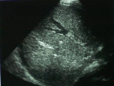
Sonogram showing hepatic vein thrombus.