Practice Essentials
Colonic atresia is a condition in which a part of the colon has not formed correctly, with the result that it is either blocked completely or missing altogether; colonic stenosis is a condition in which a part of the colon is very narrow, resulting in a partial blockage. Other obstructions of the colon that affect newborns include the following:
Although these conditions are all forms of colonic obstruction, they are different from atresia and stenosis and thus are more completely reviewed elsewhere.
Colonic atresia is a congenital anomaly that may be suggested by antenatal sonograms and is usually revealed in affected newborns shortly after birth. Patients usually present with abdominal distention and failure to pass meconium.
Stenosis of the colon is much more common. Patients usually present later in life, most often because of an identifiable event. In congenital stenosis, a narrow segment of colon is observed, but bowel continuity is maintained. A discrepancy between the colonic segments above and below the area of stenosis is present. In acquired stenosis (commonly referred to as stricture), what starts as a normal segment becomes narrowed. This is most common in premature babies who have recovered from an episode of necrotizing enterocolitis.
Colonic atresia and congenital stenosis are uncommon lesions, with a reported incidence of 4.2 per 10,000 live births. [1] Associated anomalies are common, and their severity directly affects outcome. Although the etiology of colonic atresia has traditionally been believed to be related to antenatal mesenteric vascular accidents, a 2005 study suggested that defects in the fibroblast growth factor 10 (FGF10) pathway may be involved. [2]
The combination of Hirschsprung disease and colonic atresia remains rare; however, missing the association before reconnecting the intestinal tract can lead to repeat operations, poor outcome, and increased mortality.
Surgical correction is the mainstay of therapy for atresia and stenosis. In the absence of significant comorbidity, primary resection and anastomosis constitute the recommended surgical treatment and appear to have an outcome equivalent to that of resection with creation of a colostomy and subsequent closure. [3]
Anatomy
The colon arises from the digestive tube, which is present by the end of the first month of gestation. Rapid elongation begins at 5 weeks’ gestation. Over the ensuing 5 weeks, the intestinal tube, separable into cephalad and caudal limbs (on the basis of their relation to the omphalomesenteric duct), rotates counterclockwise and returns to its familiar position in the abdomen. The proximal caudal limb is supplied by the superior mesenteric artery (SMA), whereas the distal portion is supplied by the inferior mesenteric artery (IMA). [4]
The SMA gives rise to the ileocolic, right colic, and middle colic arteries, which supply the ileocecal region, the ascending colon, and the proximal transverse colon, respectively. The left colic artery, arising from the IMA, supplies the left portion of the transverse colon and the descending colon. The middle colic and left colic arteries join near the splenic flexure to form the marginal artery. The sigmoid arteries, rectosigmoid arteries, and branches of the IMA supply the sigmoid colon.
Pathophysiology and Etiology
The feature common to both atresia and stenosis is intestinal blockage, either partial or complete.
In colonic atresia, the problem is complete bowel obstruction. Gas and stool cannot pass, and the colonic segment above the atresia becomes distended. If left untreated, this leads to perforation.
In colonic stenosis, the problem is that gas and stool try to pass through a narrow area. While the baby is passing soft baby stools, this may or may not be noticeable. When the baby’s diet changes from breast milk or formula to cereals and solid foods, the stool can become thicker and more formed. This may cause stenosis to become symptomatic, leading to distension, feeding intolerance, or failure to thrive.
In babies who have had necrotizing enterocolitis, stenosis occurs after the original episode has resolved. This may manifest in varying degrees, ranging from minor feeding intolerance and distension to near-complete bowel obstruction.
Small-bowel and colonic atresias are not believed to occur by the same process as duodenal atresia, which is suspected to be due to failure of vacuolization of the duodenum and was described by Tandler in 1900.
In 1955, Louw and Barnard hypothesized that small-bowel atresias are caused by antenatal vascular interruption. [5] The same mechanism is believed to cause colonic atresia. Thrombosis, volvulus, and herniation with strangulation are all mechanisms that may cause in-utero vascular injury and bowel necrosis with subsequent reabsorption. Fairbanks proposed a relationship between FGF10 expression, vascular development, and colonic atresia. [2]
Colonic atresia is typically classified according to the 1989 descriptions of intestinal atresia by Bland-Sutton [6] and the 1964 descriptions by Louw, [7] as follows:
-
Type 1 - The bowel and mesentery remain intact, but the bowel lumen is interrupted by a complete membrane (see the image below)
-
Type 2 - The bowel is discontinuous, with portions connected by a fibrous cord
-
Type 3 - The bowel ends are completely separated, and the mesentery has a gap
-
Stenotic lesions are characterized by intact bowel with incomplete occlusion and require no classification
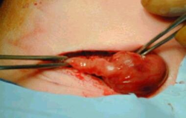 Colonic atresia, type 1 (sigmoid colon). Dilated colon abruptly tapers to unused distal segment.
In 1990, Davenport et al reviewed 118 cases of colonic atresia and reported the following distribution lesion sites [8] :
-
Ascending colon - 33 (28%)
-
Hepatic flexure - 4 (3%)
-
Transverse colon - 27 (23%)
-
Splenic flexure - 30 (25%)
-
Descending and sigmoid colon - 24 (20%)
Two thirds of colonic atresias are in the distribution of the IMA. This finding may be related to a lack of collateral blood supply or to disease processes that render this portion of the colon more susceptible to injury.
Although the colon is supplied by the SMA and the IMA, there is considerable variation among smaller named arterial branches. The parts of the colon most vulnerable to ischemia appear to be the splenic flexure and the ileocecal area—coincidentally, the areas farthest from the major trunks. However, atresia and stenosis occur throughout the colon; the variation in terminal branches, coupled with the complex rotation and fixation of the bowel, may create regions of ischemic injury in utero that result in atresia or stenosis. [9]
Any process leading to occlusion of branches of these vessels in utero may result in atresia. Compression at the umbilical ring, [10] internal hernia, intussusception, [11] choledochal cyst, [12] volvulus, and thrombosis all may initiate bowel infarction, leading to disintegration, reabsorption of dead tissue, and sealing of bowel ends. [7] Bowel content is sterile; thus, sepsis does not occur. Meconium is produced throughout the gut during the third trimester; thus, a newborn whose atresia occurs after that time may still pass meconium in the neonatal period.
Antenatal maternal use of vasoconstrictive medications (eg, cocaine, amphetamines, nicotine, or decongestants) has been suggested as a risk factor for intestinal atresia formation. [13]
Although prenatal mesenteric vascular accident has been long accepted as the most likely cause of colonic atresia, a study on the role of the FGF10 signaling pathway has brought that belief into question. In this study, mice that were unable to express FGF10 and FGF receptor 2b developed colonic atresia, even though their mesenteric vasculature was normal during periods when atresias were developing; this suggests that the etiology of colonic atresia may be more complicated than was once thought and that it may not involve a vascular issue. [2]
Congenital stenosis occurs when the bowel injury is incomplete, as may be the case when injury occurs close to the bowel wall, allowing collateral blood flow to preserve the injured tissue. Another mechanism is limited ischemia, in which the blood supply is partially or intermittently occluded, resulting in incomplete intestinal injury.
Acquired stenosis is more common than atresia or congenital stenosis. Via the same mechanism of vascular compromise, the injured bowel undergoes healing and scarring with narrowing of the affected intestine. Intense inflammatory reactions, such as those that occur in necrotizing enterocolitis and Crohn disease, may result in stricture. Tuberculosis-associated left colon stricture has also been reported. [14]
Patients who undergo bowel resection for any reason and have a segment of intestine removed and joined by anastomosis may develop stricture at the anastomotic site as a consequence of ischemia or a technical issue. Surgery is usually required to revise the joined loops.
Epidemiology
Colonic atresia is rare, with reported incidences ranging from 1 in 1500 live births [15] to 1 in 66,000. [8] Webb (1931) and Benson (1968) cited an incidence of 1 in 20,000, which most closely reflects experiences in the modern era. [16, 17] In 1982, Powell suggested that colonic atresia represents 5-15% of intestinal atresias, [18] whereas in 1966, Freeman reported a figure of 1.8%. [19] In 1953, Gross reported that colonic atresia represented 4.3% (6/140) of cases of intestinal atresia at the Boston Children’s Hospital. [20]
The 2017 National Birth Defects Prevention Network (NBDPN) annual report found rectal and large intestinal atresia/stenosis to be the most frequent gastroentestinal defect, with an incidence of 4.2 per 10,000 live births. [1]
Multiple atresias are uncommon in the colon, with a reported occurrence of 8.9%. [21] However, colonic atresia is associated with other gastrointestinal malformation in up to 80% of cases and may be overlooked when small-bowel atresia is present. [21, 22] Rare cases of familial colonic atresia have been described. Animal studies have shown an autosomal recessive pattern of inheritance in cattle. Hereditary multiple intestinal atresia affects both the large and the small intestine, whereas nonhereditary multiple intestinal atresia usually spares the colon. [23]
The incidence of colonic stenosis is not readily available, because most cases are acquired. [24] In 1953, Gross reported only one colonic lesion in 71 patients with intestinal stenosis. [20] Necrotizing enterocolitis is the most common etiology of postnatal colonic stenosis; narrowing develops in 10-25% of affected patients. [25, 26, 27]
Prognosis
With colonic atresia and stenosis, survival is related to the patient’s condition before surgery, technical difficulties with the colonic anastomosis, sepsis, and associated anomalies. [28, 22]
Whereas older series reported a high mortality for colonic atresia, subsequent series reported survival of all patients, except those with significant life-threatening comorbidities. Patients with Hirschsprung disease and colonic atresia have more complicated courses and a mortality of 10%. [29]
Colonic stenosis outcomes have also improved significantly since Gross reported the death of the single patient treated at the Boston Children’s Hospital before 1952 (see Table 1 below). [20] Improvements in resuscitation and perioperative care have resulted in survival rates of approximately 90%. [30, 31]
Table 1. Outcomes of Surgery in Colonic Atresia and Stenosis (Open Table in a new window)
Author (Year) |
No. of Patients |
Procedure |
Survival Rate |
Gross (1952) [20] |
6 |
Ostomy* |
33% |
Sturim (1966) [32] |
2 |
Ostomy |
50% |
Coran (1969) [33] |
9 |
Ostomy |
66% |
Pohlson (1988) [34] |
11 |
Ostomy (6), anastomosis (4),† resection of diaphragm (1)‡ |
73% |
Smith (1989) [35] |
2 |
Not specified |
100% |
Davenport (1990) [8] |
11 |
Ostomy (6), anastomosis (4) |
91% |
Barrack (1993) [36] |
2 |
Anastomosis |
100% |
Dalla Vecchia (1998) [22] |
21 |
Ostomy (18), anastomosis (3) |
100% |
Abu-Judeh (2001) [30] |
1 |
Anastomosis |
100% |
Dassinger (2009) [37] |
12 |
Ostomy (2), anastomosis (10) |
100% |
*Ostomy, resection and staged anastomosis months later. †Resection with primary anastomosis. ‡Cecotomy, resection of diaphragm. |
|||
Colonic atresia has been associated with abdominal wall defects and abnormalities of the genitourinary tract. [38] Nonfixation of the colon has been reported. [39] Association with anal atresia [40] and imperforate anus [41] has been reported but is extremely rare.
Colonic perforation may occur. [42] This is thought to be caused by overdistention of a closed colonic loop, with gas and stool trapped between a competent ileocecal valve proximally and the blind-ending colon distally. In 1988, Pohlson et al reported perforation of the terminal ileum in one case, clearly demonstrating that perforation can occur anywhere proximal to the obstructed bowel. [34]
Hirschsprung disease has been present in a small number of cases. [43, 44, 45, 46, 47] Additional anomalies associated with this pairing include omphalocele [48] and absence of a hand. [49] In most cases, the aganglionosis is discovered after colostomy closure when the distal bowel does not function properly.
Some authors have recommended that rectal biopsy be performed at the time of laparotomy, [50] whereas others believe that it should be reserved for children who do not pass stool readily when bowel continuity is restored after resection. [44, 46] Because colonic atresia is rare and because significant morbidity and mortality may result if the diagnosis is not made before intestinal continuity is established, it would seem prudent to perform rectal biopsy before committing to definitive repair. [29]
Cardiac conditions that require catheterization may predispose a baby to a mesenteric vascular incident resulting in colonic stenosis. Additional conditions reported in patients with colonic stenosis include the following [15, 18, 51] :
-
Cryptophthalmia syndrome (ie, cleft lip and palate, microphthalmia, dysplastic kidneys, and proximal jejunal atresia)
-
Proximal intestinal atresia
-
Neoplasm
Riley-Day syndrome (ie, familial dysautonomia) has been associated with spontaneous colon ischemia. [52] Coloboma, cataracts and facial hemihypertrophy, facial asymmetry with palsy, microphthalmia with partial iridic coloboma, exophthalmia, and bilateral optic nerve hypoplasia all have been reported. [18, 53] In 2000, Kim et al described a case of colonic atresia in monozygotic twins. [23]
-
Colonic atresia, type 1 (sigmoid colon). Dilated colon abruptly tapers to unused distal segment.
-
Contrast enema showing microcolon with dilated proximal colon (colonic atresia of sigmoid colon).
-
Contrast enema revealing colonic stenosis at hepatic flexure.
-
Abdominal radiograph of baby with colonic atresia at distal transverse colon. Colon proximal to atresia is visible as large dilated loop of intestine running obliquely across abdomen. Courtesy of Richard Glick, MD.
-
Intraoperative photograph displays proximal dilated segment in patient with type 3 atresia of distal transverse colon. Courtesy of Richard Glick, MD.
-
Small portion of bowel held on right looks like appendix but is actually distal segment for patient with type 3 atresia at distal transverse colon. Courtesy of Richard Glick, MD.
Tables
Author (Year) |
No. of Patients |
Procedure |
Survival Rate |
Gross (1952) [20] |
6 |
Ostomy* |
33% |
Sturim (1966) [32] |
2 |
Ostomy |
50% |
Coran (1969) [33] |
9 |
Ostomy |
66% |
Pohlson (1988) [34] |
11 |
Ostomy (6), anastomosis (4),† resection of diaphragm (1)‡ |
73% |
Smith (1989) [35] |
2 |
Not specified |
100% |
Davenport (1990) [8] |
11 |
Ostomy (6), anastomosis (4) |
91% |
Barrack (1993) [36] |
2 |
Anastomosis |
100% |
Dalla Vecchia (1998) [22] |
21 |
Ostomy (18), anastomosis (3) |
100% |
Abu-Judeh (2001) [30] |
1 |
Anastomosis |
100% |
Dassinger (2009) [37] |
12 |
Ostomy (2), anastomosis (10) |
100% |
*Ostomy, resection and staged anastomosis months later. †Resection with primary anastomosis. ‡Cecotomy, resection of diaphragm. |
|||


