Practice Essentials
Most airway foreign body aspirations occur in children; those aged younger than 3 years are at greatest risk. [1] See the image below.
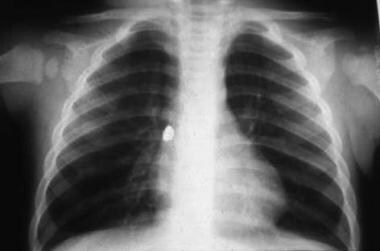 Aspirated foreign body (backing to an earring) lodged in the right main stem bronchus.
Signs and symptoms
Often, the child presents after a sudden episode of coughing or choking while eating with subsequent wheezing, coughing, or stridor. However, in numerous cases, the choking episode is not witnessed, and, in many cases, the choking episode is not recalled at the time the history is taken.
Major findings include new abnormal airway sounds, such as wheezing, stridor, or decreased breath sounds. These sounds are often, but not always, unilateral.
See Presentation for more detail.
Diagnosis
If the history and physical findings are diagnostic, no workup is needed. The child should immediately be referred for rigid bronchoscopy.
See Workup for more detail.
Management
Bronchodilators and corticosteroids should not be used to remove the foreign body, and chest physical therapy with postural drainage may dislodge the material to an area where it may cause more harm, such as at the level of the vocal cords.
Surgical therapy for an airway foreign body involves endoscopic removal, usually with a rigid bronchoscope.
See Treatment and Medication for more detail.
Background
The human body has numerous defense mechanisms to keep the airway free and clear of extraneous matter. These include the physical actions of the epiglottis and arytenoid cartilages in blocking the airway, the intense spasm of the true and false vocal cords any time objects come near the vocal cords, and a highly sensitive cough reflex with afferent impulses generated throughout the larynx, trachea, and all branch points in the proximal tracheobronchial tree. However, none of these mechanisms is perfect, and foreign bodies frequently lodge in the airways of children. [2]
Pathophysiology
Children are more prone to aspirate foreign material for several reasons. The lack of molar teeth in children decreases their ability to sufficiently chew food, leaving larger chunks to swallow. The propensity of children to talk, laugh, and run while chewing also increases the chance that a sudden or large inspiration may occur with food in the mouth. Children often examine even nonfood substances with their mouth.
More foreign body aspirations occur in children younger than 3 years than in other age groups, with a peak between the first and second birthdays. However, foreign bodies have been found in the airways of individuals of all ages and sizes. Even relatively immobile infants may aspirate foreign bodies, despite not having the ability to crawl and find things or the ability to pick up objects and put them in the mouth. They have less chewing capacity and higher respiratory rates, so any objects placed in their mouths are more likely to be aspirated than in older children. They also have well-meaning siblings, who may put the wrong foods in the baby's mouth in an attempt to help feed them.
The most common entities aspirated are small food items such as nuts, raisins, sunflower seeds, improperly chewed pieces of meat and small, smooth items such as grapes, hot dogs, and sausages. All of these should be avoided until the child is able to adequately chew them while sitting. Generally, this occurs around age 5 years, with most foreign body aspirations occurring in children younger than 3 years. Small items that are round, smooth, or both (eg, grapes, hot dogs, sausages, balloons) are more likely to cause tracheal obstruction and asphyxiation. Dried foods may cause progressive obstruction as they absorb water.
In a review of 1068 foreign body aspirations in children, the authors found 3% in the larynx, 13% in the trachea, 52% in the right main bronchus, 6% in the right lower lobe bronchus, fewer than 1% in the right middle lobe bronchus, 18% in the left main bronchus, and 5% in the left lower lobe bronchus; 2% were bilateral. [3] In a child in an upright position, the right-sided airways are direct entries from the trachea. The left main bronchus is smaller than the right main bronchus and is slightly angled. In a child in a supine position, material is more likely to enter the right main bronchus.
Epidemiology
United States statistics
Foreign body aspiration accounted for more than 17,000 emergency department visits and caused 220 deaths in children aged 14 years or younger. [4] Airway foreign bodies are the third most common cause of death due to unintentional injury in children younger than 1 year. [4]
A study by Saw-Aung et al that analyzed data from the National Electronic Injury Surveillance System showed that the incidence of foreign body aspiration among children in the United States decreased by 23.6% between 2010 and 2020. [1]
A study by Wanstreet et al used data from the Healthcare Cost and Utilization Project’s Kids’ Inpatient Database (HCUP KID) to determine that among pediatric patients (aged up to 20 years), nonfood airway foreign bodies tended to be found more frequently in older children than in younger ones. [5]
Race-, sex-, and age-related demographics
Race
No racial predilections are noted. Compared with White pediatric patients, Black children with aspirated foreign bodies had less likelihood of same-hospital admission (odds ratio [OR] = 0.8), whereas the risk of mortality was higher (OR = 9.2) in Black patients. [1]
Sex
Most reviews of foreign body aspiration in children show a slight male predominance. [6]
Age
The peak ages during which aspiration of foreign body occurs are the toddler through preschool ages, although foreign bodies have been found in the airways of people of all ages and sizes.
The results of a study by D’Souza et al suggest that 90% of episodes of pediatric nut aspiration occur in patients aged younger than 36 months. The researchers also found that the average age at aspiration of a whole nut was 24 months. [7]
Prognosis
Once the foreign material is removed, the prognosis is excellent. The sooner it is removed, the quicker and more complete the recovery.
Morbidity/mortality
Unfortunately, mortality occurs due to acute aspiration, and morbidity can occur due to acute hypoxia during the acute episode or due to chronic lung and airway damage from a long-standing aspirated foreign body. The National Safety Council estimates that 2900 deaths occur annually in the United States because of foreign body aspiration. [8]
Complications
Potential complications include the following:
-
Atelectasis due to prolonged airway obstruction
-
Bronchiectasis due to chronic infection
-
Lung abscess
-
Pneumomediastinum and pneumothorax (rare complications of foreign body removal)
-
Aspirated foreign body (backing to an earring) lodged in the right main stem bronchus.


