Continually Updated Clinical Reference
 
 
  All Sources     eMedicine     Medscape     Drug Reference     MEDLINE
 
eMedicine - Pulmonary Sequestration : Article by

Quick Find
Authors & Editors
Introduction
Clinical
Differentials
Workup
Treatment
Medication
Follow-up
Miscellaneous
Multimedia
References

Related Articles
Atelectasis, Pulmonary

Bronchiectasis

Pneumonia


Patient Education
Click here for patient education.


Author: Bruce M Schnapf, DO, Chief, Division of Pulmonology, Department of Pediatrics, Associate Professor, University of South Florida College of Medicine

Bruce M Schnapf is a member of the following medical societies: American Academy of Pediatrics, American College of Chest Physicians, and American Thoracic Society

Editors: Susanna A McColley, MD, Director of Cystic Fibrosis Center, Divisions of Pediatric Pulmonary and Critical Care, Associate Professor, Department of Pediatrics, Children's Memorial Medical Center of Chicago, Northwestern University; Mary L Windle, PharmD, Adjunct Assistant Professor, University of Nebraska Medical Center College of Pharmacy, Pharmacy Editor, eMedicine.com, Inc; Charles Callahan, DO, Professor, Deputy Chief of Clinical Services, Walter Reed Army Medical Center; Mary E Cataletto, MD, Associate Director, Division of Pediatric Pulmonology, Winthrop University Hospital; Associate Professor, Department of Clinical Pediatrics, State University of New York at Stony Brook; Michael R Bye, MD, Professor of Clinical Pediatrics, Columbia University College of Physicians and Surgeons; Acting Director, Department of Pediatric Pulmonary Medicine, Columbia University Medical Center

Author and Editor Disclosure

Synonyms and related keywords: pulmonary sequestration, extrapulmonary sequestration, intrapulmonary sequestration, sequestrum, lung bud, lung

Background

Pulmonary sequestration is an uncommon mass that is usually cystic and is composed of nonfunctioning primitive tissue that does not communicate with the tracheobronchial tree. The new nomenclature refers to this as a congenital thoracic malformation. It may present as a lung infection on chest imaging. Its blood supply is from systemic circulation rather than the pulmonary circulation. Multiple feeding vessels may be present in 15-20% of cases. The 2 forms of pulmonary sequestration are intrapulmonary, which is surrounded by normal lung tissue, and extrapulmonary, which has its own pleural investment. Other congenital malformations may be present.

Pathophysiology

The most frequently supported theory of sequestration formation involves an accessory lung bud that develops from the ventral aspect of the primitive foregut. The pluripotential tissue from this additional lung bud migrates in a caudal direction with the normally developing lung. It receives its blood supply from vessels that connect to the aorta and cover the primitive foregut. These attachments to the aorta remain to form the systemic arterial supply of the sequestration.

Early embryologic development of the accessory lung bud results in formation of the sequestration within normal lung tissue. The sequestration is encased within the same pleural covering. This is the intrapulmonary variant. In contrast, later development of the accessory lung bud results in the extrapulmonary type that may give rise to communication with the gastrointestinal tract. Both types of sequestration usually have arterial supply from the thoracic or abdominal aorta. Rarely, the celiac axis, internal mammary, subclavian, or renal artery may be involved.

Intrapulmonary sequestration occurs within the visceral pleura of normal lung tissue. Usually, no communication with the tracheobronchial tree occurs. The most common location is in the posterior basal segment, and nearly two thirds of pulmonary sequestrations appear in the left lung. Venous drainage is usually via the pulmonary veins. Foregut communication is very rare, and associated anomalies are uncommon.

Extrapulmonary sequestration is completely enclosed in its own pleural sac. It may occur above, within, or below the diaphragm, and nearly all appear on the left side. No communication with the tracheobronchial tree occurs. Venous drainage is usually via the systemic venous system. Foregut communication and associated anomalies, such as diaphragmatic hernia, are more common.

Frequency

United States

Pulmonary sequestration represents approximately 6% of all congenital pulmonary malformations. Intrapulmonary sequestrations are the most common form, and 60% of these are found in the posterior basal segment of the left lower lobe. Overall, 98% occur in the lower lobes. Bilateral involvement is uncommon. About 10% of cases may be associated with other congenital anomalies. Extrapulmonary sequestrations occur on the left in 95% of cases. Of these, 75% are found in the costophrenic sulcus on the left side. They may also be found in the mediastinum, pericardium, and within or below the diaphragm. They are associated with other congenital malformations in more than 50% of cases, such as congenital diaphragmatic hernias, congenital cystic adenomatoid malformation (CCAM) type II (hybrid lesions), and congenital heart disease. 

Mortality/Morbidity

The morbidity and mortality rates are exceedingly low if resection of the mass precedes repeated infection. Postoperative results are uniformly good.

Sex

In the extrapulmonary form, males are affected approximately 4 times more often than females. Incidence is equal in males and females in the intrapulmonary type.

Age

  • More than one half of intrapulmonary sequestrations are diagnosed in later childhood or even in adulthood. Neonates and infants are usually asymptomatic.
  • In contrast, more than one half of extrapulmonary sequestrations are diagnosed in patients younger than 1 year. Often, this is because other congenital anomalies are present, including congenital diaphragmatic hernia, cardiac malformations, and gastrointestinal malformations.


History

  • Intrapulmonary sequestration
    • Although an intrapulmonary sequestration is usually diagnosed later in childhood or adolescence, symptoms may begin early in childhood with multiple episodes of pneumonia. A chronic or recurrent cough is common. Intrapulmonary sequestration shares the visceral pleura that covers the adjacent lung tissue and is usually located in the posterobasal segment of the lower lobes. The thoracic or abdominal aorta often provides the arterial blood supply. Venous drainage is commonly provided to the left atrium via the pulmonary veins.
    • An elemental communication with other bronchi or lung parenchyma may be present, allowing infection to occur. Rarely, an esophageal bronchus may be present. Resolution of infection is usually slow and incomplete because of inadequate bronchial drainage.
    • Overdistension of the cystic mass with air can result in compression of normal lung tissue with impairment of cardiorespiratory function. Aeration probably occurs through the pores of Kohn.
    • Other congenital anomalies may appear in 10% of cases.
  • Extrapulmonary sequestration
    • Many patients present in infancy with respiratory distress and chronic cough.
    • Lesions are commonly diagnosed coincidentally during investigation of, or surgery for, an associated congenital anomaly. Therefore, clinical symptoms may be absent or minor.
    • Extrapulmonary sequestration may manifest as GI symptoms if communication with the GI tract is present. As a result, infants may have feeding difficulties. In addition, extrapulmonary sequestration may manifest as recurrent lung infection, similar to the intrapulmonary form. This type of sequestration does not contain air unless communication with the foregut is present.

Physical

  • The lung examination may reveal signs consistent with consolidation.
  • Occasionally, patients may have a systolic bruit or continuous murmur over the affected area. This is related to flow through the sequestration from the large systemic arterial supply.

Causes

  • No known causes or risk factors have been established.


Atelectasis, Pulmonary
Bronchiectasis
Pneumonia

Other Problems to be Considered

Primary lung abscess
Recurrent pneumonia
Congenital lung mass


Lab Studies

  • No laboratory studies are needed.

Imaging Studies

  • Chest radiography
    • Chest radiography findings vary depending on the size of the sequestered lung tissue and whether infection is present. If no communication between sequestration and normal lung tissue is present, radiography usually reveals a dense opacity in the posterior basal segment of the lower lobe. A cystic appearance may also be observed.
    • Lesion density often increases with secondary infection and appears as a uniform consolidation. When this area fails to clear after a course of appropriate medical therapy, the presence of a malformation, such as sequestration, should be considered, particularly if the lesion is localized to the posterior basal segment of the left lower lobe.
    • Distinguishing an intrapulmonary sequestration from extrapulmonary sequestration is difficult using plain radiography.
      • Intrapulmonary lesions tend to be heterogeneous and are not well defined.
      • Extrapulmonary masses are usually observed as solid, well defined, and retrocardiac.
  • Bronchography and arteriography are unnecessary because of current noninvasive imaging available.
  • Presence of systemic arteries revealed by chest imaging is the major diagnostic feature of pulmonary sequestration. CT scanning with contrast or magnetic resonance angiography (MRA) have been very useful. The arterial supply and venous drainage both should be outlined because of the unpredictability of vascular connections. CT angiography is helpful in identifying aberrant systemic arterial supply and 3-dimensional rendering of multidetector row CT can reveal venous drainage.
  • Real-time ultrasonography and Doppler imaging are reliable methods of demonstrating systemic origin or blood supply, as well.

Other Tests

  • Upper gastrointestinal contrast examination may be useful if communication with the gastrointestinal tract is considered.

Procedures

  • Bronchoscopy: Bronchoscopy is not necessary unless an alternative cause of the radiographic abnormalities, such as an inhaled foreign body, is suspected.
  • Balloon occlusion
    • Consider balloon occlusion or embolization of the aberrant systemic arteries at the time of catheterization.
    • Some patients may have a considerable shunt through this anomalous circuit to the extent that, once the segment has been removed, the improvement in cardiovascular status may be striking.
    • If surgical resection is necessary, risk of vascular complications is greatly reduced with this procedure.


Medical Care

  • No specific medications are useful.

Surgical Care

  • Management of an asymptomatic pulmonary sequestration with no connection to the surrounding lung is controversial; however, most references advocate resection of these lesions because of the likelihood of recurrent lung infection, the need for larger resection if the sequestration becomes chronically infected, and the possibility of hemorrhage from arteriovenous anastomoses.
  • Surgical resection is the treatment of choice for patients who present with infection or symptoms resulting from compression of normal lung tissue. 
  • Extrapulmonary lesions can usually be excised without loss of normal lung tissue.
  • Intrapulmonary lesions often require lobectomy because the margins of the sequestration may not be clearly defined. Complete thoracoscopic resection of pulmonary lobes in infants and children has been described with low mortality and morbidity.

Diet

  • No special dietary requirements are necessary.

Activity

  • Activity restriction is not necessary.


No medical therapy is indicated in the management of pulmonary sequestration, unless an infection requires the use of antibiotics.


Prognosis

  • In the absence of perioperative complications, surgical resection of the abnormal segment is usually curative.
  • Prognosis may also be determined by the presence of any accompanying anomalies.


Medical/Legal Pitfalls

  • Failure to perform surgical resection of a pulmonary sequestration likely results in recurrent pneumonia.


Media file 1:  The multicystic lesion in the left lower lobe seen on the chest radiograph of this patient was causing repeated respiratory infections.
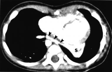
Click to see larger pictureClick to see detailView Full Size Image
Media type:  X-RAY


  • Abel RM, Bush A, Chitty LS, Harcourt J, Nicholson AG. Congenital lung disease. In: Kendig's Disorders of the Respiratory Tract in Children. 7th ed. Philadelphia, Pa: WB Saunders; 2006:301.
  • Adzick NS, Harrison MR, Crombleholme TM, et al. Fetal lung lesions: management and outcome. Am J Obstet Gynecol. Oct 1998;179(4):884-9. [Medline].
  • Albanese CT, Rothenberg SS. Experience with 144 consecutive pediatric thoracoscopic lobectomies. J Laparoendosc Adv Surg Tech A. Jun 2007;17(3):339-41. [Medline].
  • Alivizatos P, Cheatle T, de Leval M, Stark J. Pulmonary sequestration complicated by anomalies of pulmonary venous return. J Pediatr Surg. Feb 1985;20(1):76-9. [Medline].
  • Clement BS. Congenital malformations of the lungs and airways. In: Pediatric Respiratory Medicine. St Louis, Mo: Mosby; 1999:1124-5.
  • Collin PP, Desjardins JG, Khan AH. Pulmonary sequestration. J Pediatr Surg. Aug 1987;22(8):750-3. [Medline].
  • Corbett HJ, Humphrey GM. Pulmonary sequestration. Paediatr Respir Rev. Mar 2004;5(1):59-68. [Medline].
  • Deguchi E, Furukawa T, Ono S, et al. Intralobar pulmonary sequestration diagnosed by MR angiography. Pediatr Surg Int. Jul 2005;21(7):576-7. [Medline].
  • DeParedes CG, Pierce WS, Johnson DG, Waldhausen JA. Pulmonary sequestration in infants and children: a 20-year experience and review of the literature. J Pediatr Surg. Apr 1970;5(2):136-47. [Medline].
  • Flye MW, Conley M, Silver D. Spectrum of pulmonary sequestration. Ann Thorac Surg. Nov 1976;22(5):478-82. [Medline].
  • Gezer S, Tastepe I, Sirmali M, et al. Pulmonary sequestration: a single-institutional series composed of 27 cases. J Thorac Cardiovasc Surg. Apr 2007;133(4):955-9. [Medline].
  • Gustafson RA, Murray GF, Warden HE, et al. Intralobar sequestration. A missed diagnosis. Ann Thorac Surg. Jun 1989;47(6):841-7. [Medline].
  • Hadley GP, Egner J. Gastric duplication with extralobar pulmonary sequestration: an uncommon cause of "colic". Clin Pediatr (Phila). Jun 2001;40(6):364. [Medline].
  • Ikezoe J, Murayama S, Godwin JD, et al. Bronchopulmonary sequestration: CT assessment. Radiology. Aug 1990;176(2):375-9. [Medline].
  • Kang M, Khandelwal N, Ojili V, Rao KL, Rana SS. Multidetector CT angiography in pulmonary sequestration. J Comput Assist Tomogr. Nov-Dec 2006;30(6):926-32. [Medline].
  • Ko SC, Chang YC, Liaw YS, et al. Diagnosis of pulmonary sequestration by magnetic resonance imaging. J Formos Med Assoc. Mar 1998;97(3):220-3. [Medline].
  • Ko SF, Ng SH, Lee TY, et al. Noninvasive imaging of bronchopulmonary sequestration. AJR Am J Roentgenol. Oct 2000;175(4):1005-12. [Medline].
  • Laberge JM, Bratu I, Flageole H. The management of asymptomatic congenital lung malformations. Paediatr Respir Rev. 2004;5 Suppl A:S305-12. [Medline].
  • Lee EY, Siegel MJ, Sierra LM, Foglia RP. Evaluation of angioarchitecture of pulmonary sequestration in pediatric patients using 3D MDCT angiography. AJR Am J Roentgenol. Jul 2004;183(1):183-8. [Medline].
  • Levine MM, Nudel DB, Gootman N, et al. Pulmonary sequestration causing congestive heart failure in infancy: a report of two cases and review of the literature. Ann Thorac Surg. Nov 1982;34(5):581-5. [Medline].
  • Paterson A. Imaging evaluation of congenital lung abnormalities in infants and children. Radiol Clin North Am. Mar 2005;43(2):303-23. [Medline].
  • Sersar Sameh I, El Diasty M, Ibrahim Hammad R, et al. Lower lobe segments and pulmonary sequestrations. J Thorac Cardiovasc Surg. Mar 2004;127(3):898-9. [Medline].
  • Spinella PC, Strieper MJ, Callahan CW. Congestive heart failure in a neonate secondary to bilateral intralobar and extralobar pulmonary sequestrations. Pediatrics. Jan 1998;101(1 Pt 1):120-4. [Medline].
  • Telander RL, Lennox C, Sieber W. Sequestration of the lung in children. Mayo Clin Proc. Sep 1976;51(9):578-84. [Medline].
  • Torreggiani WC, Logan PM, McElvaney NG. Persistant right lower lobe consolidation. Chest. Feb 2000;117(2):588-90. [Medline].
  • Wilson RL, Lettieri CJ, Fitzpatrick TM, Shorr AF. Intralobular bronchopulmonary sequestrations associated with bronchogenic cysts. Respir Med. Apr 2005;99(4):508-10. [Medline].

Pulmonary Sequestration excerpt

Article Last Updated: Aug 15, 2007