Practice Essentials
Pulmonary hypoplasia (PH) or aplasia is a rare condition that is characterized by incomplete development of lung tissue, which can be unilateral or bilateral. It results in a reduction in the number of lung cells, airways, and alveoli that results in impaired gas exchange. See the image below.
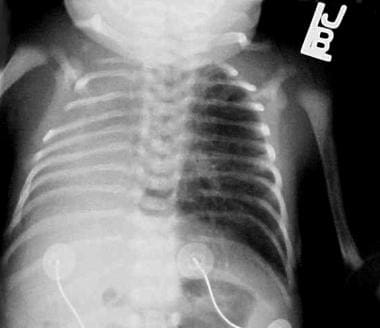 Chest radiograph of a newborn with primary pulmonary hypoplasia of the right lung showing shift of the mediastinum to the right hemithorax.
Pulmonary hypoplasia (PH) may be primary or secondary. Primary PH is extremely rare and routinely lethal. The severity of the lesion in secondary PH depends on the timing of the insult in relation to the stage of lung development. This typically occurs prior to or after the pseudoglandular stage at 6-16 weeks of gestation. In pulmonary hypoplasia, the lung consists of incompletely developed lung parenchyma connected to underdeveloped bronchi. Besides disturbances of the bronchopulmonary vasculature, there is a high incidence (approximately 50-85%) of associated congenital anomalies such as cardiac, gastrointestinal, genitourinary, and skeletal malformations. The diagnosis can result in a spectrum of respiratory complications ranging from transient respiratory distress, chronic respiratory failure, bronchopulmonary dysplasia to neonatal death in very severe cases.
Strict diagnostic criteria are not established for pulmonary hypoplasia. Various parameters such as lung weight, lung weight to body weight ratio, total lung volume, mean radial alveolar count and lung DNA assessment have been used to classify pulmonary hypoplasia. [1, 2]
Pathophysiology
For lung development to proceed normally, physical space in the fetal thorax must be adequate, and amniotic fluid must be brought into the lung by fetal breathing movements, leading to distension of the developing lung. Several studies have demonstrated that gestation age at rupture of membranes (15-28 weeks gestation), latency period (duration between rupture of membranes and birth) and the amniotic fluid index (AFI of less than 1 cm or 5 cm) can influence the development of pulmonary hypoplasia. [3]
Fetal lung fluid and oligohydramnios
Maintenance of fetal lung volume plays a major role in normal lung development. Normal transpulmonary pressure of about 2.5mm Hg allows the fetal lung to actively secrete fluid into the lumen. [4] The effect of stretch of the lung parenchyma induces and promotes lung development. Studies in sheep have demonstrated that tracheal ligation and therefore increased lung distension, accelerates lung growth whereas chronic tracheal fluid drainage has the opposite effect. [5] Cohen and colleagues have found that in-utero overexpression of the cystic fibrosis transmembrane conductance regulator (CFTR) increased liquid secretion into the lung, accelerating lung growth in a rat model. [6]
Oligohydramnios is considered to be an independent risk factor for the development of pulmonary hypoplasia. This is likely due to reduced distending forces on the lung. Studies have demonstrated that severe oligohydramnios decreased lung cell size, alters cell shape and may also negatively affect Type I cell differentiation which ultimately induces pulmonary hypoplasia.
It has been postulated that the Rho-ROCK pathway can affect the growth of the lung epithelium. Embryonic mouse models have demonstrated that ROCK protein inhibitor decreases the number of terminal lung buds. There are currently several groups studying the role of the Rho/ROCK pathway which has potential therapeutic implications in the reversal of lung hypoplasia. [7, 8]
Role of growth factors
Several growth factors such as fibroblast growth factor (FGF), epidermal growth factor (EGF), vascular endothelial growth factor (VEGF) and platelet derived growth factor (PDGF), promote cell proliferation and differentiation. Transforming growth factor family proteins like TGFß1 can oppose these effects.
Embryologically, lungs arise from the foregut. Thyroid transcription factor 1 (TTF-1) is thought to be the earliest embryologic marker associated with cells committed to pulmonary development. FGF signaling is thought to be essential in the formation of TTF-1 expressing cells and this is thought to occur even before the pseudoglandular stage of lung development. Sonic hedgehog (SHH) signaling is further responsible for branching morphogenesis and mesenchymal proliferation. Disruption of any of these pathway may result in primary pulmonary hypoplasia. [9, 10, 11, 12]
FGF7 and FGF10 promote epithelial proliferation and formation of the bronchial tree. Overexpression of FGF10 can also stimulate the formation of cysts in the rat lung. [13] EGF promotes lung branching and Type II alveolar cell proliferation. PDGF plays a crucial role in alveolarization. VEGF promotes angiogenesis and the differentiation of embryonic mesenchymal cells into endothelial cells. Bone morphogenetic protein was thought to oppose lung growth; however recent data suggests that in the presence of mesenchymal cells, BMP4 is a potent inducer of tracheal branching. [14, 15, 16, 17] Aberrant expression of these growth factor proteins in the amniotic fluid during pregnancy have been implicated in abnormal lung development. Interestingly, higher concentrations of VEGF are seen in the amniotic fluid in the second and third trimester and may be a molecular marker for hypoxia which requires further investigation. [15]
Congenital diaphragmatic hernia
The pathogenesis of PH associated with congenital diaphragmatic hernia (CDH) remains unclear. Several mechanisms have been suggested. The nitrofen model of CDH is widely accepted. Nitrofen is a human carcinogen and the retinoid acid signaling pathway is essential for the normal development of the diaphragm. Perturbation of this pathway with compounds such as nitrofen, can induce CDH and PH. Esumi and colleagues demonstrated that that administration of insulin-like growth factor 2 (IGF2) to nitrofen-induced hypoplastic lungs lead to alveolar maturation. [18, 13, 19, 20, 21, 22] Furthermore, data suggest that prenatal treatment with retinoic acid results in increased levels of placental IGF2 and promotes both placental and fetal lung growth in nitrofen induced CDH. [23]
Interestingly, erythropoietin (EPO) is a direct target of retinoic acid. A study has demonstrated decreased levels of EPO mRNA in the liver and kidney of rats which may explain modifications in the pulmonary vasculature in CDH. [22]
A study has also suggested a possible role of interleukin 6 (IL-6) in inducing catch-up growth particularly in nitrofen pre-treated explant fetal rat lungs. [24]
In cases of congenital diaphragmatic hernia (CDH) associated with pulmonary hypoplasia, hypertrophy of the contralateral lung has been demonstrated, with associated pulmonary artery hypertension. The hypoxemia in pulmonary hypoplasia stems from hypoventilation and right-to-left extrapulmonary shunting.
Etiology
The causes of primary pulmonary hypoplasia have not been identified. Sonic hedgehog (Shh) appears to play a role in pulmonary branching morphogenesis. Shh knockout transgenic mice had severely hypoplastic lungs with loss of cartilaginous rings. [9, 11] Experimental models suggest deficiencies in certain factors and/or their receptors can result in abnormal lung growth (see Table 1). These findings suggest that primary pulmonary hypoplasia likely results from disruptions in signaling during embryologic phase of lung development but needs further study.
Causes of secondary pulmonary hypoplasia include conditions that can result in small fetal thoracic volume, prolonged oligohydramnios, decreased fetal breathing movements, and congenital heart disease with poor pulmonary blood flow (see Table 2). Condition such as congenital diaphragmatic hernia, large pleural effusions and congenital pulmonary malformations also lead to the development of pulmonary hypoplasia due to mass effect. [1]
Table 1. Factors and/or Their Receptors Possibly Involved with Abnormal Lung Growth (Open Table in a new window)
Transcription Factors |
Growth Factors |
Thyroid transcription factor-1 (TTF-1) [12] |
Vascular endothelial growth factor receptors (VEGFR1 and VEGFR2) [15] |
GATA-4 [20] |
Insulinlike growth factors (IGF-1 and IGF-2) and their receptors (IGF-1R and IGF-2R) [18] |
FOG-2 [23] |
Epidermal growth factors and its receptor family (eg, ErbB receptors) [21] |
Hepatocyte nuclear factor (HNF3ß10) |
Mitogen-activated protein kinases |
|
Connective tissue growth factor [25] |
Table 2. Causes of Secondary Pulmonary Hypoplasia (Open Table in a new window)
Small Fetal Thoracic Volume |
Prolonged Oligohydramnios |
Decreased Fetal Breathing Movements |
Congenital Heart Diseases With Poor Pulmonary Blood Flow |
CDH |
Fetal renal agenesis |
Central nervous system (CNS) lesions |
|
Cystic adenomatoid malformation (CAM) |
Urinary tract obstruction |
Lesions of the spinal cord, brain stem, and phrenic nerve |
Hypoplastic right heart |
Pulmonary sequestration |
Bilateral renal dysplasia |
Neuromuscular diseases (eg, myotonic dystrophy, spinal muscular atrophy) |
Pulmonary artery hypoplasia |
Pleural effusions with fetal hydrops, hydrothorax |
Bilateral cystic kidneys |
Arthrogryposis multiplex congenital secondary to fetal akinesia |
Scimitar syndrome causing a unilateral right-sided pulmonary hypoplasia |
Thoracic neuroblastomas |
Prolonged rupture of membranes (PROM) |
Maternal depressant drugs |
Trisomies 18 and 21 |
Malformations of the thorax (eg, asphyxiating thoracic dystrophy) |
Premature PROM |
|
|
Diaphragmatic anomalies (eg, abdominal wall defects, eventration of the diaphragm) |
Potter syndrome |
|
|
Musculoskeletal disorders (eg, achondroplasia, thanatophoric dysplasia, osteogenesis imperfecta) |
|
|
|
Abdominal masses causing compression |
|
|
|
Epidemiology
United States statistics
The true incidence of pulmonary hypoplasia is unknown. The reported incidence is between 9 to 11 per 100,00 live birth which is an underestimation, as infants with lesser degrees of hypoplasia likely survive in the neonatal period. [1] Incidence also varies by etiology. Most cases are secondary to congenital anomalies (such as congenital diaphragmatic hernia and cystic adenomatous malformations) or complications related to pregnancy that inhibit lung development. These include, but are not limited to, renal and urinary tract anomalies, amniotic fluid aberrations, diaphragmatic hernia, hydrops fetalis, skeletal and neuromuscular disease and conditions like pleural effusions, chylothorax and intrathoracic masses that cause compression of the fetal thorax. [2]
The incidence of neonatal pulmonary hypoplasia in mid trimester (18-26 weeks gestation) preterm rupture of membranes ranges from 9-28%, with variability attributed to differing diagnostic criteria for pulmonary hypoplasia.
International statistics
International incidence of pulmonary hypoplasia is not known. In Canada, the estimated incidence of CAM is 1 case per 25,000-35,000 pregnancies. According to the CDH study group the incidence of CDH is 1 in every 2000-4000 births and accounts for 8% of all congenital anomalies. In Europe, the occurrence of CDH ranges from 1.7 to 5.7 cases per 10,000 live births, depending on study population and remains largely unchanged. [8, 26] However, there is no direct correlation between these predisposing lesions to the incidence of pulmonary hypoplasia.
Race- and sex-related demographics
No racial predilection has been noted.
No sex predilection has been noted.
Prognosis
Mortality has traditionally been very high. In a retrospective study of 76 premature infants less than 35 weeks’ gestation, 20 had prolonged rupture of membrane of more than 5 days and were clinically diagnosed with pulmonary hypoplasia. Of those 20 infants with pulmonary hypoplasia, 18 died. In another retrospective study of 117 infants of less than 37 weeks’ gestation who had prolonged rupture of membrane of more than 99 hours, 11 died and were considered to have pulmonary hypoplasia. The median age of death was 20 hours (range, 12-48 h), mostly commonly from respiratory failure.
While antepartum amnioinfusions for treatment of oligohydramnios have significantly reduced the risk of pulmonary hypoplasia, longitudinal follow-up studies are lacking on the long-term outcomes of these children.
Of children with pulmonary hypoplasia secondary to congenital diaphragmatic hernia (CDH), the postnatal survival rate of CDH at tertiary centers has improved, with reported rates of 70-92%. [27] However, the survival rates do not account for the cases of CDH that are stillborn, died outside a tertiary center, or died as a result of spontaneous or therapeutic abortion.
CDH survivors have a high incidence of respiratory, nutritional, musculoskeletal, neurological, and gastrointestinal morbidities. [27] In a prospective study of 41 CDH survivors, abnormal muscle tone was found in 90% at age 6 months and 51% at age 24 months. While almost half (49%) had normal scores for neurocognitive and language skills, 17% had mildly delayed and 15% had severe delayed scores. Likewise, in psychomotor testing, while 46% had normal scores, 23% and 31% scored as mildly delayed and severely delayed, respectively. Autism was present in 7%. Studies of brain maturation using MRI show delayed structural brain development and other abnormalities that may lead to long-term neurologic complications. [28]
In a retrospective follow-up study of 55 children survivors with scimitar syndrome followed at one center, a high rate of respiratory complications was observed. All (100%) of the children had right lung hypoplasia of varying degrees of severity. The median duration of follow-up was 7.2 years. Pulmonary infections were reported in 38%, and 43% of children reported wheezing episodes during the last 12 months of follow-up. A restrictive pattern of lung function was observed in the majority of patients, likely related to right-sided lung hypoplasia. Lower total lung capacity values were seen in children with the infantile form of scimitar syndrome, possibly reflective of the severity of pulmonary hypoplasia in these children. [29]
Right-sided hypoplasia, typically secondary to right sided CDH, seems to carry a higher mortality. This is likely due to higher risk of recurrent herniation, increased risk of pulmonary complications, requiring pulmonary vasodilator therapy and tracheostomy. However, no differences in neurodevelopmental outcomes was found. [30, 31]
A minimum lung volume of 45% compared with age-matched control subjects has been shown to be a predictor of survival in neonates with diaphragmatic hernia treated with extracorporeal membrane oxygenation (ECMO). Similarly, a functional residual capacity of 12.3 mL/kg, about one half the normal capacity, has been thought to be a predictor of survival in pulmonary hypoplasia with CDH.
Morbidity/mortality
In different studies, mortality rates associated with PH are reported to be as high as 71-95% in the perinatal period. [1, 2]
The following conditions increase the risk of mortality [26] :
-
Earlier gestational age at rupture of membranes, particularly at less than 25 weeks of gestation
-
Severe oligohydramnios (amniotic fluid index < 4) for more than 2 weeks
-
Earlier delivery (decreased latency period)
-
Right-sided lesion
-
Presence of genetic anomalies
To avoid mortality from severe lung hypoplasia in association with CDH or CAM, fetal surgical intervention has been attempted. Most studies report a mortality rate of 25-30% in neonates with CDH and CAM at high volume centers; mortality can be as high as 45% at peripheral care centers. However, in other cystic lung lesions, most are clinically asymptomatic and may not need aggressive management. [32] A retrospective cohort study by Cuestas et al showed that mortality was significantly higher in neonates with PH who had CDH than in those with PH secondary to renal dysplasia. [33]
Risk factors for a poor outcome include the presence of hydrops fetalis, with a mortality rate as high as 80-90%. Other indicators include the type of CAM and its size. All of these factors reflect the degree of pulmonary compromise with lesions that result in varying degrees of pulmonary hypoplasia.
A retrospective study from Barcelona that studied 60 cases of pulmonary hypoplasia between 1995 and 2014 found a mortality rate of 47% in the first 60 days of life and up to 75% in the first day of life. [34]
Complications
Complications in pediatric pulmonary hypoplasia are as follows:
-
Mortality due to acute respiratory failure in the neonatal period
-
Chronic respiratory failure or insufficiency
-
Pneumothorax, either spontaneous or as a result of ventilatory support
-
Persistent pulmonary hypertension caused by a reduced pulmonary vascular bed and worsened by hypoxia or a coexisting left-to-right intracardiac shunt
-
Chronic lung disease of infancy caused by prolonged ventilatory support
-
Airway abnormalities, including tracheobronchial compression and tracheomalacia caused by the displaced aorta and enlarged left pulmonary artery
-
Restrictive lung disease due to reduced total lung capacity
-
Recurrent respiratory infections
-
Recurrent wheezing episodes
-
Reduced exercise tolerance
-
Scoliosis in adolescent years due to abnormal thoracic cage development
-
Nutritional, musculoskeletal, neurological, and gastrointestinal comorbidities
-
Delayed growth and development
-
Chest radiograph of a newborn with primary pulmonary hypoplasia of the right lung showing shift of the mediastinum to the right hemithorax.
-
CT scan of the same patient (a newborn with primary pulmonary hypoplasia of the right lung) showing absence of the right lung. Note branching of the left lower lobe bronchus (horizontal arrow) and absence of airways in the right side (vertical arrow).
-
A posteroanterior radiograph of a 3-month-old infant with primary pulmonary hypoplasia of the right lung.
-
Lateral view of the same patient (a 3-month-old infant with primary pulmonary hypoplasia of the right lung) showing one dome of the diaphragm.
-
Bronchogram of the same patient (a 3-month-old infant with primary pulmonary hypoplasia of the right lung) showing absence of the airways in the right side and presence of the left main bronchus and its branches.
-
A chest radiograph of a 14-year-old child with primary pulmonary hypoplasia of the right side causing secondary scoliosis.
-
A chest radiograph of a newborn with achondroplasia and small chest causing hypoplasia of both lungs.
-
A chest radiograph of a newborn with diaphragmatic hernia in the right hemithorax shortly after birth.
-
CT scan of the same child (a newborn with diaphragmatic hernia in the right hemithorax shortly after birth) showing the presence of abdominal contents in the right hemithorax. Note the presence of the left lower bronchus and its main branches (horizontal arrow) and absence of the right lower lobe bronchus. The liver in the right hemithorax is indicated by the upper arrow.
-
A chest radiograph of a 10-month-old child after repair of a right diaphragmatic hernia showing loss of lung volume in the right hemithorax.
-
MRI of the same patient (a 10-month-old child after repair of a right diaphragmatic hernia) showing loss of right lung volume and smaller right pulmonary artery than the left pulmonary artery (arrow).
Tables
Transcription Factors |
Growth Factors |
Thyroid transcription factor-1 (TTF-1) [12] |
Vascular endothelial growth factor receptors (VEGFR1 and VEGFR2) [15] |
GATA-4 [20] |
Insulinlike growth factors (IGF-1 and IGF-2) and their receptors (IGF-1R and IGF-2R) [18] |
FOG-2 [23] |
Epidermal growth factors and its receptor family (eg, ErbB receptors) [21] |
Hepatocyte nuclear factor (HNF3ß10) |
Mitogen-activated protein kinases |
|
Connective tissue growth factor [25] |
Small Fetal Thoracic Volume |
Prolonged Oligohydramnios |
Decreased Fetal Breathing Movements |
Congenital Heart Diseases With Poor Pulmonary Blood Flow |
CDH |
Fetal renal agenesis |
Central nervous system (CNS) lesions |
|
Cystic adenomatoid malformation (CAM) |
Urinary tract obstruction |
Lesions of the spinal cord, brain stem, and phrenic nerve |
Hypoplastic right heart |
Pulmonary sequestration |
Bilateral renal dysplasia |
Neuromuscular diseases (eg, myotonic dystrophy, spinal muscular atrophy) |
Pulmonary artery hypoplasia |
Pleural effusions with fetal hydrops, hydrothorax |
Bilateral cystic kidneys |
Arthrogryposis multiplex congenital secondary to fetal akinesia |
Scimitar syndrome causing a unilateral right-sided pulmonary hypoplasia |
Thoracic neuroblastomas |
Prolonged rupture of membranes (PROM) |
Maternal depressant drugs |
Trisomies 18 and 21 |
Malformations of the thorax (eg, asphyxiating thoracic dystrophy) |
Premature PROM |
|
|
Diaphragmatic anomalies (eg, abdominal wall defects, eventration of the diaphragm) |
Potter syndrome |
|
|
Musculoskeletal disorders (eg, achondroplasia, thanatophoric dysplasia, osteogenesis imperfecta) |
|
|
|
Abdominal masses causing compression |
|
|
|


