Practice Essentials
Megaloblastic anemia is an aregenerative macrocytic anemia with nuclear dysmaturity, where the nucleus appears immature relative to the cytoplasm because of impaired DNA synthesis (see the image below). Megaloblastic anemia is an uncommon problem in childhood that is most frequently associated with vitamin deficiency or gastrointestinal disease. [1]
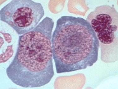 Bone marrow aspirate from a patient with untreated pernicious anemia. Megaloblastic maturation of erythroid precursors is shown. Two megaloblasts occupy the center of the slide with a megaloblastic normoblast above. Photo courtesy of Marcel E Conrad, MD.
DNA synthesis is impaired in these cases because of inadequate amounts of metabolically active folate derivatives necessary for DNA base synthesis. Megaloblastic changes affect all 3 hematopoietic cell lines. Thrombocytopenia, leukopenia, and anemia are all observed to varying extents.
The 2 most common causes of megaloblastic anemia are vitamin B-12 (cobalamin) deficiency and folic acid deficiency. Although their clinical settings differ considerably, no hematologic finding can distinguish between the 2 conditions; specific testing is necessary (see Workup). Other less common causes include the use of metabolic inhibitors such as methotrexate and 6-mercaptopurine and certain rare inborn errors such as thiamine-responsive megaloblastic anemia, [2, 3] Lesch-Nyhan syndrome, and hereditary orotic aciduria (see Etiology).
Treatment of megaloblastic anemia depends on the underlying cause. Supplemental folate or vitamin B-12 may be indicated (see Treatment).
Go to Pediatric Chronic Anemia, Anemia of Prematurity, Donath-Landsteiner Hemolytic Anemia, Pediatric Acute Anemia, and Fanconi Anemia for complete information on these topics.
Vitamin B-12 deficiency
Vitamin B-12 is commonly ingested with meat or fish. It binds to salivary haptocorrins, which are digested in the stomach, allowing the cobalamin to bind to intrinsic factor (IF). IF is produced by the parietal cells of the stomach. The IF–B-12 complex makes its way to the terminal ileum, where it binds to receptors on the enterocyte. It is transported across the cell and enters the circulation bound to a transport molecule, TC II. The B-12–TC II complex is absorbed into cells by endocytosis. In the cell, cobalamin acts as a coenzyme in several reactions, including the synthesis of methionine from homocysteine during the reduction of dihydrofolate to tetrahydrofolate and the conversion of methylmalonyl CoA to succinyl CoA. It is the role of vitamin B-12 in the reduction of folic acid derivatives that results in the megaloblastic changes seen clinically.
Vitamin B-12 deficiency can be caused by decreased ingestion (eg, poor dietary intake), impaired absorption (eg, failure to release B-12 from protein, IF deficiency, chronic pancreatic disease, competitive parasites, intrinsic intestinal disease), or impaired use (eg, congenital enzyme deficiencies, lack of transcobalamin II, administration of nitrous oxide). [4]
Inadequate vitamin B-12 dietary intake is rare in children, though it may be seen in breastfed infants whose mothers are themselves deficient. Pernicious anemia, a common cause of vitamin B-12 deficiency in adults, is rare in childhood. Deficiency of vitamin B-12 activity is usually due to malabsorption or a congenital deficiency of one of the vitamin B-12 carrier proteins. In recent years, vitamin B-12 deficiency has been described in patients with human immunodeficiency virus (HIV) infection, with or without acquired immunodeficiency syndrome (AIDS).
In addition to the hematologic manifestations of vitamin B-12 deficiency, abnormalities of the GI tract and nervous system may also be present. The underlying cause of megaloblastic anemia must be determined in each case. Failure to recognize B-12 deficiency, even in the presence of concomitant folate deficiency, may result in permanent neurologic damage. Treatment with folate alone in these cases may reverse anemia but may allow neurologic damage to progress.
Folate deficiency
Folate is ingested in the diet in many different types of food. It enters the enterocyte and is transported into the portal circulation by a carrier molecule. It circulates in the plasma mostly as 5-methyl tetrahydrofolate (THF). It enters the cell via a carrier (methotrexate competes with this carrier). In the cell, folate binds to and acts as a coenzyme with enzymes responsible for single carbon metabolism.
Folate deficiency can be caused by any of the following:
-
Decreased ingestion (eg, poor dietary intake, alcoholism, infancy)
-
Impaired absorption (eg, intestinal short circuits, celiac sprue, congenital malabsorption, certain drugs such as sulfasalazine)
-
Impaired use (eg, use of folic acid antagonists such as antiepileptic medications, sulfa antibiotics, or methotrexate)
-
Increased requirements (eg, pregnancy, infancy, hyperthyroidism, chronic hemolytic disease, cancer)
-
Increased loss (eg, hemodialysis)
Folic acid is available in a wide variety of food groups. Approximately one third of dietary folate is estimated to come from cereals and grains, another third from fruits and vegetables, and another third from meats and fish. Folic acid deficiency is commonly observed in children who are fed a severely restricted diet. This usually occurs with a diet restricted to goat's milk, which is deficient in folic acid. It may also be observed in children with celiac sprue and other malabsorption disorders that affect the proximal small intestine.
Deficiency of metabolically active folate metabolites is frequently observed in patients who receive antifolate drugs, such as sulfa antibiotics and methotrexate. A relative deficiency of metabolically active folate metabolites may also be observed in patients who are experiencing increased red cell destruction. These patients require a greater amount of folate than is usually present in the diet and develop macrocytic changes in their erythrocytes. Increased folate intake is also important during pregnancy, in which deficiencies have been associated with neural tube defects.
Workup
Hematologic testing confirms the presence of megaloblastic anemia and can exclude neoplastic and other disorders.
Measure serum vitamin B-12 levels. Methylmalonic acid and total homocysteine levels are sensitive indicators of vitamin B-12 deficiency and correlate with clinical abnormalities and therapeutic response. However, they are not specific to vitamin B-12 deficiency, and care should be taken in interpreting these results.
For folate assessment, the red blood cell (RBC) folate level is the best measure of metabolically active folate and includes 5-methyl tetrahydrofolate (THF) in the assay. Serum folate measures the circulating pool of folate but does not accurately reflect the amount of THF present in the tissues. Other tests include serum and urine assessments and the modified Schilling test.
Management
Treatment of megaloblastic anemia depends on the underlying cause. Folate deficiency due to dietary deficiency or increased demands is best treated with folate supplements. In addition, a diet rich in green, leafy vegetables is essential for normal intake of folic acid.
Folate deficiency caused by the use of sulfa drugs or other antifolate medications may be addressed by folate supplementation or by reducing or eliminating the drug. Folate deficiency due to celiac sprue requires treatment of the underlying disorder and folate supplements.
Management of vitamin B-12 deficiency is often more complex, because of the nature of B-12 deficiency in childhood. Data from small trials suggest that oral B-12 supplementation is as effective as parenteral supplementation in patients with nutritional deficiency. [5] Even in patients with intrinsic factor (IF) deficiency, oral supplements may be effective, using higher doses.
Often, however, high-dose oral B-12 supplements are unsuccessful in patients with IF deficiency or in those who have undergone intestinal surgery. These patients may require parenteral supplementation because of impaired secretion or absorption of IF.
Pathophysiology
Megaloblastic anemia is caused by various DNA synthesis defects. In folate deficiency, purine biosynthesis is affected because folic acid is essential in this process.
Folic acid is essential for purine biosynthesis. Folic acid absorbed from the diet must be activated to produce active tetrahydrofolic acid (THF). THF is necessary for single carbon transfers in the synthesis of pyrimidine nucleotides. Without adequate levels of biologically active THF, the ability to repair and replicate DNA is decreased. Vitamin B-12 is a cofactor for the activation of folic acid in a step that also converts homocysteine to methionine.
In the case of inadequate folic acid intake, THF production is depleted, and DNA synthesis is slowed. The effect on hematopoiesis is to reduce the rate of cell production, resulting in pancytopenia. In the cells that are produced, the effect created is an arrest of nuclear maturation. In other words, the cells that are produced have immature nuclei compared with the degree of maturation of the cytoplasm.
Etiology
Megaloblastic anemia is most often caused by an acquired lack of vitamin B-12 or lack of folic acid. Inherited abnormalities of the metabolism of these nutrients may be the cause. [6]
Acquired causes of insufficient vitamin B-12 include the following:
-
Inadequate intake in diet
-
Inadequate absorption (deficient IF; deficient absorption from ileum)
-
Impaired transport from the intestine
Acquired causes of insufficient folate include inadequate dietary intake and inadequate absorption from the proximal small intestine. Medications associated with folate deficiency include the following:
-
Sulfonamide antibiotics may interfere with folate metabolism, particularly when they are used on a long-term basis
-
Other antifolate antimetabolite drugs may also cause megaloblastic changes
-
Megaloblastic changes are observed with some frequency with antineoplastic agents, such as methotrexate; azathioprine (Imuran) may also cause megaloblastic changes
Increased metabolic demand (eg, chronic hemolysis, such as in sickle cell disease) or increased loss may also result in insufficient folate.
Congenital absence or deficiency of carrier proteins can cause vitamin B-12 deficiency. These deficiencies occur most commonly as autosomal recessive enzymopathies. These conditions often manifest during infancy and early childhood and are rare but important causes of megaloblastic anemia.
The Imerslund-Grasbeck syndrome of proteinuria and excretion of cobalamin and IF is a rare disorder that arises in early childhood. However, it is an important cause of B-12 deficiency.
Megaloblastic anemia did not develop in children who underwent prolonged exposure to nitrous oxide, according to a study by Duma et al, even though nitrous oxide can raise plasma homocysteine levels in pediatric patients as a result of vitamin B-12 inactivation. Children in the study, who underwent spinal surgery, were exposed to the agent for as long as 8 hours. [7]
Epidemiology
The prevalence of megaloblastic anemia in childhood has not been established. Vitamin B-12 deficiency is a worldwide problem, however, particularly in the newborn period, due to the combined effects of poor maternal diet and congenital deficiencies of transcobalamin.
Pernicious anemia is a common cause of megaloblastic anemia, especially in persons of European or African descent. Dietary vitamin B-12 deficiency is a serious problem in India, Mexico, Central America, South America, and some areas of Africa. The increase in vegetarianism is related to an increase in vitamin B-12 deficiency and is a particular concern in breastfed infants of vitamin B-12–deficient mothers.
Megaloblastic anemia is observed in all racial and ethnic groups and in both sexes. It is rarely observed in infants, but may occur in infants who breastfeed from mothers who are themselves deficient in vitamin B-12 or in infants with a congenital deficiency of one of the carrier proteins.
Prognosis
Prognosis depends on the underlying cause of the megaloblastic anemia and the degree of compliance with therapy. Folic acid deficiency is relatively easy to treat; patients usually respond to added folate in their diet. Vitamin B-12 deficiency may be a more significant concern because some patients may require vitamin B-12 injections, with which they may not readily comply. In addition, vitamin B-12 deficiency may be associated with severe neurologic abnormalities that may be long lasting and persist even with appropriate vitamin B-12 therapy. For example, Stredny et al reported on an adolescent female diagnosed with megaloblastic anemia and subacute combined degeneration, resulting from pernicious anemia–associated B-12 deficiency, who presented with emotional lability, mental status changes, hyperreflexia, and ataxia. [8]
Morbidity in megaloblastic anemia may include CNS toxicity, including dementia and loss of dorsal column function. Deficiency of vitamin B-12 is usually at the root of this. CNS dysfunction has been described in adult patients who have deficient vitamin B-12 levels in the absence of anemia. Megaloblastic anemia in pregnancy is associated with persistent learning deficits in children. [9, 10] Hyperpigmentation may also be seen.
Decreased numbers of CD4 cells and abnormal CD4/CD8 ratios as well as natural killer (NK) cell numbers have been documented in patients with pernicious anemia. These numbers normalize with cobalamin administration. [11]
Patient Education
For the new patient, education should focus on the nature of the deficiency causing the anemia and the underlying factors that produce the deficiency. Educate patients or their parents about the neurologic complications of vitamin B-12 deficiency to ensure that they understand the importance of B-12 replacement.
For patient education information, see the Esophagus, Stomach, and Intestine Center; Crohn Disease Center; and Blood and Lymphatic System Center, as well as Celiac Sprue, Crohn Disease, Diet and Nutrition in Crohn Disease, and Anemia.
-
Bone marrow aspirate from a patient with untreated pernicious anemia. Megaloblastic maturation of erythroid precursors is shown. Two megaloblasts occupy the center of the slide with a megaloblastic normoblast above. Photo courtesy of Marcel E Conrad, MD.


