Continually Updated Clinical Reference
 
 
  All Sources     eMedicine     Medscape     Drug Reference     MEDLINE
 
eMedicine - Gianotti-Crosti Syndrome : Article by

Quick Find
Authors & Editors
Introduction
Clinical
Differentials
Workup
Treatment
Medication
Follow-up
Miscellaneous
Multimedia
References

Related Articles
Atopic Dermatitis

Keratosis Pilaris


Patient Education
Click here for patient education.


Author: Howard Pride, MD, Associate Professor, Departments of Pediatrics and Dermatology, Geisinger Medical Center

Howard Pride is a member of the following medical societies: American Academy of Dermatology and Society for Pediatric Dermatology

Editors: Kevin P Connelly, DO, Clinical Assistant Professor, Department of Pediatrics, Division of General Pediatrics and Emergency Care, Virginia Commonwealth University; Medical Director, Paws for Health Pet Visitation Program of the Richmond SPCA; Pediatric Emergency Physician, Emergency Consultants Inc, Chippenham Medical Center; Mary L Windle, PharmD, Adjunct Assistant Professor, University of Nebraska Medical Center College of Pharmacy, Pharmacy Editor, eMedicine.com, Inc; Robert A Schwartz, MD, MPH, Professor and Head of Dermatology, Professor of Medicine, Professor of Pediatrics, Professor of Pathology, Professor of Preventive Medicine and Community Health, UMDNJ-New Jersey Medical School; Merrily P M Poth, MD, Professor, Department of Pediatrics and Neuroscience, Uniformed Services University of the Health Sciences; Dirk M Elston, MD, Director, Department of Dermatology, Geisinger Medical Center

Author and Editor Disclosure

Synonyms and related keywords: Gianotti-Crosti syndrome, GCS, papular acrodermatitis of childhood, papulovesicular acrolocated syndrome, hepatitis B infection, hepatitis B, lymphadenopathy, rash Epstein-Barr virus

Background

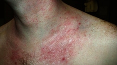
Gianotti-Crosti syndrome (GCS) is a distinct infectious exanthem with associated lymphadenopathy and acute anicteric hepatitis. Gianotti and Crosti initially described GCS as associated with a hepatitis B virus exanthem, which they termed papular acrodermatitis of childhood. A similar constellation of characteristics was later found to be associated with several infectious agents and immunizations that were called papulovesicular acrolocated syndromes. Subsequent retrospective studies have shown that these 2 entities are indistinguishable from one another, and they are now consolidated under the unifying title of GCS.

Pathophysiology

The most likely explanation for the exanthem is a local type IV hypersensitivity reaction to the offending viral or bacterial antigen within the dermis. This is based on the immunohistochemical characterization of the cutaneous inflammatory infiltrate. Findings on direct immunofluorescence examination of the skin are always negative. Electron microscopy has never revealed virus particles that suggested a reactive process other than an autoimmune phenomenon or direct infection of the skin. Inciting factors include various viral and bacterial infections, as well as recent immunizations. The rarity of GCS in adults suggests lifelong immunity to a common viral triggering agent. GCS is more common among children with atopic dermatitis, suggesting an immune mechanism. However, more information is needed in order to define the precise mechanism involved.

Frequency

United States

Because of the benign self-limited nature of GCS, most cases are not reported, and the overall incidence is unknown. Frequency probably parallels the incidence of a precipitating infection in a specific geographic region.

International

The underlying infection correlates with the endemic pathogens of a specific geographic region.

For example, in Japan and Mediterranean countries, GCS is more commonly associated with hepatitis B virus infection. With the advent of more universal hepatitis B immunization, Epstein-Barr virus is now the most common etiologic factor worldwide.

Mortality/Morbidity

  • The mere presence of a rash elicits some degree of social morbidity, depending on the age of the affected child.
  • Although typically nonpruritic, some reports document pruritus in the later stages of the rash.
  • The only significant morbidity involves the underlying infectious process, particularly the hepatitis B virus.

Race

No racial predilection has been noted; however, the underlying infection correlates with the endemic pathogens of a specific geographic region.

Sex

In the pediatric population, GCS affects males and females with equal frequency. However, affected adults have been exclusively female.

Age

GCS primarily occurs in children aged 3 months to 15 years, with a peak in children aged 1-6 years. More than 90% of patients are younger than 4 years.


History

The rash of Gianotti-Crosti syndrome (GCS) is usually of sudden onset and may be associated with an acute infectious illness or immunization. The rash is usually present for 2-4 weeks but can last up to 4 months. It may be mildly pruritic.

Physical

  • Children with GCS generally appear healthy; however, the following may occur:
    • Multiple papules erupt on the face, buttocks, and extensor surface of the extremities; papules are symmetrically distributed, discrete, and flesh-toned–to–erythematous-to-brown and flat-topped.
    • The trunk is strikingly spared, although a transient eruption can occur.
    • Individual papules are monomorphous and range in size from 1-5 mm. They may be hemorrhagic or edematous to the point of forming vesicles. They may coalesce into larger plaques.
    • The face may be the only area of involvement. One report that highlighted this clinical presentation showed that all children with an exclusively facial eruption had Epstein-Barr virus.
  • Hepatosplenomegaly and axillary or inguinal adenopathy are inconsistent findings.

Causes

  • Associated viral infections
    • Hepatitis A, B, and C
    • Rotavirus
    • Epstein-Barr virus
    • Rubella virus
    • Cytomegalovirus
    • Coxsackieviruses A16, B4, and B5
    • Adenovirus
    • Enterovirus
    • Respiratory syncytial virus
    • Parainfluenza virus
    • Parvovirus B19
    • Paravaccinia (milker's nodules)
    • Human herpesvirus 6
    • Echovirus
    • Molluscum contagiosum virus
    • HIV
  • Associated bacterial infections
  • Group A b-hemolytic streptococci
  • Mycobacterium avium-intracellulare
  • Mycoplasma pneumoniae
  • Bartonella henselae
  • Borrelia burgdorferi
  • Associated immunizations
  • Polio
  • Diphtheria
  • Influenza
  • Pertussis
  • Measles
  • Smallpox
  • Pertussis


Atopic Dermatitis
Keratosis Pilaris

Other Problems to be Considered

Insect bites
Erythema multiforme
Lichen planus
Medication reaction
Other viral exanthems
Urticaria pigmentosa
Langerhans cell histiocytosis


Lab Studies

  • In general, no laboratory studies are needed.
  • Jaundice or hepatomegaly should prompt a search for the hepatitis B virus and elevated levels of liver enzymes. However, elevated levels of liver enzymes are most likely related to Epstein-Barr virus infection.

Procedures

  • A skin biopsy may be performed to rule out other diseases in the differential diagnosis.

Histologic Findings

Skin biopsy findings are nonspecific. It is more helpful in ruling out other diseases. The epidermis shows focal spongiosis, parakeratosis, and mild acanthosis. The upper dermis has a moderately intense perivascular infiltrate of lymphocytes and histiocytes.


Medical Care

  • Gianotti-Crosti syndrome (GCS) is a benign self-limited condition that requires no treatment.
  • Topical steroids are generally not effective, although anecdotal responses have been reported.
  • Systemic treatment with antihistamines has been moderately helpful in relieving pruritus.

Consultations

  • Consultation with a dermatologist or a pediatric dermatologist should be sought in confusing cases.
  • Consultation with a pediatric gastroenterologist should be sought for the rare cases associated with the hepatitis B virus.

Activity

Activity is limited only by symptoms. The infectious period ends when the exanthem appears; therefore, the patient may participate in daycare or school at this time.


No medical therapy is necessary. Topical corticosteroids and oral antihistamines might provide some symptomatic relief of pruritus.

Drug Category: Corticosteroids, topical cream

These agents may provide relief when pruritus is present.

Drug NameTriamcinolone 0.1% cream (Aristocort)
DescriptionAntipruritic effect of this cream is fairly marginal but may be somewhat effective. Treats inflammatory dermatosis that is responsive to steroids. Decreases inflammation by suppressing the migration of polymorphonuclear leukocytes and reversing capillary permeability.
Adult DoseApply a thin film to affected areas bid prn
Pediatric DoseApply as in adults
ContraindicationsDocumented hypersensitivity; fungal, viral, and bacterial skin infections
InteractionsNone reported
PregnancyC - Safety for use during pregnancy has not been established.
PrecautionsExcessive use of topical steroids may cause skin atrophy; do not use in decreased skin circulation; potential for systemic absorption and adrenal suppression when used over large surface areas in young children

Drug Category: Antihistamines

These agents may provide some relief when pruritus is present. They are used to treat minor allergic reactions and anaphylaxis and may be used to pretreat patients with prior documentation of minor allergic reactions. These agents may control itching by blocking effects of endogenously released histamine.

Drug NameHydroxyzine (Atarax, Vistaril)
DescriptionOffers a mild degree of relief from pruritus. Antagonizes H1 receptors in periphery. May suppress histamine activity in the subcortical region of CNS.
Adult Dose25-50 mg PO q6h prn
Pediatric Dose1 mg/kg/dose PO q6h prn
ContraindicationsDocumented hypersensitivity; early pregnancy
InteractionsMay potentiate the effect of opioid analgesics, barbiturates, alcohol, or other CNS depressants
PregnancyC - Safety for use during pregnancy has not been established.
PrecautionsAssociated with clinical exacerbations of porphyria (may not be safe for patients with porphyria); ECG abnormalities (alterations in T waves) may occur; causes drowsiness


Further Outpatient Care

  • No specific follow-up is necessary for uncomplicated cases of Gianotti-Crosti syndrome (GCS); however, transaminase elevations, if present, should be monitored until normalization.
  • GCS associated with hepatitis B infection has an increased incidence of a carrier state compared with hepatitis B infection alone. In this instance, some suggest monitoring the surface antigen as an indicator of potential infectivity.

Prognosis

  • Prognosis is excellent. Lesions clear within 4-12 weeks. No long-term complications are associated with GCS.


Medical/Legal Pitfalls

  • No medical/legal pitfalls are associated with Gianotti-Crosti syndrome (GCS) because it is a self-limited condition that does not require treatment.


Media file 1:  Multiple erythematous flat-topped papules on the cheeks of an 18-month-old boy with Gianotti-Crosti syndrome.
Click to see larger pictureClick to see detailView Full Size Image
Media type:  Photo

Media file 2:  Arm of a 3-year-old boy with Gianotti-Crosti syndrome demonstrating well-defined erythematous lichenoid papules on the arm and forearm.
Click to see larger pictureClick to see detailView Full Size Image
Media type:  Photo

Media file 3:  Gianotti-Crosti syndrome. Thigh of the patient in Image 2. The trunk was strikingly spared.
Click to see larger pictureClick to see detailView Full Size Image
Media type:  Photo


  • Baleviciene G, Maciuleviciene R, Schwartz RA. Papular acrodermatitis of childhood: the Gianotti-Crosti syndrome. Cutis. Apr 2001;67(4):291-4. [Medline].
  • Brandt O, Abeck D, Gianotti R, Burgdorf W. Gianotti-Crosti syndrome. J Am Acad Dermatol. 2006;54:136-145. [Medline].
  • Caputo R, Gelmetti C, Ermacora E, et al. Gianotti-Crosti syndrome: a retrospective analysis of 308 cases. J Am Acad Dermatol. Feb 1992;26(2 Pt 1):207-10. [Medline].
  • Chuh A, Lee A, Zawar V. The diagnostic criteria of Gianotti-Crosti syndrome: are they applicable to children in India?. Pediatr Dermatol. Sep-Oct 2004;21(5):542-7. [Medline].
  • Chuh AA. Diagnostic criteria for Gianotti-Crosti syndrome: a prospective case-control study for validity assessment. Cutis. Sep 2001;68(3):207-13. [Medline].
  • Draelos ZK, Hansen RC, James WD. Gianotti-Crosti syndrome associated with infections other than hepatitis B. JAMA. Nov 7 1986;256(17):2386-8. [Medline].
  • Magyarlaki M, Drobnitsch I, Schneider I. Papular acrodermatitis of childhood (Gianotti-Crosti disease). Pediatr Dermatol. Sep 1991;8(3):224-7. [Medline].
  • Taieb A, Plantin P, Du Pasquier P, et al. Gianotti-Crosti syndrome: a study of 26 cases. Br J Dermatol. Jul 1986;115(1):49-59. [Medline].
  • Yoshida M, Tsuda N, Morihata T, et al. Five patients with localized facial eruptions associated with Gianotti-Crosti syndrome caused by primary Epstein-Barr virus infection. J Pediatr. Dec 2004;145(6):843-4. [Medline].

Gianotti-Crosti Syndrome excerpt

Article Last Updated: Dec 7, 2006