Background
Myocarditis is an inflammatory disease of the heart muscles, with most cases having a viral etiology. [1, 2, 3, 4] However, many nonviral causes are also recognized [2, 3, 4, 5] and, indeed, a number of common infectious illnesses of childhood may be associated with myocarditis in pediatric patients. [6] In addition, myocarditis can be a manifestation of hypersensitivity or it can be secondary to a drug reaction. The severity of myocarditis varies from relatively asymptomatic disease to fulminant, severe disease with a rapid, progressive, downhill course. (See Etiology, Presentation, and Workup.) [7, 8]
Epidemiologically, myocarditis appears to be sporadic. The most common cause is thought to be coxsackievirus B, but the condition can also arise from bacterial, parasitic, fungal, toxic, and drug-related causes. (See Treatment and Medication.)
Pathophysiology
When clinically significant myocarditis (regardless of cause) occurs, myocardial function is reduced in the presence of, and as a result of, extensive interstitial inflammation and/or injury. Systolic function of the left ventricle with concomitant dilatation of the heart and cardiac enlargement occurs. The overall result is an attendant reduction in cardiac output.
In addition, progressive congestive heart failure (CHF) with increased left ventricular end-diastolic volume and pressure results in an increase in left atrial pressure, which is transmitted into the pulmonary venous system. This may also result in pulmonary edema secondary to increased pulmonary arterial hydrostatic forces.
Finally, during the healing stages of myocarditis, fibroblasts may replace existing myocytes with resultant scar formation. Chronic CHF may develop. An autopsy sample that shows scarring in the ventricles is seen below.
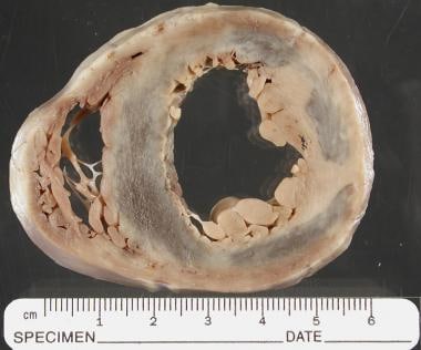 Pediatric nonviral myocarditis. This image is reveals myocarditis with scarring at autopsy. It is a short-axis gross photograph from an 8-year-old child who had clinical myocarditis, showing scarring of both ventricles, more prominent in the left. The fibrosis depicts a random distribution with epicardial, myocardial, and pericardial involvement.
Etiology
Causes of myocarditis, including nonviral causes, include the following [4, 9] :
-
Viral
-
Rickettsial: Rickettsia rickettsii, R tsutsugamushi
-
Bacterial organisms/infections: Klebsiella species, Leptospira species, Salmonella species, Brucella species, Legionella pneumophila, meningococci, clostridia, streptococci (scarlet fever), staphylococci, pneumococci, tuberculosis, syphilis, tetanus
-
Protozoa and protozoal infections: Trypanosoma cruzi (Chagas disease), toxoplasmosis, amebiasis
-
Other parasites and parasitic infections: Toxocara canis, schistosomiasis, heterophyiasis, cysticercosis, echinococci, visceral larva migrans
-
Fungal infections: Actinomycosis, coccidioidomycosis, histoplasmosis, candidiasis
-
Toxic etiology: Scorpion envenomations, diphtheria
-
Drugs: Cyclophosphamide, phenylbutazone, acetazolamide, amphotericin B, indomethacin, tetracycline, isoniazid, methyldopa, phenytoin, penicillin, sulfonamides
-
Hypersensitivity/autoimmune: Rheumatoid arthritis, rheumatic fever, ulcerative colitis, systemic lupus erythematosus (SLE), Kawasaki syndrome, dermatomyositis
-
Other: Sarcoidosis, scleroderma, idiopathic, giant cell, peripartum myocarditis
Although rare, some of the more common causes of nonviral myocarditis are discussed below in greater detail.
Chagas disease
Chagas disease is caused by a protozoal infestation with T cruzi. It is very uncommon in North America but can affect as much as half the population in endemic areas, such as South America. [10] Acute infection can cause protracted heart failure and death. Endomyocardial biopsy and microscopic examination typically demonstrate organisms, neutrophils, lymphocytes, macrophages, and eosinophils.
Giant cell myocarditis
In giant cell myocarditis, giant cells are present in the myocardium with or without granulomas. This type of myocarditis may be evidenced with tuberculosis, syphilis, rheumatoid arthritis, or rheumatic heart disease, or with fungal or parasitic infections. The characteristic cell is probably histiocytic in origin and is usually found in nonviral myocarditis. Similar cells have been noted in patients with myocarditis associated with drugs such as phenylbutazone. This type of myocarditis may not be specific [11] but, rather, may represent a final common pathologic pathway.
Systemic lupus erythematosus
Patients with SLE may have myocardial fibrinoid lesions in the connective tissue, with an accompanying cellular reaction. This reaction may also affect the cardiac valves, most notably the mitral and aortic valves. [12] Although the predominant cardiac manifestation of SLE is pericarditis, myocardial involvement with congestive heart failure (CHF) can occur. The treatment of choice is corticosteroids.
Kawasaki disease
Kawasaki disease may include myocardial involvement, which usually occurs in the acute phase; it can occur independent of any coronary artery involvement and usually resolves completely. In some cases, however, the diffuse myocarditis may be severe, and it may lead to heart failure and death. Although the pathophysiologic mechanisms that occur in Kawasaki disease are not known, a generalized autoimmune disorder is suspected.
Dermatomyositis
Dermatomyositis, a multisystem (probably autoimmune) disease, is characterized by diffuse, nonsuppurative inflammation of skeletal muscle and skin. However, cardiac involvement, including a loss of striations, fragmentation, and vascularization of muscle fibers, has been reported. The interstitium may also show a variable amount of edema. Tachycardia and conduction abnormalities are described, but heart failure is uncommon. Long-term therapy with prednisone is beneficial in most cases. Children have a good long-term prognosis, as most recover, and medication may be discontinued in 1-2 years.
Epidemiology
Myocarditis is most frequently affects younger adults aged 20 to 40 years. [3] However, when it occurs in children, this patient population appears to have a more severe presentation than that of adults, with a higher percentage requiring temporary mechanical circulatory support. In addition, the elderly may be underdiagnosed.
In general, men are affected more frequently than women, [3, 4] and certain forms of myocarditis (eg, cardiac sarcoidosis) affect black populations more than white populations in the United States. [3] Most forms of myocarditis have no known ethnic predisposition.
Although no widely available test exists that is applicable at a population level for the incidence and prevalence of myocarditis, global estimates of the burden of myocarditis have been extrapolated from population-based studies of heart muscle disease (heart disease not related to blocked arteries or abnormal heart valves), such that 2010 global data indicate about 400,000 people died of heart muscle disease (cardiomyopathy that includes myocarditis) (240,000 men, 160,000 women). [3] Expert consensus opinion suggests that up to 40% of dilated cardiomyopathy results from myocarditis, [3] and about 10% of initially unexplained cases of cardiomyopathy are caused by myocarditits [2]
Prognosis
The prognosis for myocarditis varies and depends on age, etiology, and severity at the time of presentation. [1, 2, 3, 4] The mortality rate in newborn infants is high (75%) in some reports. Older infants and children have a better prognosis, with mortality rates ranging from 10% to 25% in clinically recognizable cases. Some studies have suggested complete recovery in as many as 50% of the cases.
Perhaps as many as 25%-35% of patients who present with clinical myocarditis may be left with a significant chronic dilated cardiomyopathy and may require cardiac transplantation.
The cause of myocarditis may affect prognosis. For example, patients with conduction abnormalities or arrhythmias secondary to diphtheric myocarditis tend to have a poor prognosis. [13]
In a retrospective analysis of clinical and magnetic resonance imaging (MRI) prognostic factors from 58 pediatric myocarditis cases (nonviral and viral) over a 5-year period, Sachdeva et al found that risk factors for poor outcomes included an ejection fraction below 30% on presentation, peak brain natriuretic peptide levels above 10,000 ng/L, and late enhancements on cardiac MRI. [14]
In another retrospective analysis (2008-2012) of 75 patients aged up to 19 years admitted with a diagnosis of myocarditis and normal left ventricular systolic function at seven tertiary pediatric hospitals, factors associated with poor outcomes included significantly higher baseline levels of B-type natriuretic peptide (BNP), troponin I (TnI), and creatine kinase. [15]
-
Pediatric nonviral myocarditis. This image is reveals myocarditis with scarring at autopsy. It is a short-axis gross photograph from an 8-year-old child who had clinical myocarditis, showing scarring of both ventricles, more prominent in the left. The fibrosis depicts a random distribution with epicardial, myocardial, and pericardial involvement.