Continually Updated Clinical Reference
 
 
  All Sources     eMedicine     Medscape     Drug Reference     MEDLINE
 
eMedicine - Aortic Stenosis, Valvar : Article by

Quick Find
Authors & Editors
Introduction
Clinical
Differentials
Workup
Treatment
Medication
Follow-up
Miscellaneous
References

Related Articles
Aortic Stenosis, Subaortic

Aortic Stenosis, Supravalvar


Patient Education
Click here for patient education.


Author: Howard S Weber, MD, FAAP, FACC, FSCAI, Professor, Assistant Chief, Section of Pediatric Cardiology, Penn State University School of Medicine; Director, Pediatric Catheterization Laboratory, Milton S Hershey Medical Center

Howard S Weber is a member of the following medical societies: American Academy of Pediatrics, American College of Cardiology, and Society for Cardiac Angiography and Interventions

Coauthor(s): Paul M Seib, MD, Associate Professor of Pediatrics, University of Arkansas for Medical Sciences; Medical Director, Cardiac Catheterization Laboratory, Co-Medical Director, Cardiovascular Intensive Care Unit, Arkansas Children's Hospital

Editors: Juan Carlos Alejos, MD, Associate Clinical Professor, Department of Pediatrics, Division of Cardiology, University of California at Los Angeles; Mary L Windle, PharmD, Adjunct Assistant Professor, University of Nebraska Medical Center College of Pharmacy, Pharmacy Editor, eMedicine.com, Inc; John W Moore, MD, MPH, Professor of Clinical Pediatrics, Division of Pediatric Cardiology, Mattel Children's Hospital of University of California at Los Angeles; Gilbert Herzberg, MD, Assistant Professor, Department of Pediatrics, Section of Pediatric Cardiology, New York Medical College; Steven R Neish, MD, SM, Director of Pediatric Cardiology Fellowship Program, Department of Pediatrics, Baylor College of Medicine

Author and Editor Disclosure

Synonyms and related keywords: valvar aortic stenosis, left ventricular outflow tract obstruction, aortic valve stenosis, supravalvar aortic stenosis, subvalvular aortic stenosis, valvular aortic stenosis

Background

Congenital heart disease occurs in 8 of 1000 live births. Aortic valve stenosis results from minor to severe degrees of aortic valve maldevelopment. This stenosis causes mild to severe obstruction of left ventricular outflow that may be associated with other left heart obstructive lesions or extracardiac malformations, including genetic disorders. This article focuses on presentation, clinical features, and therapeutic options associated with aortic valve stenosis.

Pathophysiology

Pure aortic valve stenosis results in compensatory ventricular hypertrophy proportional to the degree of obstruction. Mild degrees of obstruction are usually well tolerated, with minimal hypertrophy and normal left ventricular function. As stenosis progresses, often in association with periods of rapid somatic growth, hypertrophy increases and reduces wall stress. With severe hypertrophy and valvar obstruction, myocardial ischemia may result from the combination of limited cardiac output, reduced coronary perfusion, and increased myocardial oxygen consumption. Fibrosis may occur in areas of the myocardium damaged by ischemia. Infants with severe obstruction in utero may develop endomyocardial fibroelastosis, causing reduced ventricular function. A small, fixed cross-sectional area of the aortic valve can limit the ability to increase cardiac output with exercise. This may result in exercise-induced syncope or sudden death.

Development of significant aortic insufficiency in the presence of stenosis is less common and may result in ventricular dilation, adding a volume load to the already stressed myocardium and increasing wall stress. Rarely, left ventricular dysfunction and symptomatic congestive heart failure occur unless stenosis is reduced, insufficiency is relieved, or both.

Frequency

United States

Aortic valve stenosis accounts for 3-5% of all congenital heart defects. Authorities estimate a bicuspid aortic valve is present in up to 1% of the general population, though accurate figures regarding prevalence of this abnormality are difficult to obtain because many of these valves function well for many decades.

International

The incidence of aortic valve stenosis is fairly consistent in most studies reported from around the world.

Mortality/Morbidity

Mortality is higher in patients presenting with aortic valve stenosis during the first year of life, specifically in the neonatal period. Mortality is in part influenced by associated congenital cardiac anomalies, which occur in as many as 20% of patients. These include patent ductus arteriosus, coarctation of aorta, ventricular septal defect, mitral valve abnormalities, and left ventricular hypoplasia. Undetected, severe aortic valve stenosis is a known cause of sudden death and accounts for approximately 1% of all causes of sudden death in young people.

Race

Some studies have documented a higher prevalence in white children than in black and Hispanic children.

Sex

There is a strong male sex predilection: the male-to-female ratio is 4:1.

Age

An estimated 10-15% of patients with aortic valve stenosis present with the condition when they are younger than 1 year. The remainder of patients present later in childhood or in adulthood. Adult patients with bicuspid aortic valves may develop significant stenosis only after they develop valve calcification in the fourth, fifth, or sixth decade of life.
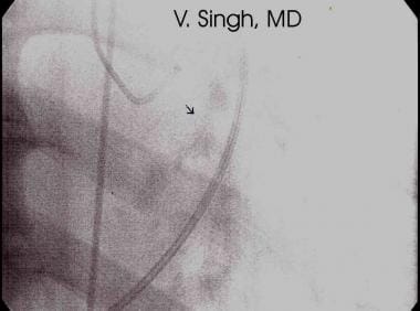
 Bicuspid aortic valves without stenosis or insufficiency in adult patients can be associated with pathologic enlargement of the aortic root, which infrequently leads to surgical aortic root replacement.


History

Presentations of aortic valve stenosis vary, ranging from critical aortic stenosis in neonates to no symptoms in children who have only a systolic ejection click on physical examination, either with or without a murmur to suggest aortic stenosis. Rarely, aortic valve stenosis causes sudden death during exercise or can present later in life from calcific aortic stenosis. Consider the presentation of aortic valve stenosis in the neonatal period, childhood, and in adolescent or adult patients.

  • Neonatal aortic valve stenosis
    • Neonatal aortic valve stenosis sometimes presents as congestive heart failure in the first week of life. These patients may have left the hospital initially stable, with a patent ductus arteriosus. Once the ductus arteriosus begins to close, clinical signs of heart failure occur that mimic sepsis, and a cardiac murmur may be unimpressive in the setting of low cardiac output. There may also be significant mitral valve insufficiency that adds to the congestive heart failure symptoms. Patients with this presentation often require emergency resuscitation and possibly the administration of prostaglandin E1, if the patient presents within the first 24-48 hours of age, to reestablish adequate systemic blood flow.
    • Often, neonates with aortic stenosis are asymptomatic but present with a systolic murmur, which leads to cardiology referral. Subsequent progression of aortic valve stenosis can be highly variable in rapidity and the degree of stenosis. Yetman et al (1995) have described patients in whom rapid progression occurs within 6 months of diagnosis. These patients often have well-preserved ventricular function but, during rapid growth, can exhibit fairly dramatic increases in the aortic valve gradient, requiring intervention. The absence of symptoms, which in fact may be difficult to discern in this age group, does not correlate with the severity of aortic valve obstruction.
  • Childhood aortic valve stenosis
    • Older children most often present with a systolic murmur as the first sign of aortic valve stenosis. These children usually are asymptomatic and have a systolic murmur detected during a sports physical or at a preschool entrance examination. If symptoms occur, a sense of easy fatigability may be reported. A history of syncope or anginal-type chest pain related to exertion should prompt an immediate evaluation and intervention by a pediatric cardiologist. Most often, these patients have pure aortic valve stenosis, though, occasionally, a patient presents with predominantly aortic valve insufficiency and minimal stenosis.
    • Another group of patients monitored through childhood are those who have undergone previous intervention in the neonatal period and have stable degrees of palliated aortic valve stenosis, insufficiency, or both. Detection of symptoms on the basis of history alone is often misleading, because exercise tolerance is often well preserved until aortic stenosis becomes more severe. Careful questioning with regard to exercise tolerance, levels of activity, avoidance of strenuous activities, and the presence of dyspnea, chest pain, presyncope, or excessive nap taking may reveal subtle signs of progressive aortic valve stenosis.
  • Adolescent or adult aortic valve stenosis
    • Adolescents and adults diagnosed for the first time with aortic valve stenosis often have a bicuspid aortic valve with mild degrees of stenosis or insufficiency. Many of these patients remain free of symptoms or problems for many years unless endocarditis occurs or valve calcification develops. Some of these patients have had prior intervention for aortic stenosis that was more severe at a younger age and have undergone either surgical or balloon valvotomy for palliation. In rare cases, a bicuspid aortic valve without stenosis or insufficiency presents as pathologic aortic root dilation resulting in spontaneous dissection.

Physical

  • Neonatal aortic valve stenosis
    • Neonates who present with critical aortic stenosis and low cardiac output have reduced or absent pulses and poor peripheral perfusion. They are tachycardiac and tachypneic, may have significantly increased work of breathing, and appear distressed. A systolic murmur may be unimpressive because of low cardiac output secondary to left ventricular dysfunction. A precordial thrill is somewhat rare in the neonate. A click may be heard, although this is often difficult to discern because the degree of tachycardia and poor excursion of the valve may make the click less noticeable. The peripheral pulses usually are low volume and are reduced symmetrically, unlike the differential pulses that are present in the setting of critical coarctation of the aorta. Severe aortic valve insufficiency in the neonatal period is rare; however, if it is present, consider the diagnosis of an aortic–left ventricular tunnel.
  • Childhood aortic valve stenosis
    • A systolic ejection murmur is present at the left middle and the right upper sternal border. A thrill in the suprasternal notch is common even with modest levels of aortic valve stenosis and helps localize pathology to the aortic valve. An ejection click is noted along the aortic axis and often is audible at the apex when it is not heard elsewhere. A precordial thrill is less common, but, when present, it usually is indicative of severe aortic valve stenosis. The apical impulse may be normal or increased in intensity. As with other semilunar valve pathologies, the severity of obstruction is proportional to the length and grade of the systolic murmur, assuming normal ventricular function and cardiac output. A fourth heart sound, when present, usually indicates significant left ventricular hypertrophy. The peripheral pulses may be normal or reduced, depending on the degree of obstruction. Bounding pulses (water-hammer pulses) indicate significant aortic valve insufficiency.
  • Adolescent or adult aortic valve stenosis
    • Aortic valve stenosis in adolescents and adults is similar to that seen in children, although older patients are more likely to have aortic valve insufficiency. A systolic ejection click is sometimes less noticeable with associated calcification of the aortic valve, which results in diminished valve excursion.
    • Maneuvers to improve auscultation include having larger patients lean forward or assume the left lateral decubitus position. Having the patient squat may accentuate murmur of aortic insufficiency.

Causes

Causes of aortic valve stenosis are multifactorial, although studies suggest a higher rate of recurrence of left ventricular outflow tract obstructive lesions than other forms of congenital heart disease. The recurrence risk in offspring of an affected father is approximately 3%, but it is approximately 15% in offspring of an affected mother. Abnormal fetal hemodynamics are theorized to contribute to development of aortic valve stenosis. Similarly, other forms of left heart obstructive disease may occur repeatedly within families (eg, hypoplastic left heart syndrome in a child whose older sibling had coarctation of the aorta). A definite genetic defect for aortic valve stenosis has not been identified, but the presence of a bicuspid aortic valve has been documented in multiple family members.


Aortic Stenosis, Subaortic
Aortic Stenosis, Supravalvar


Lab Studies

  • The evaluation for sepsis in infants presenting with shock includes blood, urine, and CSF cultures.
  • Pulse oximeter assessment is performed both preductally and postductally.

Imaging Studies

  • Echocardiography
    • In the neonate, transthoracic echocardiography usually provides complete diagnostic and hemodynamic information. Essential considerations are the details of valve anatomy, anulus size, distribution of valve tissue, degree of left ventricular hypertrophy, and left ventricular systolic function. The presence or absence of associated lesions, such as coarctation of the aorta or subaortic stenosis, can also be well delineated. Patients presenting with critical aortic valve obstruction and poor left ventricular systolic function may have echodense endocardium typical of endocardial fibroelastosis. These findings usually indicate that the severe obstruction was present for a significant amount of time prenatally.
    • In older patients, transthoracic echocardiography is usually diagnostic, although, rarely, a large adolescent or adult patient may require transesophageal echocardiography to clearly delineate the left ventricular outflow tract and to detail the valve anatomy.
    • Doppler echocardiography is used to estimate the severity of aortic valve stenosis. The peak instantaneous systolic gradient often overestimates the transvalvular peak-to-peak gradient obtained during cardiac catheterization. Mean Doppler gradients correlate well with mean gradients measured during cardiac catheterization. Significant individual variation exists in how closely the peak or mean Doppler-derived gradient predicts the peak-to-peak gradient measured at the time of cardiac catheterization. In the presence of poor left ventricular systolic function and low cardiac output, the Doppler-derived peak instantaneous gradient may be negligible and may not be indicative of the severity of obstruction.

Other Tests

  • Compared with echocardiography, MRI is rarely used to assess the details of aortic valve anatomy and is much more difficult to use in neonates, who have faster heart rates and more motion artifacts. New developments in gated MRI for assessing ventricular function may make MRI increasingly useful in adult patients.
  • Chest radiography may demonstrate cardiomegaly with pulmonary venous congestion primarily in neonates who present with critical stenosis and symptoms of congestive heart failure.
  • Electrocardiography in older patients may demonstrate left ventricular hypertrophy with or without a strain pattern, but it is less useful in neonates or young children with significant aortic valve disease.

Procedures

  • Exercise stress testing
    • Exercise stress testing can usually be performed in children aged 6 years or older and is helpful in eliciting symptoms that may not be evident from routine history. Doppler studies can be helpful in determining whether exercise restrictions are necessary by measuring the change in aortic valve gradient from rest to immediately after maximal exercise. Stress echocardiography is also useful in delineating the response of the left ventricle to increasing afterload during exercise in the setting of significant aortic valve disease. The exercise stress test establishes a baseline against which to compare subsequent study results, especially if the patient's symptoms change or the Doppler-derived gradient worsens.
    • Exercise stress testing may also provide some risk stratification if intervention is delayed or contemplated. Factors such as heart rate, blood pressure response to exercise, exercise duration, provocable arrhythmias or ECG ischemic changes, and measured oxygen consumption provide useful data on which to base decisions regarding timing of intervention.
  • Cardiac catheterization
    • Cardiac catheterization is usually performed in infants, children, and older adolescents in anticipation of balloon aortic valvuloplasty. In adult patients with calcific changes, the valve is usually less compliant and amenable to balloon dilation, with a higher risk for aortic valve insufficiency. Occasionally, the peak systolic gradient measured in the catheterization laboratory with the patient under conscious sedation is significantly less than that estimated by Doppler echocardiography, causing intervention to be deferred.
    • Other indications for catheterization may include the need for accurate hemodynamic assessment in patients with multiple levels of obstruction, such as mitral stenosis or subaortic stenosis in combination with aortic valve stenosis. In the latter instance, high-fidelity catheters capable of discriminating between multiple levels of obstruction in close proximity are probably preferable but are significantly more difficult to use, especially in young patients.

Histologic Findings

Pathologically, the severity of stenosis is secondary to thickening and increased rigidity of the valve tissue, with varying degrees of diminished commissural separation. Most commonly, the valve is bicuspid with a single fused commissure and an eccentrically placed orifice. A third or rudimentary commissure may sometimes be apparent. Less commonly, the valve is unicuspid and dome shaped. Rarely, the valve has 3 unseparated cusps, with the stenosis being centrally located. Secondary calcification of the valve is extremely rare, and, at times, the aortic valve anulus may also be underdeveloped or hypoplastic, adding to the severity of left ventricular outflow tract obstruction.

The left ventricular myocardium hypertrophies concentrically in the presence of significant obstruction. Critically ill neonates may have extensive endocardial fibroelastosis, especially in the presence of a dilated nonhypertrophied left ventricle. Patients with chronic aortic stenosis and significantly elevated left ventricular systolic pressure may exhibit fibrotic changes in the myocardium. Left ventricular volume may be reduced, normal, or increased. One study identified mitral valve abnormalities in 7.5% of 200 infants undergoing surgical valvotomy. Ischemia or infarction can result in papillary muscle dysfunction and mitral valve regurgitation.


Medical Care

  • Neonatal aortic stenosis
    • Patients with critical aortic stenosis and low cardiac output require resuscitation and institution of prostaglandin E1 at a dose of 0.01-0.1 mcg/kg/min. Establishing patency of the ductus arteriosus can restore adequate systemic blood flow and the perfusion of vital organs. Inotropic drugs such as dopamine, dobutamine, and epinephrine are indicated in cases of reduced cardiac output. With critical aortic stenosis, avoid drugs that cause significant vasodilation, as they may cause significant hypotension in the presence of a small cross-sectional aortic valve area. Patients with increased work of breathing and pulmonary edema benefit from intubation, positive pressure ventilation, and diuretic therapy.
    • Asymptomatic neonates with aortic valve stenosis whose valve gradients do not yet warrant intervention require conservative cardiac follow-up care and prophylaxis for subacute bacterial endocarditis (SBE) when indicated. Frequent follow-up care for neonatal patients in the first 6 months of life is recommended until the rate of progression of aortic valve stenosis with growth is determined. The exact gradient at which intervention is required is controversial and should be guided by additional echocardiographic variables.
    • Older children, as well as adolescents and adults, require ongoing medical follow-up care to detect evidence of progressive aortic stenosis, left ventricular hypertrophy, or new-onset aortic insufficiency. Pay careful attention to good dental hygiene and dental care, as well as to SBE prophylaxis. Occasionally, patients who develop progressive aortic insufficiency may benefit from afterload reduction. Patients with significant aortic stenosis who have developed worsening systolic function should undergo reduction of the stenosis. Similarly, patients with aortic valve insufficiency who exhibit a significant decline in left ventricular systolic performance, even within the range of normal, may need aortic valve replacement.

Surgical Care

Early reports of transcatheter balloon dilation in the 1980s were encouraging, although morbidity related to aortic valve insufficiency and femoral artery compromise were considered limitations of the procedure. With the advent of improved catheter technology, percutaneous balloon valvuloplasty has become the procedure of choice in most centers for severe congenital aortic valve stenosis and can be safely performed with virtually no mortality and minimal morbidity.

  • Balloon aortic valvuloplasty
    • Balloon aortic valvuloplasty is a good initial treatment in most patients with aortic valve stenosis. Patients with severely dysplastic valves may have a less favorable result with balloon aortic valvuloplasty, but, in most patients, the results are similar to those obtained with surgical valvotomy. The overall goal, especially in neonates and infants, is to relieve the aortic valve obstruction sufficiently without development of significant valve insufficiency, thereby resulting in normalization of left ventricular systolic function. Achievement of this goal typically entails performing a conservative balloon valvuloplasty by reducing the peak-to-peak systolic gradient by 50%. Balloon diameters are usually 80-100% of the aortic valve anulus dimension. In critically ill patients, surgical backup or circulatory support in the form of an extracorporeal membrane oxygenator (ECMO) should be available.
    • Neonates with critical aortic stenosis who are maintained on prostaglandin E1 should be sedated and intubated before the procedure is begun, to help maintain hemodynamic stability during the catheterization procedure. Several catheter techniques have been described over the past 20 years, with each one having its own advantages and disadvantages. There is currently no consensus regarding which approach is optimal in the neonate with critical aortic valve stenosis. The techniques include retrograde catheterization via the femoral artery, the right subscapular artery, the umbilical artery, or the right carotid artery, as well as antegrade transvenous catheterization through the atrial septum. The advantages of a transvenous, subscapular artery, carotid artery, or umbilical artery approach is preservation of the femoral arteries for later intervention and reduced risk of femoral arterial occlusion, which may still occur despite the availability of very low profileballoondilationcatheters.Crossing the aortic valve in neonates via the umbilical artery, however, can be quite challenging. The transvenous antegrade approach can also be difficult, especially in the presence of a small, hypertrophied left ventricle, and can result in injury to the mitral valve apparatus. Crossing the aortic valve in a retrograde manner via the right carotid artery is technically easier, although a surgical cutdown and repair of the vessel are necessary. This particular procedure can in fact be performed at the bedside with the aid of continuous transesophageal echocardiographic guidance, which provides continuous hemodynamic assessment preintervention and postintervention and eliminates the need for repeated angiograms to assess for aortic valve insufficiency and for transporting a sick neonate to and from the catheterization laboratory.

      Sheaths as small as 4F can be used for femoral arterial access in neonates or infants in whom umbilical vessels are closed. A prograde transseptal approach has occasionally been successful. Another option includes transventricular balloon valvotomy from the left ventricular apex.

  • Surgical aortic valvotomy
    • Although surgical aortic valvotomy was once believed to carry an extremely high risk of morbidity and mortality, it is now considered to be relatively safe and effective, though it has largely been displaced by balloon aortic valvuloplasty during cardiac catheterization. Occasionally, however, a significantly unstable neonatal patient who has a small aortic anulus or who requires attention to other associated lesions may be referred for surgical aortic valvotomy. Turley et al (1990) reported good results with surgical aortic valvotomy, though advances in catheter and balloon technology have made it possible to perform balloon aortic valvuloplasty during cardiac catheterization, even in small preterm infants.
    • Data from the Pediatric Cardiac Care Quality Assurance Consortium have shown a marked decline in surgical aortic valvotomy as balloon aortic valvuloplasty has become more widely used. Patients who require enlargement of an aortic anulus, resection of subaortic stenosis, or repair of supravalvar aortic stenosis or who have a small left ventricle are candidates for surgical aortic valvotomy in addition to other indicated procedures.
  • Surgical aortic valve replacement
    • The surgical replacement of an aortic valve is primarily reserved for patients in whom balloon valvuloplasty or surgical valvotomy has failed and in whom severe stenosis exists or significant aortic valve insufficiency has developed in association with left ventricular dilation or deterioration of left ventricular systolic function. Three options for aortic valve replacement are described below:
      • Mechanical prosthetic aortic valves are highly dependable and long lasting but require anticoagulation to prevent thromboembolic complications. Anticoagulation necessitates that the patient avoid collision sports and other activities that may result in significant bleeding. Warfarin adds significant complexity to the management of pregnancy, but the issues that arise are not insurmountable. Delaying surgical valve replacement as long as possible to allow maximum growth of children without compromising ventricular function is an important goal of presurgical management. Such delay allows insertion of the largest possible prosthetic valve and reduces the need for valve replacement solely because of patient growth.
      • Bioprosthetic aortic valve replacement includes a variety of available bioprosthetic materials that generally do not last as long as mechanical prosthetic valves. Bioprosthetic valves may be used in patients with contraindications to mechanical valves, in women contemplating pregnancy in the near future, in patients who want to pursue collision sports or other activities with a high risk of trauma, and in patients who may be unable to receive or comply with anticoagulation therapy.
      • The Ross procedure (pulmonary autograft) or autotransplantation of a pulmonary valve to the aortic position is favored by some surgeons because of the potential for growth of the pulmonary autograft valve through childhood. Such growth has been documented, as has adequate performance of a pulmonary valve in the aortic position. Problems with this procedure in children include development of early stenosis or insufficiency of a pulmonary homograft placed in the pulmonary position. This can necessitate multiple interventional catheterizations or surgical reoperations during childhood to alleviate the obstruction or insufficiency of the homograft. Significant enlargement of the neoaortic root, especially within the sinuses, may occur, and aortic insufficiency occasionally develops. This procedure does avoid the need for anticoagulation, especially in active, injury-prone children.

Consultations

Patients with dysmorphic features may require a genetic evaluation. Genetic counseling with regard to the risk of left ventricular outflow tract obstruction in subsequent pregnancies also may be indicated. A neonatologist may be consulted to assist with management of critically ill neonates, especially those born prematurely.

Diet

No specific dietary restrictions are necessary.

Activity

Activity limitations depend on the degree of severity of aortic valve stenosis and, in older children, the results noted on exercise stress testing. Strenuous isometric sports should be avoided; specific recommendations regarding sports participation have been published by the American Heart Association.


Ongoing treatment aims to preserve ventricular function and allow the left ventricle to function with only mild to moderate obstruction. If ventricular dysfunction is detected in patients with significant aortic stenosis, medical therapy should be used only for stabilization; such patients are likely to benefit from interventions to reduce the degree of stenosis. Patients with significant aortic valve insufficiency in combination with mild to moderate stenosis may be treated carefully with afterload reduction, although hypotension may occur. Patients with small aortic valve areas have a limited capacity for increased cardiac output with activity and may develop syncope or ischemic chest pain with exercise.

Antibiotics are required for endocarditis prophylaxis in patients undergoing procedures that may cause bacteremia. For more information, see Antibiotic Prophylactic Regimens for Endocarditis.

Drug Category: Prostaglandins

Alprostadil (PGE1) is used for treatment of ductal-dependent cyanotic congenital heart disease caused by decreased pulmonary blood flow. Patients with critical aortic stenosis and low cardiac output require resuscitation with prostaglandin E1. Establishing patency of the ductus arteriosus can restore systemic blood flow and the perfusion of vital organs. Alprostadil is first-line palliative therapy to maintain patency of the ductus arteriosus temporarily, before surgery. It produces vasodilation and increases cardiac output. Each 1 mL ampule contains 500 mcg/mL.

Drug NameAlprostadil Pediatric (Prostin VR Pediatric Injection)
DescriptionEffective in relaxing smooth muscle of ductus arteriosus. Beneficial in infants with congenital defects that restrict pulmonary or systemic blood flow and who, in order to get adequate oxygenation and lower body perfusion, depend on a patent ductus arteriosus.
Pediatric Dose0.01-0.1 mcg/kg/min IV into large vein or umbilical cord
ContraindicationsDocumented hypersensitivity; hyaline membrane disease or respiratory distress syndrome
InteractionsLimited data exist; caution with concurrent use of antiplatelet drugs or anticoagulants
PregnancyX - Contraindicated in pregnancy
PrecautionsLong-term infusions may cause cortical proliferation of the long bones in neonates; due to the inhibitory effects of prostaglandins in platelet aggregation, exercise caution when administering to neonates with bleeding tendencies
Adverse effects and toxicity include apnea, seizures, fever, hypotension, leukocytosis, and pulmonary overcirculation; neonates are usually intubated prophylactically because of potential risk of apnea (10-12%); prolonged use occasionally is necessary (in transplant candidates with hypoplastic left heart syndrome) and may be associated with third spacing of fluid; monitor blood oxygenation and arterial pressure


Further Inpatient Care

  • Manage congestive heart failure.
  • Monitor for arrhythmias and initiate antiarrhythmic therapy as needed.
  • Monitor postprocedural or postsurgical recovery.
  • Provide patient education.

Further Outpatient Care

  • Manage congestive heart failure (CHF).
  • Provide physical rehabilitation.
  • Perform exercise stress testing to determine response to therapy or activity restrictions.
  • Provide patient education.

In/Out Patient Meds

  • Use SBE prophylaxis for indicated procedures.
  • Prescribe other medicines as indicated for treatment of specific conditions such as CHF and arrhythmias.

Transfer

  • Transfer to a center capable of providing specialized care for infants and children with aortic stenosis.
  • In those cases of aortic stenosis diagnosed prenatally, the mother should be referred for delivery at a center capable of providing specialized care for newborns with severe aortic stenosis, which includes the initiation of prostaglandin E1.

Deterrence/Prevention

  • Fetal echocardiography is indicated for all subsequent pregnancies following the birth of a child with congenital aortic valve stenosis. When aortic stenosis is identified prenatally, delivery should take place in a tertiary center prepared to care for a neonate with severe aortic stenosis. Identified genetic abnormalities (eg, Turner syndrome or monosomy X) indicate the need for screening of parents to look for evidence of translocation.

Complications

  • Congestive heart failure
  • Myocardial injury and fibrosis
  • Syncope
  • Endocarditis
  • Calcific aortic stenosis
  • Progressive aortic insufficiency
  • Systemic embolization
  • Arrhythmias
  • Sudden death

Prognosis

  • The prognosis for patients with congenital aortic valve stenosis depends on the valve anatomy and its response to intervention. The severity of the disorder is quite variable, ranging from no symptoms in children with only a bicuspid aortic valve and minimal stenosis to critical illness in neonates who face the prospect of serial valvotomies and eventual aortic valve replacement. The main goal is to preserve the native aortic valve and ventricular function for as long as possible until aortic valve replacement is necessary.

Patient Education

  • Education is indicated for affected families regarding signs of progressive aortic stenosis, CHF, and low cardiac output. Education regarding good dental care and avoidance of risk factors for endocarditis is imperative. In addition, careful attention to SBE prophylaxis for indicated procedures is mandatory. Other goals of parent education include setting reasonable goals for exercise participation that allow for a healthy lifestyle but downplaying long-term goals for competitive athletics at a high level until complete relief of aortic valve disease is accomplished by aortic valve repair or replacement. Similarly, emphasize that treatment of aortic valve disease is a lifelong process and is best introduced early and reinforced often to ensure that patients with aortic valve stenosis continue to receive ongoing follow-up care as they make the transition from an adolescent lifestyle at home to the independent lifestyle of young adulthood.


Medical/Legal Pitfalls

  • Failure to provide good patient education to help parents understand the severity and long-term nature of aortic valve disease and the long-term commitment to surveillance and appropriate intervention
    • Failure to explain potential risks of interventions—in particular, potential complications of surgical or balloon valvotomy
    • Failure to emphasize risks of anesthesia and bypass for surgical valvotomy or for balloon valvotomy, cardiac perforation, acute low cardiac output from aortic insufficiency, and acute myocardial dysfunction

Special Concerns

  • Choice of an aortic valve for aortic valve replacement may be more complicated in female patients contemplating pregnancy.
  • Choice of a mechanical valve, a bioprosthetic valve, or a Ross procedure should be reviewed at length for all patients, particularly those contemplating valve replacement.
  • Teratogenic effects of warfarin, management issues of anticoagulation during pregnancy, and need for reoperation, as well as durability of various valve options, should be reviewed at length.


  • Alekyan BG, Petrosyan YS, Coulson JD, et al. Right subscapular artery catheterization for balloon valvuloplasty of critical aortic stenosis in infants. Am J Cardiol. Nov 15 1995;76(14):1049-52. [Medline].
  • Artman M, Boucek RJ Jr, Hammon J, Graham TP Jr. Emergency palliation of critical valvular aortic stenosis. A new application of prostaglandin E1. Am J Dis Child. Apr 1983;137(4):339-40. [Medline].
  • Beekman RH, Rocchini AP, Gillon JH, Mancini GB. Hemodynamic determinants of the peak systolic left ventricular-aortic pressure gradient in children with valvar aortic stenosis. Am J Cardiol. Mar 15 1992;69(8):813-5. [Medline].
  • Berger S, Dhala A, Friedberg DZ. Sudden cardiac death in infants, children, and adolescents. Pediatr Clin North Am. Apr 1999;46(2):221-34. [Medline].
  • Blaufox AD, Lai WW, Lopez L, et al. Survival in neonatal biventricular repair of left-sided cardiac obstructive lesions associated with hypoplastic left ventricle. Am J Cardiol. Nov 1 1998;82(9):1138-40, A10. [Medline].
  • Chambers J. Exercise testing to guide surgery in aortic stenosis [editorial]. Heart. Jul 1999;82(1):7-8. [Medline].
  • Egito ES, Moore P, O''Sullivan J, et al. Transvascular balloon dilation for neonatal critical aortic stenosis: early and midterm results. J Am Coll Cardiol. Feb 1997;29(2):442-7. [Medline].
  • Fedderly RT. Left ventricular outflow obstruction. Pediatr Clin North Am. Apr 1999;46(2):369-84. [Medline].
  • Fischer DR, Ettedgui JA, Park SC, et al. Carotid artery approach for balloon dilation of aortic valve stenosis in the neonate: a preliminary report. J Am Coll Cardiol. Jun 1990;15(7):1633-6. [Medline].
  • Fixler DE, Pastor P, Sigman E, Eifler CW. Ethnicity and socioeconomic status: impact on the diagnosis of congenital heart disease. J Am Coll Cardiol. Jun 1993;21(7):1722-6. [Medline].
  • Glick BN, Roberts WC. Congenitally bicuspid aortic valve in multiple family members. Am J Cardiol. Feb 15 1994;73(5):400-4. [Medline].
  • Hausdorf G, Schneider M, Schirmer KR, et al. Anterograde balloon valvuloplasty of aortic stenosis in children. Am J Cardiol. Feb 15 1993;71(5):460-2. [Medline].
  • Hubbell MM Jr, Gowdamarajan R. Aortic stenosis. In: Moller JH, ed. Surgery of Congenital Heart Disease: The Pediatric Cardiac Care Consortium 1984-1995. Armonk, NY:. Futura Publishing Co Inc;1998:125-41.
  • Kim KS, Maxted W, Nanda NC, et al. Comparison of multiplane and biplane transesophageal echocardiography in the assessment of aortic stenosis. Am J Cardiol. Feb 15 1997;79(4):436-41. [Medline].
  • Kiraly P, Kapusta L, Thijssen JM, Daniels O. Left ventricular myocardial function in congenital valvar aortic stenosis assessed by ultrasound tissue-velocity and strain-rate techniques. Ultrasound Med Biol. Apr 2003;29(4):615-20. [Medline].
  • Latson LA. Aortic stenosis: valvar, supravalvar, and fibromuscular Subvalvar. In: Garson A Jr, Bricker JT, Fisher DJ, Neish SR, eds. The Science and Practice of Pediatric Cardiology. 2nd ed. Philadelphia, Pa: Williams & Wilkins;1998:1257-76.
  • Lemler MS, Valdes-Cruz LM, Shandas RS, Cape EG. Insights into catheter/Doppler discrepancies in congenital aortic stenosis. Am J Cardiol. May 15 1999;83(10):1447-50. [Medline].
  • Magee AG, Nykanen D, McCrindle BW, et al. Balloon dilation of severe aortic stenosis in the neonate: comparison of anterograde and retrograde catheter approaches. J Am Coll Cardiol. Oct 1997;30(4):1061-6. [Medline].
  • McLean KM, Lorts A, Pearl JM. Current treatments for congenital aortic stenosis. Curr Opin Cardiol. May 2006;21(3):200-4. [Medline].
  • Meliones JN, Beekman RH, Rocchini AP, Lacina SJ. Balloon valvuloplasty for recurrent aortic stenosis after surgical valvotomy in childhood: immediate and follow-up studies. J Am Coll Cardiol. Apr 1989;13(5):1106-10. [Medline].
  • Moore P, Egito E, Mowrey H, et al. Midterm results of balloon dilation of congenital aortic stenosis: predictors of success. J Am Coll Cardiol. Apr 1996;27(5):1257-63. [Medline].
  • Nowlen TT, Ayres NA, Kearney DL, et al. Premature closure of the foramen ovale associated with aortic stenosis, left ventricular dilation with thrombus, and early mortality. Am J Cardiol. May 1 2000;85(9):1159-61, A9. [Medline].
  • Parsons MK, Moreau GA, Graham TP Jr, et al. Echocardiographic estimation of critical left ventricular size in infants with isolated aortic valve stenosis. J Am Coll Cardiol. Oct 1991;18(4):1049-55. [Medline].
  • Puntel RA, Webber SA, Ettedgui JA, Tacy TA. Rapid enlargement of neoaortic root after the Ross procedure in children. Am J Cardiol. 1999;84:747-9. [Medline].
  • Rajdev S, Nanda NC, Patel V, et al. Live/real-time three-dimensional transthoracic echocardiographic assessment of combined valvar and supravalvar aortic stenosis. Am J Geriatr Cardiol. May-Jun 2006;15(3):188-90. [Medline].
  • Rhodes LA, Colan SD, Perry SB, et al. Predictors of survival in neonates with critical aortic stenosis [published erratum appears in Circulation 1995 Oct 1;92(7):2005]. Circulation. Dec 1991;84(6):2325-35. [Medline].
  • Roberts CS, Roberts WC. Dissection of the aorta associated with congenital malformation of the aortic valve. J Am Coll Cardiol. Mar 1 1991;17(3):712-6. [Medline].
  • Rosenfeld HM, Landzberg MJ, Perry SB, et al. Balloon aortic valvuloplasty in the young adult with congenital aortic stenosis. Am J Cardiol. Jun 1 1994;73(15):1112-7. [Medline].
  • Sandhu SK, Silka MJ, Reller MD. Balloon aortic valvuloplasty for aortic stenosis in neonates, children, and young adults. J Interv Cardiol. Oct 1995;8(5):477-86. [Medline].
  • Simpson JM, Sharland GK. Natural history and outcome of aortic stenosis diagnosed prenatally. Heart. Mar 1997;77(3):205-10. [Medline].
  • Starnes VA, Luciani GB, Wells WJ, et al. Aortic root replacement with the pulmonary autograft in children with complex left heart obstruction. Ann Thorac Surg. Aug 1996;62(2):442-8; discussion 448-9. [Medline].
  • Turley K, Bove EL, Amato JJ, et al. Neonatal aortic stenosis. J Thorac Cardiovasc Surg. Apr 1990;99(4):679-83; discussion 683-4. [Medline].
  • Waller BF, McKay C, VanTassel JW, et al. Catheter balloon valvuloplasty of stenotic aortic valves. Part I: Anatomic basis and mechanisms of balloon dilation. Clin Cardiol. Oct 1991;14(10):836-46. [Medline].
  • Waller BF, Dorros G, Lewin RF, et al. Catheter balloon valvuloplasty of stenotic aortic valves--Part II: Balloon valvuloplasty during life subsequent tissue examination. Clin Cardiol. Nov 1991;14(11):924-30. [Medline].
  • Weber HS, Mart CR, Myers JL. Transcarotid balloon valvuloplasty for critical aortic valve stenosis at the bedside via continuous transesophageal echocardiographic guidance. Catheter Cardiovasc Interv. Jul 2000;50(3):326-9. [Medline].
  • Weber HS. Catheter management of aortic valve stenosis in neonates and children. Catheter Cardiovasc Interv. Jun 2006;67(6):947-55. [Medline].
  • Yetman AT, Rosenberg HC, Joubert GI. Progression of asymptomatic aortic stenosis identified in the neonatal period. Am J Cardiol. Mar 15 1995;75(8):636-7. [Medline].
  • Zeevi B, Keane JF, Castaneda AR, et al. Neonatal critical valvar aortic stenosis. A comparison of surgical and balloon dilation therapy. Circulation. Oct 1989;80(4):831-9. [Medline].
  • de Kort E, Thijssen JM, Daniels O, et al. Improvement of heart function after balloon dilation of congenital valvar aortic stenosis: a pilot study with ultrasound tissue Doppler and strain rate imaging. Ultrasound Med Biol. Jul 2006;32(7):1123-8. [Medline].

Aortic Stenosis, Valvar excerpt

Article Last Updated: Aug 21, 2006