You are in: eMedicine Specialties >
Pediatrics: Genetics and Metabolic Disease > Metabolic Diseases
Glycogen-Storage Disease Type I
Article Last Updated: Jun 4, 2007
AUTHOR AND EDITOR INFORMATION
Section 1 of 11
Author: Karl S Roth, MD, Chair, Professor, Department of Pediatrics, Creighton University School of Medicine
Karl S Roth is a member of the following medical societies: Alpha Omega Alpha, American Academy of Pediatrics, American College of Nutrition, American Pediatric Society, American Society for Clinical Nutrition, American Society of Nephrology, Association of American Medical Colleges, Medical Society of Virginia, New York Academy of Sciences, Sigma Xi, Society for Pediatric Research, and Southern Society for Pediatric Research
Editors: Edward Kaye, MD, Vice President of Clinical Research, Genzyme Corporation; Mary L Windle, PharmD, Adjunct Assistant Professor, University of Nebraska Medical Center College of Pharmacy, Pharmacy Editor, eMedicine.com, Inc; Hagop Youssoufian, MSc, MD, Medical Director, Adjunct Associate Professor, Clinical Discovery Department, Bristol-Myers Squibb; Paul D Petry, DO, FACOP, FAAP, Clinical Assistant Professor of Pediatrics, University of North Dakota, School of Medicine and Health Sciences; Consulting Staff, Altru Health System; Bruce A Buehler, MD, Professor, Department of Pathology and Microbiology, Director, Hattie B Munroe Center for Human Genetics, Chairman, Department of Pediatrics, University of Nebraska Medical Center
Author and Editor Disclosure
Synonyms and related keywords:
glucose-6-phosphatase deficiency, glucose-6-phosphate translocase deficiency, von Gierke disease, glycogenosis, GSD, type I GSD, GSD type I, GSD I, GSD Ia, GSD Ib, glycogen storage disease type I, glycogen-storage disease type I, excessive glycogen, nosebleeds, fasting hypoglycemia, seizures, cyanosis, apnea, acidosis, hyperuricemia, nephrolithiasis, von Willebrand disease, von Willebrand’s disease, defective platelet aggregation, apoptosis, biotinidase deficiency, hepatic adenoma, hepatocellular carcinoma, progressive renal insufficiency, hyperuricemic nephrocalcinosis, hyperlipidemic xanthomas, short stature
Background
In 1929, von Gierke provided the initial description of glycogen-storage disease type I (GSD I) from autopsy reports of 2 children whose large livers contained excessive glycogen. He also reported similar findings in the kidneys. Both children had frequent nosebleeds before their deaths, consistent with histories documented in current patients. In 1952, Cori and Cori reported 6 similar patients.1 Two of the patients had almost total deficiency of hepatic glucose-6-phosphatase, whereas the remaining 4 had normal enzyme activity. These authors recognized that defects in the enzymology of hepatic GSD may cause a heterogeneous group of disorders. However, the mystery of patients with these clinical symptoms (despite normal phosphatase activity) remained unsolved until 1978, when Narisawa et al identified a defect in intracellular transport of the enzyme substrate.2 In recognition of the original clinical description of the disease, the type I Cori classification has been preserved; GSD type Ia (GSD Ia) designates the true enzyme defect, and GSD type Ib (GSD Ib) designates the intracellular transport defect. Because free glucose is the product of the hepatic glucose-6-phosphatase reaction, either type leads to accumulation of liver glycogen, accompanied by fasting hypoglycemia. Hepatomegaly, the natural consequence of glycogen accumulation, is the clinical hallmark of the disease.
Pathophysiology
The liver loses its capacity as a glucose-homeostatic organ because of a fundamental inability to release free glucose. GSD I (subtypes Ia and Ib) is one of the few genetic-biochemical causes of hypoglycemia in newborns. The usual homeostatic mechanism cannot halt the rapid drop in blood glucose levels that normally occurs during the first several hours after birth (reflecting consumption of maternal glucose), and the decrease continues. This decrease in circulating glucose can be precipitous, resulting in no measurable blood level. Seizures, cyanosis, and apnea may ensue. In older children, repeated episodes of hypoglycemia may result in brain damage, as measured on performance testing and assessment of brainstem auditory-evoked potentials. In the hepatocyte, the glycogen catabolic machinery normally responds to stimuli caused by hypoglycemia (eg, neural, hormonal), ending in a flood of glucose-6-phosphate that cannot be released from the cell. However, glucose-6-phosphate is also the substrate for glycolysis and produces lactate. Lactate exits the hepatocyte, causing clinically significant lactic acidemia in proportion to the degree of stimulus for glycogen breakdown. The accumulation of lactic acid in blood can cause true acidosis with a large anion gap, a characteristic of GSD I. The immense increase in the intracellular phosphorylated intermediate compounds of glycolysis concurrently inhibits rephosphorylation of adenine nucleotides, activating the nucleic acid degradation pathway and resulting in increased uric acid, the end product. Hyperuricemia can reach levels that require use of xanthine oxidase inhibitors to prevent nephrolithiasis. Nephrolithiasis secondary to increased uric acid is a constant threat to patients with poorly controlled disease. Severe hypoglycemia stimulates epinephrine secretion, which activates lipoprotein lipase and the release of free fatty acids. These fatty acids are transported to the liver, where they are used for triglyceride synthesis and are exported as very-low-density lipoprotein (VLDL), which is elevated in these patients. Paradoxically, even in the face of hypoglycemia, patients with GSD I do not develop significant ketosis because the abundance of acetyl coenzyme A (CoA) derived from glycolysis activates the acetyl CoA carboxylase enzyme that produces malonyl CoA in the first step of fatty acid synthesis. Because malonyl CoA inhibits transport of fatty acid into the mitochondrion, beta-oxidation of fatty acids for energy to support the hypoglycemic cells does not occur. This causes a continuing drop in blood glucose levels and explains the absence of ketone bodies.
The lipid abnormalities, which include hypercholesterolemia (decreased high-density lipoprotein [HDL] cholesterol and increased low-density lipoprotein [LDL] cholesterol), together with the characteristic hypertriglyceridemia do not cause premature atherosclerotic lesions in affected individuals. Recent evidence suggests that sera from patients with GSD Ia are able to more efficiently promote scavenger receptor class B type I–mediated cellular cholesterol efflux and that an increased antioxidant effect of these sera is directly related to the increased urate concentration.3 Nosebleeds experienced by the patients in von Gierke's report probably resulted from the bleeding tendency characteristic of GSD Ia and Ib. This tendency resembles von Willebrand disease and suggests alterations in membrane glycoprotein synthesis. Although such changes have been found, no definitive explanation addresses how these alterations actually cause defective platelet aggregation. Patients with GSD Ib are susceptible to gram-positive infections. Neutrophils from patients with GSD Ib have a significantly impaired respiratory-burst response to stimuli compared with type Ia neutrophils. Because the respiratory burst generates superoxide, which is a major defense against gram-positive organisms, a defect in this response would be expected to render the neutrophils susceptible, causing neutropenia and diminishing the individual's resistance to infection. Evidence suggests that microsomal transport of glucose-6-phosphate has a role in antioxidant protection of the neutrophil; therefore, a genetic defect in the transporter could impair cellular function and lead to apoptosis. Growth is generally impaired in patients with GSD I, although growth can be improved with good dietary therapy in most patients. However, growth remains unimproved by treatment in some patients; the endocrine parameters of growth in this group are not measurably different from the larger number of patients. Several studies have also documented a decreased bone mineral density in some patients with GSD I. One such investigation reported no correlation between bone mineral density and turnover markers, indicating an uncoupling of bone turnover in patients with GSD.4 The cause of severe anemia in the absence of renal function compromise in children with GSD I has remained unclear. Some have recently proposed that hepcidin production by hepatic adenomas plays central in patients with GSD I. Hepcidin is a peptide hormone that is also a key regulator of the egress of cellular iron; in excess, it may interfere with intestinal iron transport as well as iron release from macrophages. Although extremely rare, GSD subtypes Ic and Id have occurred. Individuals with these forms probably have unusual mutations in the translocase gene (11q23).
Frequency
United States
The lack of newborn screening precludes reliable estimates of the incidence. The only estimates are for GSDs as a group, which suggest an incidence of 1 case per 20,000-25,000 births. GSD I is unlikely to occur more frequently than 1 case in 50,000 infants. In an odd quirk, clinical symptoms of biotinidase deficiency in patients with underlying GSD Ia have been associated with marked elevations in biotinidase levels on serum assay. However, mass screening for biotinidase deficiency does not reveal elevated activity.
Mortality/Morbidity
Affected newborns are at risk for all neonatal hypoglycemic complications. (Older children under treatment may experience symptoms identical to those listed below with impending hypoglycemia.)
- Hypoglycemic complications are entirely nonspecific and include the following:
- Twitching
- Cyanosis
- Seizures
- Irritability
- Apathy
- Hypotonia
- Hypothermia
- Apnea
- Coma (may be secondary to cerebral edema from combined hypoglycemia and hypoxia secondary to hypoglycemic seizures)
- Long-term consequences of glycogen storage include the following:
- Hepatic adenomas
- Hepatocellular carcinoma
- Progressive renal insufficiency
- Hyperuricemic nephrocalcinosis
- Hyperlipidemic xanthomas
- Short stature
- Hypoglycemic brain damage
- In GSD Ib, secondary consequences of neutrophilic abnormalities include multiple and recurrent infections, brain abscess, and pseudocolitis.
Sex
GSD Ia and Ib occur with equal frequency in both sexes.
Age
As genetic disorders, both types are present at conception, with clinical onset at birth.
History
- Initial symptoms of neonatal hypoglycemia occur shortly after birth, and patients do not respond to glucagon administration. Symptoms include the following:
- Tremors
- Irritability
- Cyanosis
- Seizures
- Apnea
- Coma
- Older infants may present with the following:
- Frequent lethargy
- Difficult arousal from overnight sleep
- Tremors
- Overwhelming hunger
- Poor growth
- Apparent increase in abdominal girth, although extremities appear thin
- A doll-like facial appearance caused by adipose tissue deposition in the cheeks
- Young children with glycogen-storage disease type Ia (GSD Ia) may experience nosebleeds.
- Young children with GSD Ib may have frequent otitides, gingivitis, and boils.
- Symptoms of severe hypoglycemia in patients of all ages are likely to follow any illness that causes mild anorexia or fasting (eg, viral gastroenteritis).
- In middle childhood, patients may manifest evidence of rickets and anemia.
- Patients with GSD Ib at all ages may be affected by a Crohnlike ileocolitis (pseudocolitis). The severity of the primary disorder is not correlated with the intestinal symptoms.
Physical
- Affected infants are healthy at birth, although some are born with an enlarged liver.
- Careful abdominal examination is mandatory for any neonate with hypoglycemia.
- Abdominal protuberance develops early because of massive hepatomegaly.
- Splenomegaly does not occur.
- The liver is firm and uniform in consistency in early life but may become nodular with the development of adenoma.
- The patient may present with poor growth, short stature, and rachitic changes.
- Gingivitis and compromised dentition may be present.
- Xanthoma may be found on extensor surfaces, such as the elbows and knees.
- Ultrasonography may reveal large kidneys in patients of all ages. Proteinuria may accompany this finding.
- In addition to hypoglycemia, an increased plasma lactate value is a characteristic laboratory finding in a symptomatic newborn with GSD I. The increased lactate originates from the flooding of the glycolytic pathway by glucose-6-phosphate, which is derived from breakdown of glycogen, cannot be cleaved to free glucose, and is released into blood.
Causes
- GSD Ia and Ib are autosomal recessive genetic traits caused by mutations at loci 17q21 and 11q23, respectively.
-
- GSD Ia is caused by deficient activity of the enzyme glucose-6-phosphatase, representing at least 14 distinct allelic variants.
-
- GSD Ib is caused by deficiency of glucose-6-phosphate translocase, which is responsible for importing glucose-6-phosphate from the cytosol to the interior of the microsome, thus bringing substrate into contact with enzyme. To date, allelic variation in this disorder has not been explored.
-
Biotinidase Deficiency
Constitutional Growth Delay
Crohn Disease
Disorders of Bone Mineralization
Epistaxis
Fructose 1-Phosphate Aldolase Deficiency (Fructose Intolerance)
Glycogen-Storage Disease Type I
Glycogen-Storage Disease Type II
Glycogen-Storage Disease Type III
Glycogen-Storage Disease Type IV
Glycogen-Storage Disease Type V
Glycogen-Storage Disease Type VI
Growth Failure
Growth Hormone Deficiency
Hepatoblastoma
Hepatocellular Carcinoma
Liver Tumors
Other Problems to be Considered
Niemann-Pick disease, type A (classic infantile form)
Type 1 hyperlipoproteinemia
Lab Studies
- The initial laboratory workup should include measuring blood glucose levels with electrolytes. If the blood glucose concentration is low, measurement of electrolyte levels may permit calculation of an increased anion gap, which suggests lactic acidemia.
- Measurement of liver function, plasma uric acid levels, and urinary creatinine clearance are essential.
- Perform a CBC count and differential.
- In patients with glycogen-storage disease type Ia (GSD Ia), the WBC count is usually within reference ranges because the defect does not affect leukocyte function.
- In contrast, GSD Ib causes chronic granulocytopenia due to the impaired function of the neutrophils, particularly in relation to gram-positive organisms.
- Perform a coagulation profile to include bleeding time tests.
Imaging Studies
- Ultrasonography of liver and kidneys may be useful.
- Abdominal ultrasonography can enable reasonable estimates of liver and kidney size. These estimates are especially useful during treatment of patients with chronic GSD because the relative size of the liver is expected to diminish with growth of the child.
- Ultrasonography may also be useful late in the clinical course to provide a baseline for evaluating nodules.
- Bone-density studies may be useful in middle childhood to identify patients who may have diminished bone density in adolescence.
Other Tests
- Glucagon administration produces no hyperglycemic response, although it markedly increases the plasma lactic acid level.
- Oral (PO) administration of galactose (1.75 g/kg) causes no change in blood glucose levels, although plasma lactic acid levels markedly increase.
Procedures
- Always directly measure glucose-6-phosphatase activity in the liver before and after detergent treatment of the homogenate, especially when activity appears to be within the reference ranges before treatment.
-
- Open biopsy is usually needed to obtain sufficient tissue for this assay; therefore, measure all other parameters before the procedure is performed.
-
Histologic Findings
Although histology should be routinely performed on liver samples obtained during surgery, neither light nor electron microscopy permit differentiation of the underlying reasons for glycogen storage. Clinically significant lipid storage occurs with both types of GSD I, which does little to clarify the picture.
Medical Care
- Diagnostic evaluation is most safely performed in a hospital, especially in infants, because of the potential for severe hypoglycemia. Many untreated children are admitted by a hematologist or gastroenterologist for the diagnosis of massive hepatomegaly.
- Young infants require continuous nasogastric (NG) tube feedings to sustain blood sugar levels.
- Older children can usually be switched to raw cornstarch feedings, which sustain blood glucose values for 4-6 hours.
- Large quantities of raw cornstarch may be necessary, because overall use of this material is impaired in patients compared with healthy control subjects.
- Pay scrupulous attention to the dental and oral health of patients with glycogen-storage disease type Ib (GSD Ib) to reduce incidence of infection.
- Any intercurrent infection that causes decreased intake requires intravenous (IV) glucose support until resolution.
- Currently, efforts are underway in animal models to develop gene therapy in patients with both forms of GSD I
Surgical Care
- Surgery is usually unnecessary after initial diagnosis using open liver biopsy.
- Promptly attend to any skin infection in patients with GSD Ib because deep-tissue extension requiring surgical debridement and plastic reconstruction may develop.
- In older children with GSD Ib, cautiously evaluate abdominal pain for pseudocolitis.
- Perform ultrasonography annually to evaluate for hepatic adenomas, which may require surgical removal.
- Hepatic transplantation has been attempted in a few cases with modified success
Consultations
- Biochemical genetics specialist
-
- Nephrologist
-
Diet
- Diet, the mainstay of therapy for both types of GSD I, requires close monitoring and adjustment by a highly specialized nutritionist.
- The fundamental principle of diet management for these patients is maintenance of a steady-state balance between circulating glucose and existing glycogen stores. Consequently, a chief aim is to avoid excessive carbohydrates and calories while supplying adequate calories and protein for growth.
- Because of the triglyceridemia characteristic in this disorder, counsel the patient to avoid high lipid intake.
- Most biochemical parameters can be substantially normalized and liver size can be reduced by approaching glucose homeostasis by means of overnight feeding.
- Overnight NG feedings should be administered only by a pump equipped with an alarm in case of flow interruption.
- When pancreatic amylase reaches sufficient activity in children older than 2-3 years, overnight feeding is usually replaced by raw cornstarch at bedtime and early morning hours.
Activity
- Instruct the patient to avoid all contact sports because of the propensity for infection and bleeding, as well as the potential for liver damage.
-
- Encourage the patient to engage in all other physical activities up to individual limits. Personal limitations should be the basis for participation in school activities.
-
Glycogen-storage disease type Ia (GSD Ia) has no specific medication requirement beyond prophylactic PO iron and prompt treatment of intercurrent infections (which may interrupt PO intake). Weekly administration of granulocyte colony–stimulating factor (GCSF), in addition to prophylactic PO iron and prompt treatment of intercurrent infections, is critical in patients with GSD Ib. GCSF administration is now standard therapy to prevent or reduce incidences of serious infection. GCSF may also delay or prevent pseudocolitis symptoms.
Drug Category: Trace elements
These are inorganic substances found in small amounts in the tissues and required for various metabolic processes.
| Drug Name | Iron sulfate (Feosol) |
| Description | Nutritionally essential inorganic substance. |
| Adult Dose | 60 mg (as elemental iron) PO qd |
| Pediatric Dose | 1-2 mg/kg/d (as elemental iron) PO; not to exceed 15 mg/d |
| Contraindications | Documented hypersensitivity; hemochromatosis, hemosiderosis, or hemolytic anemias |
| Interactions | Ascorbic acid enhances absorption; interferes with tetracycline absorption; milk, cereal, tea, coffee, eggs, dietary fiber, and antacids impair absorption |
| Pregnancy | A - Safe in pregnancy
|
| Precautions | Gastrointestinal upset; iron toxicity observed with ingestion of large amount and can be fatal, especially in children; parenteral (IV) administration may cause several reactions including headaches, malaise, fever, generalized lymphadenopathy, arthralgia, and urticaria; can cause severe anaphylaxis, phlebitis at infusion site |
Drug Category: Colony stimulating factors
These agents act as hematopoietic growth factors that stimulate the development of granulocytes. They are used to treat or prevent neutropenia when patients are receiving myelosuppressive cancer chemotherapy and to reduce neutropenia associated with bone marrow transplantation. These drugs are also used to mobilize autologous peripheral blood progenitor cells for bone marrow transplantation and to manage chronic neutropenia.
| Drug Name | Filgrastim (G-CSF, Neupogen) |
| Description | GCSF that activates and stimulates production, maturation, migration, and cytotoxicity of neutrophils. |
| Adult Dose | 5 mcg/kg SC qwk |
| Pediatric Dose | Administer as in adults |
| Contraindications | Documented hypersensitivity |
| Interactions | Synergistic effects with interleukin-3 (increase megakaryocyte and platelet production) |
| Pregnancy | C - Safety for use during pregnancy has not been established.
|
| Precautions | Do not use 12-24 h before or 24 h after cytotoxic chemotherapy because increases sensitivity of rapidly dividing myeloid cells to cytotoxic chemotherapy; do not admix with sodium chloride; caution in gout and psoriasis; adverse effects include fever, bone pain, and flu-like symptoms |
Further Inpatient Care
- Conditions that reduce PO intake require IV glucose to maintain blood sugar and to avoid complications of severe hypoglycemia.
-
Further Outpatient Care
- Close nutritional and biochemical genetic follow-up is critical, especially during initial and pubertal growth periods.
-
- Patients should be seen by subspecialists at least every 6 months.
-
In/Out Patient Meds
- No medications are required for glycogen-storage disease type Ia (GSD Ia).
- Patients with GSD Ib require GCSF on a weekly basis.
Transfer
- Consider transferring any patient who is admitted for any reason other than routine IV fluid administration for blood glucose support.
-
Complications
- Severe hypoglycemia, cerebral edema, coma, death
- Hepatic adenoma, adenocarcinoma, or both
- Glomerular hyperfiltration and glomerulosclerosis
- Brain damage
- Severe anemia
- Growth failure
Prognosis
- Patients receiving proper treatment should have a reasonable life span.
-
Patient Education
- Teach parents of infants how to insert an NG feeding tube.
-
- Teach family members how to test blood glucose levels.
-
- Teach family members and older children how to recognize signs of impending hypoglycemia.
-
- Provide intensive nutritional education to patients so they can assist in their own dietary control as early as possible.
-
- For excellent patient education resources, visit eMedicine's Ear, Nose, and Throat Center. Also, see eMedicine's patient education article Nosebleeds.
-
Medical/Legal Pitfalls
- Percutaneous liver biopsy in an unrecognized patient with postoperative hemorrhage
- Administration of steroid or other symptomatic therapy in a hypoglycemic newborn who suffers hypoglycemic damage after discharge
- Failure to distinguish glycogen-storage disease type Ia (GSD Ia) from Ib, resulting in administration of GCSF to a patient with GSD Ia, which can cause life threatening or disfiguring infection
- Failure to maintain adequate surveillance for hepatic adenomas
| Media file 1:
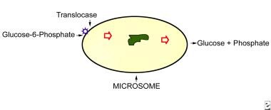
Microsome is shown in relation to the substrate, glucose-6-phosphate, which has been released from cytosolic glycogen. This substrate is transferred across the microsomal membrane by the protein translocase, where by glucose-6-phosphatase acts on it to release free glucose and inorganic phosphate. Patients with glycogen-storage disease type Ia are genetically deficient in glucose-6-phosphate activity, while those affected with glycogen-storage disease type Ib lack translocase. |
 | View Full Size Image | |
Media type: Graph
|
- Cori GT, Cori CF. Glucose-6-phosphatase of the liver in glycogen storage disease. J Biol Chem. Dec 1952;199(2):661-7. [Medline].
- Narisawa K, Igarashi Y, Otomo H, Tada K. A new variant of glycogen storage disease type I probably due to a defect in the glucose-6-phosphate transport system. Biochem Biophys Res Commun. Aug 29 1978;83(4):1360-4. [Medline].
- Nguyen AD, Pan CJ, Weinstein DA, Chou JY. Increased scavenger receptor class B type I-mediated cellular cholesterol efflux and antioxidant capacity in the sera of glycogen storage disease type Ia patients. Mol Genet Metab. Nov 2006;89(3):233-8. [Medline].
- Cabrera-Abreu J, Crabtree NJ, Elias E, et al. Bone mineral density and markers of bone turnover in patients with glycogen storage disease types I, III and IX. J Inherit Metab Dis. 2004;27(1):1-9. [Medline].
- Ambruso DR, McCabe ER, Anderson D, et al. Infectious and bleeding complications in patients with glycogenosis Ib. Am J Dis Child. Jul 1985;139(7):691-7. [Medline].
- Baker L, Dahlem S, Goldfarb S, et al. Hyperfiltration and renal disease in glycogen storage disease, type I. Kidney Int. Jun 1989;35(6):1345-50. [Medline].
- Bashan N, Hagai Y, Potashnik R, Moses SW. Impaired carbohydrate metabolism of polymorphonuclear leukocytes in glycogen storage disease Ib. J Clin Invest. May 1988;81(5):1317-22. [Medline].
- Bianchi L. Glycogen storage disease I and hepatocellular tumours. Eur J Pediatr. 1993;152 Suppl 1:S63-70. [Medline].
- Bodamer OA, Feillet F, Lane RE, et al. Utilization of cornstarch in glycogen storage disease type Ia. Eur J Gastroenterol Hepatol. Nov 2002;14(11):1251-6. [Medline].
- Chen YT, Coleman RA, Scheinman JI, et al. Renal disease in type I glycogen storage disease. N Engl J Med. Jan 7 1988;318(1):7-11. [Medline].
- Chen YT, Cornblath M, Sidbury JB. Cornstarch therapy in type I glycogen-storage disease. N Engl J Med. Jan 19 1984;310(3):171-5. [Medline].
- Chen YT, Scheinman JI, Park HK, et al. Amelioration of proximal renal tubular dysfunction in type I glycogen storage disease with dietary therapy. N Engl J Med. Aug 30 1990;323(9):590-3. [Medline].
- Chou JY, Mansfield BC. Gene therapy for type I glycogen storage diseases. Curr Gene Ther. Apr 2007;7(2):79-88. [Medline].
- Greene HL, Slonim AE, O'Neill JA Jr, Burr IM. Continuous nocturnal intragastric feeding for management of type 1 glycogen-storage disease. N Engl J Med. Feb 19 1976;294(8):423-5. [Medline].
- Heyne K, Henke-Wolter J. Glycogen storage disease Ib: modification of alpha 1-antitrypsin glycoprotein microheterogeneity. Eur J Pediatr. Jan 1989;148(4):341-3. [Medline].
- Koeberl DD, Kishnani PS, Chen YT. Glycogen storage disease types I and II: treatment updates. J Inherit Metab Dis. Apr 2007;30(2):159-64. [Medline].
- Leuzzi R, Bánhegyi G, Kardon T, et al. Inhibition of microsomal glucose-6-phosphate transport in human neutrophils results in apoptosis: a potential explanation for neutrophil dysfunction in glycogen storage disease type 1b. Blood. Mar 15 2003;101(6):2381-7. [Medline]. [Full Text].
- Martin AP, Bartels M, Schreiber S, Buehrdel P, Hauss J, Fangmann J. Successful staged kidney and liver transplantation for glycogen storage disease type Ib: A case report. Transplant Proc. Dec 2006;38(10):3615-9. [Medline].
- Melis D, Parenti G, Della Casa R, et al. Brain damage in glycogen storage disease type I. J Pediatr. May 2004;144(5):637-42. [Medline].
- Melis D, Parenti G, Della Casa R, et al. Crohn's-like ileo-colitis in patients affected by glycogen storage disease Ib: two years' follow-up of patients with a wide spectrum of gastrointestinal signs. Acta Paediatr. Dec 2003;92(12):1415-21. [Medline].
- Mundy HR, Hindmarsh PC, Matthews DR, et al. The regulation of growth in glycogen storage disease type 1. Clin Endocrinol (Oxf). Mar 2003;58(3):332-9. [Medline].
- Rake JP, Visser G, Huismans D, et al. Bone mineral density in children, adolescents and adults with glycogen storage disease type Ia: a cross-sectional and longitudinal study. J Inherit Metab Dis. 2003;26(4):371-84. [Medline].
- Roe TF, Coates TD, Thomas DW, et al. Brief report: treatment of chronic inflammatory bowel disease in glycogen storage disease type Ib with colony-stimulating factors. N Engl J Med. Jun 18 1992;326(25):1666-9. [Medline].
- Roe TF, Thomas DW, Gilsanz V, et al. Inflammatory bowel disease in glycogen storage disease type Ib. J Pediatr. Jul 1986;109(1):55-9. [Medline].
- Roy CN, Weinstein DA, Andrews NC. 2002 E. Mead Johnson Award for Research in Pediatrics Lecture: the molecular biology of the anemia of chronic disease: a hypothesis. Pediatr Res. Mar 2003;53(3):507-12. [Medline]. [Full Text].
- Talente GM, Coleman RA, Alter C, et al. Glycogen storage disease in adults. Ann Intern Med. Feb 1 1994;120(3):218-26. [Medline]. [Full Text].
- Veiga-da-Cunha M, Gerin I, Van Schaftingen E. How many forms of glycogen storage disease type I?. Eur J Pediatr. May 2000;159(5):314-8. [Medline].
- Visser G, Rake JP, Labrune P, et al. Consensus guidelines for management of glycogen storage disease type 1b - European Study on Glycogen Storage Disease Type 1. Eur J Pediatr. Oct 2002;161 Suppl 1:S120-3. [Medline].
- Visser G, Rake JP, Labrune P, et al. Granulocyte colony-stimulating factor in glycogen storage disease type 1b. Results of the European Study on Glycogen Storage Disease Type 1. Eur J Pediatr. Oct 2002;161 Suppl 1:S83-7. [Medline].
- Wolf B, Freehauf CL, Thomas JA, et al. Markedly elevated serum biotinidase activity may indicate glycogen storage disease type Ia. J Inherit Metab Dis. 2003;26(8):805-9. [Medline].
Glycogen-Storage Disease Type I excerpt Article Last Updated: Jun 4, 2007
|