Background
Before discussing the fascinating congenital heart defect of ventricular inversion, a definition of terms is essential, particularly in reference to the two ventricles. This article refers to right and left ventricles based on their specific anatomic characteristics and not on their spatial relationships (ie, on the right and left sides of the body).
The right ventricle is tubular, with its inflow portion separated from its outflow portion by the muscular crista supraventricularis (conus). Its ventricular septal surface is coarsely trabeculated; the inflow (ie, tricuspid) valve has three leaflets and associated papillary muscles, including a papillary muscle attached to the conus. The left ventricle is cone shaped with its inlet valve and outlet valve in continuity. Its ventricular septal surface is finely trabeculated; the inflow (ie, mitral) valve has two leaflets and two papillary muscles with no septal attachments. Therefore, a ventricle is named right or left only on the basis of its anatomy. The reader must keep this essential point in mind.
The atrioventricular (AV) valves derive embryologically, in significant part, from the wall of the ventricle into which they enter. An AV valve entering a right ventricle has the morphology of a tricuspid valve, and an AV valve entering a left ventricle has the morphology of a mitral valve. For the purposes of this article, transposition of the great arteries refers to their anteroposterior (AP) interrelationship. See the image below.
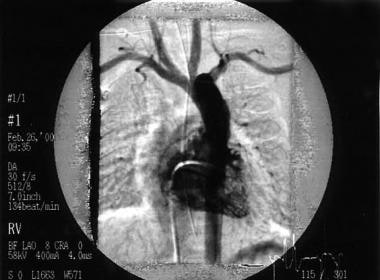 This right ventricular angiogram shows a patient with transposition of the great arteries. The aorta arises directly from the right-sided anterior right ventricle (10° left anterior oblique [LAO]).
The aorta lies anterior, and the pulmonary trunk lies posterior. This definition is neither comprehensive nor uniformly accepted. Similarly, the right and left atria can be identified solely on the basis of their anatomic characteristics. This article refers to these structures on this basis.
Definition
Ventricular inversion refers to a specific congenital heart defect in which the ventricles are exchanged in position so that the left atrium enters the right ventricle and the right atrium enters the left ventricle.
In regard to the person's body and in the absence of other abnormalities affecting chamber position, the left ventricle lies primarily to the right of the right ventricle. The term ventricular inversion includes understanding that the aorta arises from the right ventricular outflow tract in a position anterior to the pulmonary trunk that arises from the left ventricle (ie, the commonly accepted elementary definition of transposition of the great arteries).
As described below, a natural consequence of ventricular inversion results in the aorta arising from the right ventricular outflow tract anterior to the pulmonary trunk that arises from the left ventricle; in other words, the great arteries are transposed. Just as the term normal heart includes the pulmonary trunk anterior from the right ventricular outflow tract and the aorta posterior from the left ventricle, the term ventricular inversion includes an aorta anterior from the right ventricular outflow tract and a pulmonary trunk posterior from the left ventricle. Transposition of the great arteries is inherent in ventricular inversion and does not represent an additional abnormality.
Embryology
At the beginning of 4 weeks' gestation, the embryonic heart includes the primary heart tube that eventually forms the ventricles, their outflow tracts, and the proximal great arteries. This initially straight structure, fixed at its ends, grows more rapidly than the pericardial cavity in which it lies and, as a result, it must bend. This results in a C-shaped loop with the convexity directed anteriorly and rightward. It places the left side of systemic venous flow entering the heart tube in relation to the initial portion of the heart tube, the component that forms the left ventricle.
With division of the AV canal, the right side of the entering systemic venous flow (ie, right atrium) aligns with the second portion of the heart tube (ie, the portion that forms the right ventricle). Normal septation of the distal portion of the heart tube aligns the aorta with the left ventricle and the pulmonary trunk with the right ventricle. Critical to this process is bending of the heart tube to the right, so-called d-looping. This is not a random process. The precise definition of what controls d-looping remains obscure but is probably genetically determined.
Ventricular inversion results from bending of the heart tube to the left, so-called L-looping. If cardiac development otherwise proceeds in usual fashion, the right side of systemic venous inflow (ie, right atrium) aligns with the initial portion of the heart tube (ie, left ventricle). With division of the AV canal, the left atrium aligns with the second portion of the heart tube (ie, right ventricle).
The process of division of the distal portion of the primary heart tube (ie, truncus arteriosus) into the aorta and pulmonary trunk remains somewhat controversial, particularly in regard to the development of isolated, simple transposition of the great arteries. However, all agree that d-looping with subsequent normal cardiac development results in the pulmonary trunk arising from the right ventricle anteriorly to the aorta that arises from the left ventricle. Simple transposition of the great arteries in d-looping results in the aorta arising from the right ventricle anteriorly to the pulmonic trunk that arises from the left ventricle.
In L-looping, in the ordinary course of events, the aorta arises anteriorly from the right ventricle, and the pulmonary trunk arises posteriorly from the left ventricle. Although this condition satisfies the definition of transposition mentioned above and though it results in discordance of the normal ventricular-arterial relationship, septation of the truncus arteriosus in this manner appears to be normal in L-looping. In other words, otherwise normal development of the heart after L-looping includes transposition of the great arteries; therefore, it should not be considered an additional abnormality.
Anatomy
The anatomy of the atria in ventricular inversion remains normal. The right atrium enters the left ventricle through a mitral valve that is anatomically normal. Left ventricular internal anatomy is normal, and the mitral valve is in fibrous continuity with its outlet valve, the pulmonary valve. The pulmonary trunk is central and posterior in position. The left atrium enters the right ventricle through a tricuspid valve that almost always demonstrates at least minor anatomic abnormalities. The outflow area of the right ventricle (ie, infundibulum) leads to the aortic valve located at the left upper heart border. The anterior ascending aorta rises more or less straight toward the midline and then passes to the left of the trachea, branching normally.
Because of the abnormal relationship of the atria to the ventricles, the position of the AV node is abnormal (ie, above and to the left of its normal position). An accessory AV node may also be present. The bundle of His and the right and left bundle branches are inverted, a condition that substantially lengthens the main bundle.
The coronary arteries usually have normal morphology but are distributed and named in accordance with the ventricles. Therefore, the left coronary artery arises from right posterior sinus and the right coronary arises from the left posterior sinus.
Pathophysiology
In the absence of an additional heart defect the circulation in ventricular inversion is normal. Systemic venous blood passes from the right atrium to the left ventricle and then to the pulmonary arteries. Pulmonary venous blood enters the left atrium, passes into the right ventricle, and then enters the aorta. This physiology accounts for the terms congenitally corrected transposition and physiologically corrected transposition. However, these names emphasize transposition rather than ventricular inversion as the primary abnormality. Moreover, this distraction from the primary embryologic abnormality of looping makes understanding this defect more complex and more difficult than it otherwise is.
The heart with ventricular inversion may have no other clinically significant abnormality. Therefore, the individual's cardiac function remains normal. However, this situation is the exception, occurring in less than 1% of all persons with ventricular inversion. The vast majority of hearts with ventricular inversion have associated defects. Any cardiac defect that can occur in a normal d-looped heart can occur in the heart with ventricular inversion.
Some defects are more common than others. A ventricular septal defect (VSD) occurs in 80% of cases, and pulmonic stenosis, usually subvalvar, occurs in 50%. Anatomic abnormality of the tricuspid valve occurs in almost all cases although functional abnormality, usually tricuspid regurgitation, occurs in approximately one third. Some type of AV conduction abnormality is observed in one third of persons with ventricular inversion. Positional abnormalities of the ventricular mass, mesoversion, and dextroversion are encountered fairly often; in these cases, ensure that the patient does not have a heterotaxy syndrome such as asplenia or polysplenia because these conditions usually prevent accurate definition of left and right cardiac anatomy. [1]
The most rare and most interesting associated cardiac defect observed in ventricular inversion is that which causes the pulmonary artery to arise anteriorly from the right ventricle and the aorta posteriorly from the left ventricle. In other words, the great arteries are reversed from their expected position for the heart with ventricular inversion.
This condition causes systemic venous blood to travel from right atrium to the left ventricle and then to the aorta (ie, back to the systemic circulation); pulmonary venous blood enters the pulmonary artery returning to the lungs. Therefore, this situation is physiologically identical to simple transposition of the great arteries in D-looping. Some use the terms isolated ventricular inversion or ventricular inversion without transposition to describe these cases in which the pulmonary artery is anterior and the aorta posterior even though the physiology is that of simple transposition. One can make the argument that these hearts demonstrate two independent developmental abnormalities: ventricular inversion and transposition of the great arteries. In this case, the usual definition of transposition (ie, aorta anterior, pulmonary trunk posterior) does not apply.
If transposition is defined as pulmonary artery posterior and aorta anterior, an admittedly useful definition, the heart with ventricular inversion, anterior pulmonary artery, and posterior aorta cannot be labeled as demonstrating transposition, though the aorta and pulmonary artery are reversed in relation to their expected positions. Some authorities call this heart atrial-ventricular discordance with ventricular-arterial concordance, but this term tends to obscure the combination of defects that appears to be present. Given the controversies in the terminology, it is prudent and important to concentrate on understanding this interesting defect and not to dwell on the issue of naming it.
Natural history
The natural history of ventricular inversion depends on the associated heart defect, if any. Many defects are stable. On the contrary, regurgitation of the tricuspid (left-sided AV valve) tends to progress and may cause right ventricular dysfunction more rapidly than a similar degree of mitral regurgitation in a noninverted heart causes left ventricular dysfunction. Data from a substantial multicenter study suggest that individuals with ventricular inversion have a long-term risk of developing both right ventricular myocardial dysfunction and congestive heart failure that is not directly related to the degree of tricuspid valve regurgitation. Furthermore, other reports indicate that right ventricular coronary blood-flow reserve is decreased in the absence of ischemic symptoms.
Etiology
The cause of ventricular inversion is not known. In a single case-control study, 36 patients with ventricular inversion were compared with 3495 population-based live-born infant control subjects. [2] An increased incidence of this defect was associated with parental exposure to toxic chemicals from the air and from hazardous-waste sites.
Note the following:
-
The looping that occurs in the development of the embryonic heart tube is not a random event and must be under the same control as other developmental processes. The cause for l-looping remains unknown. The fact that other congenital heart defects occur in the vast majority of hearts that exhibit L-looping implies that the process predisposes individuals to additional abnormalities.
-
No familial factors have been determined.
-
No teratogenic factors have been identified with certainty. Data from a single study suggest that parental exposure to toxic chemicals increases the risk of ventricular inversion. [2]
-
See Pathophysiology for associated defects.
Epidemiology
United States data
Ventricular inversion accounts for approximately 0.5% of all congenital heart defects. Ventricular inversion occurs with VSD in 80% of cases and occurs with pulmonic stenosis (usually subvalvar) in 50%. Almost all patients have an associated anatomic abnormality of the tricuspid valve. Functional abnormality, usually tricuspid regurgitation, is present in approximately one third. Ventricular inversion is associated with some type of AV conduction abnormality in one third of patients. Associated positional abnormalities of the ventricular mass, mesoversion, and dextroversion are encountered fairly often. More complex defects (eg, single ventricle of the double-inlet left ventricle [DILV] type, often with coarctation of the aorta) also occur.
International data
As in the United States, ventricular inversion accounts for approximately 0.5% of all congenital heart defects.
Race-, sex-, and age-related demographics
No significant racial influences have been identified.
The prevalence is higher in males than in females, with a male-to-female ratio of approximately 2:1.
Ventricular inversion is a congenital abnormality and therefore is present at birth. It may be diagnosed in persons of any age, including young infants. The defect cannot be acquired. It most commonly comes to attention because of associated heart defects, including complete heart block. Ventricular inversion without an associated defect may escape detection indefinitely.
Ventricular inversion may be discovered on an ECG recorded during investigation of an innocent heart murmur. In rare cases, ventricular inversion is recognized on a plain posterior-anterior chest radiograph. Fetal echocardiography can identify ventricular inversion.
Patient Education
Patient restrictions regarding exercise and other lifestyle issues depend on the associated cardiac defect.
Instruct parents and older patients regarding the symptoms of cardiac conduction abnormality and specifically the symptoms of sudden-onset of Adams-Stokes syndrome, which can cause complete heart block.
When children with ventricular inversion reach maturity, they must be educated regarding the importance of the defect and of any associated abnormalities.
-
This right ventricular angiogram shows a patient with transposition of the great arteries. The aorta arises directly from the right-sided anterior right ventricle (10° left anterior oblique [LAO]).


