You are in: eMedicine Specialties > Pediatrics: General Medicine > Allergy and Immunology Purine Nucleoside Phosphorylase DeficiencyArticle Last Updated: Jan 15, 2008AUTHOR AND EDITOR INFORMATIONAuthor: Alan P Knutsen, MD, Professor of Pediatrics, Allergy and Immunology, Director of Pediatric Clinical Immunology Laboratory, Department of Pathology, St Louis University Health Sciences Center Alan P Knutsen is a member of the following medical societies: American Academy of Allergy Asthma and Immunology and Clinical Immunology Society Editors: Ann O'Neill Shigeoka †, MD, Former Clinical Associate Professor, Department of Pediatrics, Division of Immunology-Rheumatology, University of Utah School of Medicine; Mary L Windle, PharmD, Adjunct Assistant Professor, University of Nebraska Medical Center College of Pharmacy, Pharmacy Editor, eMedicine.com, Inc; David J Valacer, MD, Consulting Staff, Hoffman La Roche Pharmaceuticals; David Pallares, MD, Clinical Assistant Professor, Department of Pediatrics, Division of Allergy and Immunology, University of Louisville; Harumi Jyonouchi, MD, Associate Professor, Department of Pediatrics, Division of Pulmonary Allergy/Immunology and Infectious Diseases, UMDNJ-New Jersey Medical School Author and Editor Disclosure Synonyms and related keywords: purine nucleoside phosphorylase deficiency, PNP deficiency, severe combined immunodeficiency, SCID, adenosine deaminase deficiency, ADA deficiency, purine metabolism, combined immunodeficiency, CID, common variable immunodeficiency, CVID, lymphopenia, autoimmune hemolytic anemia, immune thrombocytopenia, neutropenia, thyroiditis, lupus, failure to thrive, lymphoma, oral candidiasis, Pneumocystis carinii pneumonia, developmental delay, hypertonia, herpes, recurrent herpes zoster, idiopathic thrombocytopenic purpura INTRODUCTIONBackgroundTwo immunodeficiency disorders are associated with 2 enzyme deficiencies of the purine salvage pathway that result in severe combined immunodeficiency (SCID). One of the disorders is known as adenosine deaminase (ADA) deficiency, and the other is purine nucleoside phosphorylase (PNP) deficiency. ADA deficiency results in a T-, B-, and natural killer (NK)–cell form of SCID with marked lymphopenia. PNP deficiency causes decreased numbers of T cells and lymphopenia. Serum immunoglobulin (Ig) levels are normal to near-normal, but antibodies are deficient. ADA and PNP deficiency are autosomal recessive disorders. In both disorders, the enzyme deficiencies result in accumulation of toxic metabolites. In ADA deficiency, the toxic metabolites block T-, B-, and NK-cell development; whereas in PNP deficiency, the metabolites are toxic to T-cell development. PNP deficiency is also associated with neurologic symptoms, including mental retardation and muscle spasticity, in 67% of patients. In addition, PNP deficiency is associated with increased risk of autoimmune disorders, such as autoimmune hemolytic anemia, immune thrombocytopenia, neutropenia, thyroiditis, and lupus. PathophysiologyPNP is an enzyme in the purine salvage pathway that metabolizes inosine and guanosine to hypoxanthine. In the preceding step of the pathway, ADA metabolizes adenosine to inosine. ADA deficiency causes an SCID that accounts for approximately 20% of all SCID cases. In both metabolic disorders, the enzyme deficiencies cause the accumulation of metabolites that are toxic to T and B cells. In ADA deficiency, adenosine and adenine accumulate in the plasma. ATP accumulates in erythrocytes, and ADP, guanosine triphosphate (GTP), and ATP accumulate in lymphocytes. Deoxy-ATP (dATP) can reach toxic levels that inhibit ribonucleotide reductase, an enzyme essential for synthesis of DNA precursors. In PNP deficiency, similar changes occur in elevated deoxy-GTP (dGTP) levels. dATP and dGTP predominantly accumulates in lymphoid tissue. dGTP inhibits ribonucleotide reductase, which is needed for synthesis of deoxynucleotides. In both ADA and PNP deficiencies, thymocytes are thought to be selectively destroyed because of elevated levels of dATP and dGTP. In a further description of the mechanism of T-cell depletion in PNP deficiency in a murine model, Arpaia et al reported increased in vivo apoptosis of T cells and increased in vitro sensitivity to gamma irradiation.1 The immune deficiency in PNP deficiency may be the result of inhibited mitochondrial DNA repair due to the accumulation of dGTP in the mitochondria. The end result is increased sensitivity of T cells and thymocytes to spontaneous mitochondrial damage, leading to T-cell depletion due to apoptosis. With ADA deficiency, destruction of resting T and B cells is increased. In comparison, PNP deficiency results in selective destruction of T cells, with little effect on B cells. Numerous mutations of the ADA gene (on chromosome 20) and PNP genes (on band 14q13) have been identified. PNP is a trimer with molecular weight of 84-94 kDa. Most identified mutations are missense mutations, but deletion is also described. All reported patients with homozygous mutations of PNP have been symptomatic. Because only small amounts of ADA are necessary for competent immunity, some patients with ADA mutations may still have 8-42% ADA activity and no profound immunodeficiency. FrequencyUnited StatesPNP deficiency is rare. PNP deficiency accounts for approximately 4% of all cases of combined immunodeficiency (CID), and ADA deficiency accounts for approximately 20% of all cases of CID. InternationalThe prevalence of primary immunodeficiency ranges from approximately 1 case per 54,000 population in Switzerland to 1 case per 200,000 population in Japan. CID accounts for 11-13% of all primary immunodeficiency disorders. Mortality/MorbidityPatients with PNP deficiency are at risk for life-threatening recurrent viral, bacterial, fungal, mycobacterial, and protozoal infections. In addition, failure to thrive eventually ensues.
SexPNP deficiency is an autosomal recessive disorder with equal incidence in boys and girls. AgeAlthough symptoms typically appear in the first year of life in patients with PNP deficiency, gradual deterioration of the T-cell immune system may delay the onset of symptoms until the second year of life. CLINICALHistoryMost patients have a history of recurrent viral, bacterial, fungal, mycobacterial, and protozoal infections, similar to patients with severe T- and B-cell immunodeficiency. Oral candidiasis that is recalcitrant to therapy occurs in approximately 85% of patients with severe T-cell immunodeficiency. In addition, the presenting infections are often those caused by opportunistic microorganisms, such as Pneumocystis carinii pneumonia. Neurologic problems are commonly associated with purine nucleoside phosphorylase (PNP) and adenosine deaminase (ADA) deficiencies and have therapeutic implications. More than 50% of patients with PNP deficiency have neurologic impairments that may predate the onset of infections. Neurologic problems include developmental delay, hypertonia, spasticity, and tremors. Patients with ADA deficiency may also have neurologic problems, principally neurodevelopmental delays. Of importance, polyethylene glycol (PEG) ADA therapy does not correct the neurodevelopmental problems in ADA deficiency, whereas immune reconstitution does. Likewise, bone marrow transplantation does not correct neurological deficits in both PNP and ADA deficiencies.
PhysicalPhysical examination reveals a paucity of peripheral lymphoid tissue, such as lymph nodes, tonsillar tissue, and adenoids. The liver and spleen are usually normal in size but can be enlarged in patients with accompanying hemolytic anemia or lymphoma. In neonates, the thymic shadow is typically small on chest radiography. Neurologic symptoms, consisting of developmental delay, hypertonia, spasticity, and tremors, may be present. Patients may fail to thrive. CausesPNP deficiency is a genetic disorder caused by a deficiency of the enzyme PNP. The PNP gene has been localized to band 14q13. Missense mutations have been identified in some patients. The PNP protein is a trimer with a molecular weight of 84-94 kDa, with the highest levels in lymphoid tissue. The mechanism by which PNP deficiency causes neurologic disease is unknown. DIFFERENTIALSSevere Combined Immunodeficiency
| ||||||||||||||||||||||||||||||||||||||||||||||||||||||||||||||||||||||||||||||||||||||||||||||||||||||||||||||||||||||||||||||||||||||||||||||||||||||||||||||||||||||||||||||||||||||||||||||||||||||||||||||||||||||||||||||||
| Study | Infantile Onset | Late Onset | Adult Onset |
|---|---|---|---|
| Lymphopenia | Markedly decreased | Decreased | Decreased |
| CD3+ cells | Absent or trace | Markedly reduced | Markedly reduced |
| CD4/CD8 ratio | Too few to test | <1 | <1 |
| Phytohemagglutinin response | Absent | Reduced | Reduced |
| Antigen response | Absent | Trace | Trace |
| Mixed lymphocyte culture response | Reduced | ... | ... |
| Ig response | Absent | Low to absent | Normal (low IgG2) |
| IgE | Low | Elevated | Elevated |
| Antibody response | Absent | Absent to low | Low to polysaccharides antigens |
| Eosinophilia | Rare | Common | Common |
| Infections | Predominantly viral, fungal, opportunistic, bacterial | Bacterial sinopulmonary | Bacterial sinopulmonary, varicella-zoster, herpes simplex, candidal |
Genetic studies to examine mutations of genes that encode for ADA and PNP are readily available and should be performed. In PNP deficiency, Grunebaum et al (2004) identified “hot spots” at chromosomes 58 and 234 with increased frequency of mutations in the gene that encodes PNP.2
If thymic biopsy is performed (which is usually not necessary), the results demonstrate marked loss of corticomedullary differentiation; absence of Hassall corpuscles; and depletion of thymocytes, especially in the thymic cortex and medulla.
Histopathology of lymphoid tissue reveals abnormalities, predominantly in T-cell dependent areas. The thymus is markedly reduced in size, with depleted thymocytes. Hassall corpuscles are present but poorly formed. By comparison, Hassall corpuscles are usually absent in patients with SCID. In the lymph nodes and spleen, paracortical regions are reduced or absent. Germinal centers are reduced; however, plasma cells can be identified.
No surgical care is needed.
Consult a hematologist or an immunologist skilled in bone marrow transplantation.
No special diet is required.
Because patients with PNP are susceptible to viral, fungal, and bacterial infections, limit these patients' exposure to other persons.
Replacement therapy with intravenous immunoglobulin in patients with primary immune deficiencies
Regarding replacement therapy with intravenous Ig (IVIG) in patients with primary immune deficiencies, the overall consensus among clinical immunologists is that a dose of IVIG 400-600 mg/kg/mo or a dose that maintains trough serum IgG levels of more than 500 mg/dL is desirable. Patients with meningoencephalitis (X-linked agammaglobulinemia) require higher doses (eg, 1 g/kg) and perhaps intrathecal therapy.
Preinfusion, or trough, serum IgG levels are measured every 3 months until a steady state is achieved and then every 6 months if the patient is stable. These levels may be helpful in adjusting the dose of IVIG to achieve adequate serum levels. For persons with high catabolism of infused IgG, more frequent infusions (eg, every 2-3 wk) of smaller doses may maintain the serum level in the reference range. The rate of IgG elimination may be increased during active infection; therefore, measuring serum IgG levels and adjusting to higher doses or shorter intervals may be required.
Precautions and adverse reactions to intravenous immunoglobulin therapy
The US Food and Drug Administration (FDA) advises against exceeding the recommended doses and infusion rates and suggest the use of minimal practical concentrations in patients at risk for renal failure (eg, those with preexisting renal insufficiency, diabetes, volume depletion, sepsis, paraproteinemia; those older than 65 y; and those using nephrotoxic drugs).
Initial treatment should be administered under the close supervision of experienced personnel. The risk of adverse reactions with initial treatments is high, especially in patients with infections and in those who form immune complexes. In patients with active infection, infusion rates may need to be reduced and the dose halved (ie, 200-300 mg/kg), with the remaining dose given the next day to achieve a full dose. Treatment should not be discontinued. After normal serum IgG levels are achieved, adverse reactions are uncommon unless patients have active infections.
With the new generation of IVIG products, adverse effects are reduced. Adverse effects include tachycardia, chest tightness, back pain, arthralgia, myalgia, hypertension or hypotension, headache, pruritus, rash, and low-grade fever. More serious reactions are dyspnea, nausea, vomiting, circulatory collapse, and loss of consciousness. Patients with profound immunodeficiency or active infections have reactions more severe than those of other patients.
The adverse reactions are thought to be related to the anticomplementary activity of IgG aggregates in the IVIG and the formation of immune complexes. The formation of oligomeric or polymeric IgG complexes that interact with Fc receptors and that trigger the release of inflammatory mediators is another cause. Most adverse reactions are rate related. Slowing the infusion rate or discontinuing therapy until symptoms subside may diminish the reaction. Pretreatment with ibuprofen (5-10 mg/kg every 6-8 h), acetaminophen (15 mg/kg/dose), diphenhydramine (1 mg/kg/dose), and/or hydrocortisone (6 mg/kg/dose, maximum 100 mg) 1 hour before the infusion may prevent adverse reactions. In some patients with a history of severe adverse effects, doses of analgesic and antihistamine may be repeated.
Acute renal failure is a rare but significant complication of IVIG treatment. Reports suggest that IVIG products that contain sucrose as a stabilizer are associated with an increased risk for this renal complication. Acute tubular necrosis, vacuolar degeneration, and osmotic nephrosis suggest osmotic injury to the proximal renal tubules. The infusion rate for sucrose-containing IVIG should not exceed 3 mg/kg/min based on sucrose content. Risk factors for this adverse reaction are preexisting renal insufficiency, diabetes mellitus, dehydration, age older than 65 years, sepsis, paraproteinemia, and concomitant use of nephrotoxic agents. For patients at increased risk, BUN and creatinine levels should be monitored before the start of treatment and before each infusion. If renal function deteriorates, the product should be discontinued.
IgE antibodies to immunoglobulin A (IgA) have been reported to cause severe transfusion reactions in patients with IgA deficiency. The literature has a few reports of true anaphylaxis in patients with selective IgA deficiency and CVID who developed IgE antibodies to IgA after treatment with Ig. In actual experience, however, this is rare. In addition, this is not a problem for patients with X-linked agammaglobulinemia (Bruton disease) or SCID. Caution should be exercised in patients with IgA deficiency (<7 mg/dL) who need IVIG because of IgG-subclass deficiencies. IVIG preparations with low concentrations of contaminating IgA are advised (see Table 2).
Comparison of intravenous immunoglobulin products
For replacement therapy in patients with primary immune deficiency, all brands of IVIG are probably equivalent, although their viral inactivation processes (eg, solvent detergent vs pasteurization and liquid vs lyophilized) may differ (Table 2). The choice may depend on the formulary, local availability, and/or cost. The dose, manufacturer, and lot number should be recorded for each infusion to review for adverse events or other consequences. Recording all adverse effects that occur during the infusion is crucial. Periodic monitoring of liver and renal function about 3-4 times yearly, is also recommended.
Table 2. Intravenous Immunoglobulin
| Brand (Manufacturer) | Manufacturing Process | pH | Additives* | Parenteral Form and Final Concentration | IgA Content (mcg/mL) |
|---|---|---|---|---|---|
| Carimune NF (ZLB Behring) | Kistler-Nitschmann fractionation; pH 4.0, nanofiltration | 6.4-6.8 | 6% solution: 10% sucrose <20 mg NaCl/g protein | Lyophilized powder 3%, 6%, 9%, 12% | Trace |
| Flebogamma (Grifols USA) | Cohn-Oncley fractionation, PEG precipitation, ion-exchange chromatography, pasteurization | 5.1-6.0 | Sucrose-free, contains 5% D-sorbitol | Liquid 5% | <50 |
| Gammar-P IV (ZLB Behring) | Cohn-Oncley fraction II/III; ultrafiltration; pasteurization | 6.4-7.2 | 5% solution: 5% sucrose, 3% albumin, 0.5% NaCl | Lyophilized powder 5% | <20 |
| Gamunex (Talecris Biotherapeutics) | Cohn-Oncley fractionation, caprylate-chromatography purification, cloth and depth filtration, low pH incubation | 4.0-4.5 | Contains no sugar, contains glycine | Liquid 10% | 46 |
| Iveegam EN (Baxter Bioscience) | Cohn-Oncley fraction II/III; ultrafiltration; pasteurization | 6.4-7.2 | 5% solution: 5% glucose, 0.3% NaCl | Lyophilized powder 5% | <10 |
| Gammagard S/D, Polygam S/D (Baxter Bioscience for the American Red Cross) | Cohn-Oncley cold ethanol fractionation, cation and anion exchange chromatography, solvent detergent treated, nanofiltration, low pH incubation | 6.4-7.2 | 5% solution: 0.3% albumin, 2.25% glycine, 2% glucose | Lyophylized powder 5%, 10% | <1.6 (5% solution) |
| Gammagard Liquid 10% (Baxter Bioscience) | Cohn-Oncley cold ethanol fractionation, cation and anion exchange chromatography, solvent detergent treated, nanofiltration, low pH incubation | 4.6-5.1 | 0.25M glycine | Ready-for-use Liquid 10% | 37 |
| Octagam (Octapharma USA) | Cohn-Oncley fraction II/III; ultrafiltration; low pH incubation; S/D treatment pasteurization | 5.1-6.0 | 10% maltose | Liquid 5% | 200 |
| Panglobulin (Swiss Red Cross for the American Red Cross) | Kistler-Nitschmann fractionation; pH 4.0, trace pepsin, nanofiltration | 6.6 | Per gram of IgG: 1.67 g sucrose, <20 mg NaCl | Lyophilized powder 3%, 6%, 9%, 12% | 720 |
Prophylactic treatment for P carinii pneumonia is TMP-SMZ. Administer IVIG therapy to provide functional antibodies.
| Drug Name | SMZ-TMP (Bactrim, Septra, Cotrim) |
|---|---|
| Description | Inhibits bacterial growth by inhibiting synthesis of dihydrofolic acid; for prophylaxis of P carinii pneumonia. |
| Adult Dose | 160 mg TMP/800 mg SMZ PO qd |
| Pediatric Dose | TMP 150 mg/m2/d and SMZ 750 mg/m2/d PO bid on 3 consecutive d/wk; total daily dose not to exceed TMP 320 mg and SMZ 1600 mg |
| Contraindications | Documented hypersensitivity; megaloblastic anemia due to folate deficiency |
| Interactions | PT may increase with warfarin (perform coagulation tests and adjust dose accordingly); coadministered dapsone may increase blood levels of both drugs; coadministered diuretics increases incidence of thrombocytopenic purpura in elderly patients; phenytoin levels may increase with coadministration; may potentiate effects of methotrexate in bone marrow depression; hypoglycemic response to sulfonylureas may increase with coadministration; may increase levels of zidovudine |
| Pregnancy | C - Fetal risk revealed in studies in animals but not established or not studied in humans; may use if benefits outweigh risk to fetus |
| Precautions | Discontinue at first sign of rash or adverse reaction; frequently obtain CBC counts; discontinue if significant hematologic changes occur; goiter, diuresis, and hypoglycemia may occur with sulfonamides; prolonged infusion or high doses may cause bone marrow depression (if signs occur, give 5-15 mg/d leucovorin); caution in folate deficiency (eg, persons with chronic alcoholism, elderly patients, those receiving anticonvulsant therapy, persons with malabsorption syndrome); hemolysis may occur in G-6-PD deficiency; patients with AIDS may not tolerate or respond; caution in renal or hepatic impairment (perform urinalyses and renal function tests during therapy); give fluids to prevent crystalluria and stone formation |
| Drug Name | Fluconazole (Diflucan) |
|---|---|
| Description | Synthetic PO antifungal (broad-spectrum bistriazole) that selectively inhibits fungal cytochrome P-450 and sterol C-14 alpha-demethylation, which prevents conversion of lanosterol to ergosterol, thereby disrupting cellular membranes. Has little affinity for mammalian cytochromes, which is believed to explain its low toxicity. Available as tablets for PO administration, as a powder for PO suspension, and as a sterile solution for IV use. Indicated for fungal prophylaxis during immunosuppression |
| Adult Dose | 200 mg PO/IV qd |
| Pediatric Dose | 6 mg/kg PO/IV qd |
| Contraindications | Documented hypersensitivity |
| Interactions | CYP450 2C19 and 3A4 inhibitor; levels may increase with hydrochlorothiazide; fluconazole levels may decrease with chronic coadministration of rifampin; may increase concentrations of theophylline, phenytoin, tolbutamide, cyclosporine, glyburide, and glipizide; effects of anticoagulants may increase with fluconazole coadministration |
| Pregnancy | C - Fetal risk revealed in studies in animals but not established or not studied in humans; may use if benefits outweigh risk to fetus |
| Precautions | Adjust dose for renal insufficiency; closely monitor if rashes develop, and discontinue drug if lesions progress; may cause clinical hepatitis, cholestasis, and fulminant hepatic failure (including death) when taken with underlying medical conditions (eg, AIDS, malignancy) or while taking multiple concomitant medications; not recommended for breastfeeding mothers Convenience and efficacy of single dose regimen for treatment of vaginal yeast infections should be weighed against difficulties resulting from higher incidence of adverse reactions reported with PO fluconazole versus intravaginal agents |
The optimal combination of an ablative agent (ie, busulfan) with immunosuppressive agents (eg, antithymocyte globulin, cyclophosphamide) has not been systematically studied and should be the focus of future clinical trials. The risks of this preparative regimen are sterility, liver, heart and lung toxicity, and malignancy.
| Drug Name | Busulfan (Myleran) |
|---|---|
| Description | Potent cytotoxic drug; causes profound myelosuppression at recommended dose. As alkylating agent, mechanism of action of active metabolites may involve cross-linking of DNA, which may interfere with growth of normal and neoplastic cells. |
| Pediatric Dose | <2 years: 1.25 mg/kg PO q6h on days -9 through -6 before transplantation (day 0) >2 years: 1 mg/kg PO q6h on days -9 through -6 before transplantation Dose of busulfan adjusted on basis of first-dose kinetics (steady-state level of 400-600 ng/mL) |
| Contraindications | Documented hypersensitivity |
| Interactions | CYP3A3/4 enzyme substrate; acetaminophen, cyclophosphamide, itraconazole, and thioguanine may increase toxicity; phenytoin may decrease levels |
| Pregnancy | D - Fetal risk shown in humans; use only if benefits outweigh risk to fetus |
| Precautions | Regularly examine hematologic profile (particularly neutrophils and platelets) to monitor for hematopoietic suppression; may cause pulmonary fibrosis; if WBC count is high, use hydration and allopurinol to prevent hyperuricemia |
| Drug Name | Cyclophosphamide (Cytoxan, Neosar) |
|---|---|
| Description | Chemically related to nitrogen mustards. As alkylating agent, mechanism of action of active metabolites may involve cross-linking of DNA, which may interfere with growth of normal and neoplastic cells. |
| Pediatric Dose | 50 mg/kg IV on days -5 through -2 before transplantation (day 0) |
| Contraindications | Documented hypersensitivity |
| Interactions | Allopurinol may increase risk of bleeding or infection and enhance myelosuppressive effects; may potentiate doxorubicin-induced cardiotoxicity; may reduce digoxin serum levels and antimicrobial effects of quinolones; chloramphenicol may increase half-life and decrease metabolite concentrations; may increase effect of anticoagulants; coadministration with high doses of phenobarbital may increase rate of metabolism and leukopenic activity; thiazide diuretics may prolong cyclophosphamide-induced leukopenia and neuromuscular blockade by inhibiting cholinesterase activity |
| Pregnancy | D - Fetal risk shown in humans; use only if benefits outweigh risk to fetus |
| Precautions | Regularly examine hematologic profile (particularly neutrophils and platelets) to monitor for hematopoietic suppression; regularly examine urine for RBCs, (may precede hemorrhagic cystitis) |
| Drug Name | Lymphocyte immune globulin (Atgam) |
|---|---|
| Description | May modify T-cell function and might eliminate antigen-reactive T-lymphocytes in peripheral blood. |
| Pediatric Dose | 30 mg/kg IV on days -3 through -1 before transplantation (day 0) |
| Contraindications | Documented hypersensitivity |
| Interactions | None reported |
| Pregnancy | C - Fetal risk revealed in studies in animals but not established or not studied in humans; may use if benefits outweigh risk to fetus |
| Precautions | To reduce risk of phlebitis, administer only IV; emergency resources should be immediately available to manage rash, dyspnea, hypotension, or anaphylaxis |
Cyclosporine and corticosteroids are administered to prevent acute GVHD.
| Drug Name | Cyclosporine (Sandimmune, Neoral) |
|---|---|
| Description | Cyclic polypeptide; suppresses some humoral immunity and moreso cell-mediated immune reactions (eg, delayed hypersensitivity, allograft rejection, experimental allergic encephalomyelitis, GVHD) in many organs. Base dose on ideal body weight. |
| Pediatric Dose | 5-6 mg/kg/d IV infusion beginning on day -2 before transplantation (day 0); adjust dose to maintain whole blood level of 250-350 ng/mL; continue until days 100-365 after transplantation |
| Contraindications | Documented hypersensitivity; uncontrolled hypertension or malignancies; concomitant PUVA or UVB radiation for psoriasis (may increase risk of cancer) |
| Interactions | Substrate of CYP3A4; CYP3A4 inducers (eg, carbamazepine, phenytoin, isoniazid, rifampin, phenobarbital) may decrease concentrations; CYP3A4 inhibitors (eg, azithromycin, itraconazole, nicardipine, ketoconazole, fluconazole, erythromycin, verapamil, grapefruit juice, diltiazem, aminoglycosides, acyclovir, amphotericin B, clarithromycin) may increase toxicity; concurrent lovastatin may increase risk of acute renal failure, rhabdomyolysis, myositis, and myalgia |
| Pregnancy | C - Fetal risk revealed in studies in animals but not established or not studied in humans; may use if benefits outweigh risk to fetus |
| Precautions | Evaluate renal and liver functions often by measuring BUN, serum creatinine, serum bilirubin, and liver enzyme levels; may increase risk of infection and lymphoma |
| Drug Name | Methylprednisolone (Solu-Medrol) |
|---|---|
| Description | Decreases inflammation by suppressing migration of polymorphonuclear leukocytes and reversing increased capillary permeability. |
| Pediatric Dose | Posttransplantation days 5-7: 10 mg/kg/d IV Posttransplantation days 8-10: 5 mg/kg/d IV Posttransplantation days 11-13: 3 mg/kg/d IV; then 10% weekly reduction taper; typically discontinued by day 60 |
| Contraindications | Documented hypersensitivity; viral, fungal, or tubercular skin infections |
| Interactions | Coadministered digoxin may increase digitalis toxicity due to hypokalemia; estrogens may increase levels; phenobarbital, phenytoin, and rifampin may decrease levels (adjust dose); monitor for hypokalemia in patients taking concurrent diuretics |
| Pregnancy | C - Fetal risk revealed in studies in animals but not established or not studied in humans; may use if benefits outweigh risk to fetus |
| Precautions | Hyperglycemia, edema, osteonecrosis, peptic ulcer disease, hypokalemia, osteoporosis, euphoria, psychosis, growth suppression, myopathy, and infections |
| Media file 1: 
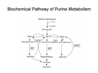
Biochemical pathway of purine metabolism. AMP = adenosine monophosphate, APRT = adenine phosphoribosyltransferase, GMP = guanosine monophosphate, HGPRT = hypoxanthine-guanine phosphoribosyltransferase, IMP = inosine monophosphate, NP = nucleoside phosphorylase, PPriboseP = 5-phosphorylribose-1-pyrophosphate. | |
View Full Size Image | Media type: Image |
Purine Nucleoside Phosphorylase Deficiency excerpt
Article Last Updated: Jan 15, 2008