| Patient Education |
|
Click here for patient education.
|
|
You are in: eMedicine Specialties >
Pediatrics: General Medicine > Nephrology
Potter Syndrome
Article Last Updated: Mar 27, 2006
AUTHOR AND EDITOR INFORMATION
Section 1 of 10
Author: Vellore K Sairam, MBBS, Assistant Professor, Department of Nephrology, Sri Ramachandra Medical College and Research Institute, India
Coauthor(s):
Luther Travis, MD, William W Glauser Professor of Pediatrics and Pediatric Nephrology, Department of Pediatrics, Divisions of Nephrology and Diabetes, University of Texas Medical Branch and Children's Hospital
Editors: Laurence Finberg, MD, Clinical Professor, Department of Pediatrics, University of California at San Francisco and Stanford University; Mary L Windle, PharmD, Adjunct Assistant Professor, University of Nebraska Medical Center College of Pharmacy, Pharmacy Editor, eMedicine.com, Inc; Frederick J Kaskel, MD, PhD, Director of the Division and Training Program in Pediatric Nephrology, Vice Chair, Department of Pediatrics, Montefiore Medical Center and Albert Einstein School of Medicine; Howard Trachtman, MD, Program Director, Pediatrics Research, Schneider Children's Hospital, Department of Pediatrics, Division of Nephrology, Professor, Albert Einstein College of Medicine; Craig B Langman, MD, The Isaac A Abt, MD, Professor of Kidney Diseases, Feinberg School of Medicine, Northwestern University; Division Head of Kidney Diseases, Children's Memorial Hospital, Chicago
Author and Editor Disclosure
Synonyms and related keywords:
Potter syndrome, Potter's syndrome, Potter disease, Potter's disease, Potter facies, Potter's facies, Potter sequence, oligohydramnios secondary to renal disease
Background
Potter syndrome, which causes a typical physical appearance, is the result of oligohydramnios secondary to renal diseases such as bilateral renal agenesis. Other causes of Potter syndrome include obstructive uropathy, autosomal recessive polycystic kidney disease, and renal hypoplasia. The causes of Potter syndrome, other than renal agenesis, result in less severe forms of deformation that are usually referred to as the Potter sequence.
Pathophysiology
The fetus continuously swallows amniotic fluid, which is reabsorbed by the gastrointestinal tract and then reintroduced into the amniotic cavity by the kidneys. Oligohydramnios occurs if the volume of amniotic fluid is less than normal for the corresponding period of gestation. This may be due to decreased urine production secondary to bilateral renal agenesis, obstruction of the urinary tract, or, occasionally, prolonged rupture of membranes. The resulting oligohydramnios is the cause of the deformities observed in Potter syndrome.
Genetics
During nephrogenesis, genes, transcription factors, and growth factors control the essential interaction between the ureteric bud and the metanephric mesenchyme. For example, the L1M1 and PAX2 transcription factors are essential for the formation of the mesonephric duct, from which the ureteric bud develops. L1M1-deficient mice have complete renal agenesis. If the PAX2 gene is deficient, deletion of the caudal portion of the mesonephric duct and, therefore, renal agenesis, results. WT-1, a zinc-finger transcription factor expressed in the metanephric mesenchyme, is essential for ureteric bud outgrowth.
Homozygous null-mutants for WT-1 have complete renal agenesis. Similarly, transcription factor Eya-1 from the metanephric mesenchyme is required for ureteric bud outgrowth, and deficiency of this protein is shown to cause branchiootorenal syndrome. The glial cell line–derived neurotrophic factor (GDNF) from the metanephric mesenchyme binds to the C-ret receptor on the branching ureteric bud and is responsible for the branching and elongation of the ureteric bud. Inactivation of either GDNF or the C-ret receptor leads to renal agenesis. Heterozygotes may have unilateral renal abnormality while the other kidney has normal development. Limb deformity (ld) gene codes for 4 different spliced formin genes, which are expressed in the mesonephric duct and branching ureteric ducts. Mutation of the ld gene leads to limb deformity with renal agenesis. Mutation of the formin IV gene leads only to kidney abnormality.
Homozygous mutation of the alpha-8 integrin subunit produces abnormalities with phenotypes similar to ld mutation with deformities such as renal aplasia, dysplasia, or hypoplasia. Transcription factors such as EMX-2, BF-2, fibroblast growth factor 7 (FGF 7), epithelial growth factor receptor (EGF-R), GDNF, retinoic acid receptor alpha, and beta 2 are involved in the branching of the ureteric bud. A heterozygous mutation defect of the growth factor bone morphogenetic protein 4 (bmp 4) leads to genitourinary abnormalities, such as hypoplasia or dysplasia, ureterovesicular junction obstruction, hydronephrosis, or the bifid/duplex kidney. This is a defect of ureteric branching and not induction of the ureteric bud; thus, renal aplasia does not occur.
Various human abnormalities are associated with renal agenesis or dysplastic kidney abnormalities, such as renal coloboma syndrome, Kallmann syndrome, and branchiootorenal syndrome.
Frequency
United States
Bilateral renal agenesis occurs in 1 per 3000 births and is responsible for 20% of Potter syndrome cases. The frequency of other causes of Potter sequence is not known.
Mortality/Morbidity
Potter syndrome is usually fatal in the first few days of the patient's life; most often, the cause is pulmonary failure. Neonates with the Potter sequence have an increased morbidity rate because of respiratory failure, pneumothorax, and acute renal failure during the neonatal period. During early childhood, patients may have chronic lung disease or chronic renal failure.
Race
No racial predilection is known.
Sex
Males have an increased incidence of the Potter sequence because they have a higher rate of Eagle-Barrett (prune belly) syndrome and obstructive uropathy secondary to posterior urethral valves.
Age
Patients present as neonates.
History
Features of the history may include the following:
- Antepartum period (Findings of either of the historical factors below requires close follow-up of the neonate during the prenatal and neonatal periods).
- History of oligohydramnios
- History of prenatal sonograms depicting renal agenesis or evidence of hydronephrosis (obstructive uropathy) or other renal disorders
- Neonatal period
- Absence or paucity of urine output during the neonate's initial 48 hours life
- Respiratory distress
- Lack of proper force in the urinary stream in neonates with posterior urethral valves
Physical
Findings at physical examination may include the following:
- Potter facies: Affected infants have a flattened nose, recessed chin, prominent epicanthal folds, and low-set abnormal ears.
- Pulmonary hypoplasia: The degree of pulmonary hypoplasia depends on the degree and duration of oligohydramnios, as well as the stage of lung development at which oligohydramnios occurs.
- Features of Eagle-Barrett (prune belly) syndrome: This is an occasional cause of the Potter sequence. Neonates have a deficient abdominal wall, undescended testes, dilated ureters, and a renal pelvis.
- Skeletal malformations: Hemivertebrae, sacral agenesis, and limb anomalies may be present.
- Ophthalmologic malformations: Cataract, angiomatous malformation in the optic disc area, prolapse of the lens, and expulsive hemorrhage may be present.
- Cardiovascular malformations: Ventricular septal defect, endocardial cushion defect, tetralogy of Fallot, and patent ductus arteriosus may be present.
Causes
Causes of Potter syndrome may include the following:
- Bilateral renal agenesis
- Infantile polycystic kidney disease
- Cystic kidney disease
- Posterior urethral valves
- Early rupture of membranes
Multicystic Renal Dysplasia
Patau Syndrome
Polycystic Kidney Disease
Posterior Urethral Valves
Prader-Willi Syndrome
Other Problems to be Considered
Bilateral renal agenesis Trisomy 7
Lab Studies
- Obtain serum electrolyte tests to evaluate electrolyte problems such as hyponatremia, hypernatremia, hyperkalemia, hypocalcemia, hyperphosphatemia, and/or metabolic acidosis, which may be present in neonates with renal failure.
- Serum creatinine levels are used to assess renal function and the glomerular filtration rate (GFR). The GFR can be calculated by using various formulas, such as that reported by Schwartz and colleagues, as follows:
- In low birth weight (LBW) neonates, the formula is (0.33 X height in cm)/serum creatinine level.
- In term infants, the formula is (0.45 X height in cm)/serum creatinine level.
- The serum blood urea nitrogen result is not a good indicator of renal function.
- Obtain a CBC count with differential to evaluate for anemia secondary to erythropoietin deficiency.
- Urinalysis is used to reveal either microhematuria or proteinuria.
- If sepsis is suspected, obtain cultures of the urine, blood, and cerebrospinal fluid.
- Chromosomal analysis is obtained if the physical examination findings suggest the presence of an associated genetic disorder, such as trisomy 7 or trisomy 13 (Patau syndrome).
- Other tests, such as evaluations of the urine sodium level, urine creatinine level, urine osmolality, and serum osmolality, are indicated if the neonate has renal failure.
Imaging Studies
- Prenatal imaging studies
- Both abdominal and transvaginal ultrasonography are effectively used in pregnant mothers with oligohydramnios. The absence of the bladder and kidneys in the fetus implies bilateral renal agenesis.
- Prenatal ultrasonographic findings may suggest the presence of other conditions, such as multicystic dysplastic kidney, polycystic renal disease, and Prader-Willi syndrome.
- Doppler ultrasonography can be helpful in depicting fetal pulmonary hypoplasia by revealing poor angiogenesis in the lung and enabling the measurement of the blood-flow velocity waveform of the pulmonary artery.
- Neonatal imaging studies
- Abdominal ultrasonography is used to confirm the renal disease detected in the prenatal period.
- Sonograms also provide useful information related to the bladder and ureters, and they are useful in depicting obstructive uropathy.
- Chest radiography is used to reveal spontaneous pneumothorax and pulmonary hypoplasia, which has a known association with the Potter sequence.
- Other examinations that may be indicated include voiding cystourethrography and renal scanning.
Other Tests
- In neonates who die from this condition, an autopsy is recommended.
Procedures
- Chest tube placement may be required in neonates with spontaneous pneumothorax.
- A peritoneal dialysis line or a central venous line may be placed in children who have renal failure and require dialysis.
Medical Care
The renal function and respiratory status of neonates born with Potter syndrome must be assessed. Associated anomalies of the gastrointestinal, cardiovascular, and musculoskeletal systems should also be evaluated. Once the long-term prognosis of survival is determined, resuscitation and management plans should be addressed.
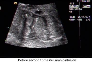
- In neonates with bilateral renal agenesis, severe neonatal respiratory distress due to associated pulmonary hypoplasia, and spontaneous pneumothorax, further treatment may not be indicated. The decision should be made after discussion with the parents and all consultants involved.
- Children with Potter sequence due to conditions such as infantile polycystic kidney disease, multicystic dysplastic kidney, hypoplastic kidney, Prader-Willi syndrome, and rupture of membranes during gestation have a higher survival rate than children with Potter syndrome due to other conditions.
- Children who survive the disease require management of the following:
- Pulmonary hypoplasia: Mechanical ventilation and chest tube placement may be indicated for ventilatory support and for the treatment of spontaneous pneumothorax.
- Renal function: This is assessed with imaging studies and calculation of the GFR by using the serum creatinine concentration.
- Management of renal failure may be required.
- Nutrition: Adequate nutrition is required. Nasogastric feeding may be indicated in infants.
- Growth: The use of growth hormone is indicated in children with a low GFR who do not grow at a healthy rate.
- Electrolyte abnormalities such as hypocalcemia and hyperphosphatemia can be treated with medications, including calcium carbonate and vitamin D.
- Anemia is treated with oral or parenteral iron and erythropoietin.
- Children may have hypertension from either fluid-related causes or activation of the renin-angiotensin system. Antihypertensives that may be given include diuretics, beta-blockers, calcium-channel blockers, and angiotensin-converting enzyme inhibitors.
Surgical Care
A peritoneal dialysis catheter or a central venous line may be placed for dialysis, if indicated.
Consultations
A neonatologist, pediatric nephrologist, pediatric pulmonologist, geneticist, and pediatric surgeon should be consulted as needed.
Diet
Appropriate restriction of fluid during renal failure is indicated. Nutrition with adequate protein and caloric intake is indicated. Children with hypertension must avoid excessive salt intake.
Activity
No restriction of physical activity is needed for children who survive the neonatal period.
Medications are typically indicated when acute or chronic renal failure occurs. Growth hormone may be required because of inadequate growth in children with renal failure. Treatment regimens for hypertension are designed to reduce blood pressure and other risk factors of coronary heart disease. Erythropoietin is essential for red blood cell production and may be required because of decreased erythropoietin levels in renal failure. Vitamin D analogs are essential to provide homeostasis for calcium regulation.
Diuretic agents help relieve fluid overload associated with renal failure. Additional pharmacotherapy for hypertension associated with renal failure should be individualized based on the patient's age, race, known pathophysiologic variables, and concurrent conditions. Treatment goals are not only to lower blood pressure safely and effectively but also to prevent or reverse hyperlipidemia, glucose intolerance, and left ventricular hypertrophy. For complete information, see the pediatric topics Hypertension and Neonatal Hypertension.
Drug Category: Pituitary hormones
The anterior lobe of the pituitary gland is responsible for the secretion of adrenocorticotrophic hormone (corticotropin); gonadotrophic hormones (gonadotropins), including follicle-stimulating hormone and luteinizing hormone; growth hormone (somatropin); lactogenic hormone (prolactin); and thyroid-stimulating hormone (thyrotropin). The secretion of anterior pituitary hormones is regulated by a complex interaction between stimulatory and inhibitory neural and hormonal influences. Hypothalamic releasing factors stimulate the release of anterior pituitary hormones into the systemic circulation. Some pituitary hormones (eg, growth hormone) are controlled with a system of double regulation (ie, the hypothalamus secretes a release-inhibiting factor).
| Drug Name | Human growth hormone (Genotropin, Humatrope, Nutropin, Serostim, Saizen) |
| Description | Used to treat inadequate growth in children with chronic renal failure. Stimulates growth of linear bone, skeletal muscle, and organs. Stimulates erythropoietin, which increases red blood cell mass. |
| Pediatric Dose | 0.15-0.3 mg/kg/wk SC initially, divided into daily or 6 times/wk doses for infections; individualize dose according patient and specific product recommendations |
| Contraindications | Documented hypersensitivity; closed epiphyses; actively growing intracranial tumor; any underlying intracranial lesion |
| Interactions | Corticosteroids may decrease growth-promoting effects |
| Pregnancy | C - Safety for use during pregnancy has not been established.
|
| Precautions | Caution in diabetes mellitus; reconstitute with sterile water for injection in neonates |
Drug Category: Erythropoietin
Glycoprotein is normally produced in the kidneys. It is responsible for the stimulation of red blood cell production. Anemia occurs because of deficient erythropoietin production during renal failure.
| Drug Name | Epoetin alfa (Epogen, Procrit) |
| Description | Derived via recombinant DNA techniques. The amino acid sequence is identical to that of endogenous erythropoietin. Stimulates division and differentiation of committed erythroid progenitor cells; induces reticulocyte release from bone marrow into blood stream. |
| Adult Dose | 50-150 U/kg IV/SC 3 times/wk |
| Pediatric Dose | Administer as in adults |
| Contraindications | Documented hypersensitivity; uncontrolled hypertension |
| Interactions | May increase heparin requirements in dialysis patients |
| Pregnancy | C - Safety for use during pregnancy has not been established.
|
| Precautions | Caution in porphyria, hypertension, history of seizures; decrease dose if increase in hematocrit level exceeds 4 U in any 2-wk period |
Drug Category: Diuretic agents
Diuretic agents promote the excretion of water and electrolytes by the kidneys. They are used to treat heart failure or hepatic, renal, or pulmonary disease when sodium and water retention results in edema or ascites. They may be used as monotherapy or in combination to treat hypertension. In renal failure, hypertension is due to fluid overload.
| Drug Name | Furosemide (Lasix) |
| Description | Increases excretion of water by interfering with chloride-binding cotransport system, which in turn inhibits sodium and chloride reabsorption in the ascending loop of Henle and distal renal tubule. Dose must be individualized to the patient. |
| Adult Dose | 20-80 mg/d PO/IV/IM; titrate to 600 mg/d for severe edematous states |
| Pediatric Dose | 1-2 mg/kg/dose PO; not to exceed 6 mg/kg/dose; do not administer more frequently than q6h 1 mg/kg IV; administer slowly under close supervision; not to exceed 6 mg/kg/d |
| Contraindications | Documented hypersensitivity; hepatic coma; anuria; severe electrolyte depletion |
| Interactions | Metformin decreases concentrations; interferes with hypoglycemic effect of antidiabetic agents and antagonizes muscle-relaxing effect of tubocurarine; coadministration with aminoglycosides appears to increase auditory toxicity; hearing loss of varying degrees may occur; anticoagulant activity of warfarin may be enhanced when taken concurrently; increased plasma lithium levels and toxicity are possible when taken concurrently |
| Pregnancy | C - Safety for use during pregnancy has not been established.
|
| Precautions | Obtain frequent serum electrolyte, CO2, glucose, creatinine, uric acid, calcium, and BUN determinations during the first few months of therapy and periodically thereafter |
Drug Category: Vitamin D analogs
These agents regulate serum calcium via their actions on calcium and phosphorus metabolism at intestinal, renal, and skeletal sites. The kidney appears to play a central role in this system. It produces calcitriol (ie, 1,25-dihydroxyvitamin D, the primary active metabolite of vitamin D3), which acts on distal organs; at the same time, it is the target organ of PTH; calcitonin; and, possibly, calcitriol.
| Drug Name | Calcitriol (Rocaltrol) |
| Description | Vitamin D analog used in the treatment of vitamin D deficiency. Increases calcium levels by promoting the absorption of calcium in the intestines and its retention in kidneys |
| Adult Dose | 0.25 mcg/d PO initially; may increase by 0.25 mcg at 4- to 8-wk intervals prn; typical maintenance dose is 0.5-1 mcg/d |
| Pediatric Dose | Initial: 0.015 mcg/kg/d PO Maintenance: 0.005-0.04 mcg/kg/d PO |
| Contraindications | Documented hypersensitivity; hypercalcemia; malabsorption syndrome |
| Interactions | Cholestyramine and colestipol decrease the absorption; magnesium-containing antacids and thiazide diuretics can increase effects |
| Pregnancy | C - Safety for use during pregnancy has not been established.
|
| Precautions | Adequate response to calcitriol depends on adequate dietary calcium intake; maintain adequate fluid intake |
Further Inpatient Care
- Affected neonates should be admitted to the neonatal intensive care unit.
Further Outpatient Care
- Depends on the underlying renal condition and renal function
- Close follow-up with a pediatric nephrologist needed
- Involves careful monitoring of renal function
- Requires careful monitoring of respiratory function
- Includes careful monitoring of medications and their adverse effects.
In/Out Patient Meds
- Medications that are used in the treatment of hypertension include beta-blockers, calcium-channel blockers, angiotensinogen-converting enzyme inhibitors, and diuretics.
- Diuretics can be used in the treatment of fluid overload and related hypertension.
- Calcium carbonate is used to treat hypocalcemia and hyperphosphatemia.
- Vitamin D is used to treat hyperparathyroidism.
- Erythropoietin is used in conditions associated with its deficiency that occur with renal failure.
- Growth hormone is used in children with growth-hormone deficiency associated with renal failure.
- Oral or parenteral iron may be required to treat anemia associated with the disease.
Transfer
- Transfer the patient to a center where pediatric subspecialists are available for consultation.
Deterrence/Prevention
- No preventive measures are known for any causes listed above.
Complications
- Associated pulmonary complications include the following:
- Spontaneous pneumothorax due to pulmonary hypoplasia
- Neonatal respiratory distress due to pulmonary hypoplasia
- Associated renal complications include the following:
- Hypertension that requires antihypertensive drug therapy
- Hyperkalemia
- Hypercalcemia
- Hyperphosphatemia
- Hyponatremia
- Acute renal failure
Prognosis
Patient Education
- Prenatal care should be provided with the help of a perinatologist.
- Parents should be fully aware of and educated about oligohydramnios and its long-term consequences.
Medical/Legal Pitfalls
- Failure to consider medical termination of pregnancy for bilateral renal agenesis associated with oligohydramnios
- Failure to obtain serial fetal ultrasonography in pregnant women with suspected Potter syndrome
- Failure to refer the parents to a perinatologist for counseling
- Failure to inform the parents of risks associated with the condition
- Failure to perform the delivery in a high-risk center
- Failure to recognize Potter syndrome in a neonate
- Barratt TM. Pediatric Nephrology. 4th ed. 1999.
- Behrman R. Nelson's Textbook of Pediatrics. 16th ed. 2000.
- Chernick V, Boat TF, eds. Kendig's Disorders of the Respiratory Tract in Children. 6th ed. 1998.
- Dicker D, Samuel N, Feldberg D, Goldman JA. The antenatal diagnosis of Potter syndrome (Potter sequence). A lethal and not-so-rare malformation. Eur J Obstet Gynecol Reprod Biol. Sep 1984;18(1-2):17-24. [Medline].
- Fantel AG, Shepard TH. Potter syndrome. Nonrenal features induced by oligoamnios. Am J Dis Child. Nov 1975;129(11):1346-7. [Medline].
- Garne E, Loane M, Dolk H. Prenatal diagnosis of severe structural congenital malformations in Europe. Ultrasound Obstet Gynecol. Jan 2005;25(1):6-11. [Medline].
- Ginsberg J, Buchino JJ, Menefee M, et al. Multiple congenital ocular anomalies with bilateral agenesis of the urinary tract. Ann Ophthalmol. Jul 1979;11(7):1021-9. [Medline].
- Greenwood RD, Rosenthal A, Nadas AS. Cardiovascular malformations associated with congenital anomalies of the urinary system. Observations in a series of 453 infants and children with urinary system malformations. Clin Pediatr (Phila). Dec 1976;15(12):1101-4. [Medline].
- Insunza A, Gonzalez F, Guzman E, et al. [Potter syndrome caused by bilateral renal agenesis and duodenal atresia]. Rev Chil Obstet Ginecol. 1993;58(6):477-80. [Medline].
- Kadhim HJ, Lammens M, Gosseye S, et al. Brain defects in infants with Potter syndrome (oligohydramnios sequence). Pediatr Pathol. Jul-Aug 1993;13(4):519-36. [Medline].
- Kovacs T, Csecsei K, Toth Z, Papp Z. Familial occurrence of bilateral renal agenesis. Acta Paediatr Hung. 1991;31(1):13-21. [Medline].
- Pramanik AK, Altshuler G, Light IJ, Sutherland JM. Prune-belly syndrome associated with Potter (renal nonfunction) syndrome. Am J Dis Child. Jun 1977;131(6):672-4. [Medline].
- Preus M, Kaplan P, Kirkham TH. Renal anomalies and oligohydramnios in the cerebro-oculofacio-skeletal syndrome. Am J Dis Child. Jan 1977;131(1):62-4. [Medline].
- Prouty LA, Myers TL. Oligohydramnios sequence (Potter''s syndrome): case clustering in northeastern Tennessee. South Med J. May 1987;80(5):585-92. [Medline].
- Rachdi R, Kaabi M, M''Hamdi H. [Potter''s reno-facial syndrome]. Tunis Med. Jul 2004;82(7):690-7. [Medline].
- Schmidt W, Kubli F. Early diagnosis of severe congenital malformations by ultrasonography. J Perinat Med. 1982;10(5):233-41. [Medline].
- Strauss A, Hasbargen U, Paek B, et al. [Prenatal diagnosis of kidney parenchyma diseases]. Z Geburtshilfe Neonatol. Mar-Apr 2001;205(2):71-5. [Medline].
- Vanderheyden T, Kumar S, Fisk NM, et al. Fetal renal impairment. Semin Neonatol. Aug 2003;8(4):279-89. [Medline].
- Wiesel A, Queisser-Luft A, Clementi M, et al. Prenatal detection of congenital renal malformations by fetal ultrasonographic examination: an analysis of 709,030 births in 12 European countries. Eur J Med Genet. Apr-Jun 2005;48(2):131-44. [Medline].
- Yoshimura S, Masuzaki H, Miura K, et al. Diagnosis of fetal pulmonary hypoplasia by measurement of blood flow velocity waveforms of pulmonary arteries with Doppler ultrasonography. Am J Obstet Gynecol. Feb 1999;180(2 Pt 1):441-6. [Medline].
Potter Syndrome excerpt Article Last Updated: Mar 27, 2006
|