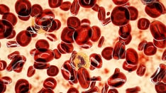
Continually Updated Clinical Reference
 
 
  All Sources     eMedicine     Medscape     Drug Reference     MEDLINE
 
eMedicine - Polycythemia Vera : Article by

Quick Find
Authors & Editors
Introduction
Clinical
Differentials
Workup
Treatment
Medication
Follow-up
Miscellaneous
References

Related Articles
Acute Lymphoblastic Leukemia

Acute Myelocytic Leukemia

Leukocytosis

Myelofibrosis

Polycythemia

Polycythemia of the Newborn

Polycythemia Vera

Splenomegaly

Thrombocytosis


Patient Education
Click here for patient education.


Author: Josef T Prchal, MD, FRCP, Professor of Medicine (Hematology), Adjunct Professor of Pathology and Genetics, University of Utah School of Medicine

Josef T Prchal is a member of the following medical societies: American College of Physicians, American Society of Human Genetics, and Southern Society for Clinical Investigation

Coauthor(s): Scott J Samuelson, MD, Fellow in Hematology and Oncology, Huntsman Cancer Institute, University of Utah School of Medicine

Editors: Sharada A Sarnaik, MD, Director of Sickle Cell Program, Department of Pediatrics, Professor, Children's Hospital of Michigan and Wayne State University; Mary L Windle, PharmD, Adjunct Assistant Professor, University of Nebraska Medical Center College of Pharmacy, Pharmacy Editor, eMedicine.com, Inc; James L Harper, MD, Associate Professor, Department of Pediatrics, Division of Hematology/Oncology and Bone Marrow Transplantation, Associate Chairman for Education, Department of Pediatrics, University of Nebraska Medical Center; Assistant Clinical Professor, Department of Pediatrics, Creighton University; Director, Continuing Medical Education, Children's Memorial Hospital; Pediatric Director, Nebraska Regional Hemophilia Treatment Center; Helen SL Chan, MBBS, FRCP(C), FAAP, Senior Scientist, Research Institute; Professor, Division of Hematology/Oncology, Department of Pediatrics, The Hospital for Sick Children, University of Toronto, Canada; Robert J Arceci, MD, PhD, King Fahd Professor of Pediatric Oncology, Department of Oncology, Division of Pediatric Oncology, Johns Hopkins University School of Medicine

Author and Editor Disclosure

Synonyms and related keywords: PV, primary polycythemia, polycythemia rubra vera, PRV, Osler-Vaquez disease, Osler disease, Osler's disease, Vaquez disease, Vaquez' disease, erythremia, splenomegalic polycythemia, erythrocytosis megalosplenica, cryptogenic polycythemia, erythropoietin independence, Epo independence, primary familial and congenital polycythemia, PFCP, Chuvash polycythemia

Background

Polycythemia vera is a disorder of the multipotent hematopoietic stem cell that manifests as excess production of normal erythrocytes and variable overproduction of leukocytes and platelets. It is grouped with the Philadelphia Chromosome Negative Myeloproliferative Disorders and can usually be differentiated from them by the predominance of erythrocyte production.

Pathophysiology

Key biologic features

Two key aspects of the biology of polycythemia vera can be scrutinized to identify this disorder:

  1. Clonality
  2. Erythropoietin (Epo) independence

Clonality

In polycythemia vera, a single clonal population of erythrocytes, granulocytes, B cells, and platelets arises when a hematopoietic stem cell gains a proliferative advantage over other stem cells. T lymphocytes and natural killer cells remain polyclonal in polycythemia vera; this situation is related to their longevity. Clonality can currently be tested only in female patients. The test is done by examining X-chromosomal polymorphisms and by taking advantage of inactivation of the X chromosome.

Epo independence

Epo independence is the ability of erythroid colonies formed from the hematopoietic stem cell in the setting of polycythemia vera to grow without Epo. Although the colonies do not require Epo, they remain responsive to it. In fact, the Epo receptor (EpoR) is normal, without defects in function or quantity. Experiments in which antibodies are used to neutralize Epo or block the EpoR do not abolish the formation of Epo-independent erythroid colonies.

Molecular mechanisms

Our understanding of the molecular mechanisms underlying polycythemia vera was greatly enhanced in the last 2 years. Genome-wide scanning, in which clonal polycythemia vera cells and nonclonal cells from the same individual were compared, revealed a loss of heterozygosity in chromosome 9p. Such a loss is found in approximately 30% of patients with polycythemia vera. This is not a classic chromosomal deletion, but rather a duplication of a portion of a chromosome and the loss of the corresponding parental region. The process is called uniparental disomy. The 9p arm contains a gene encoding for the JAK2 tyrosine kinase. The JAK family of kinases is critical in cytokine receptor signaling and transmits the activating signal in the Epo-EpoR signaling pathway. Inhibition of JAK2 eliminates the Epo independence of erythroid progenitors.

Following these observations was the identification of a loss-of-function somatic mutation in an autoinhibitory JAK2 domain. The mutation essentially produces a gain-of-function mutation affecting the kinase. This occurs when a point mutation leads to a valine-to-phenylalanine mutation at codon 617 of the JAK2 gene. The JAK2V617F mutation leads to constitutive phosphorylation activity and the recruitment of signal transducer and activator of transcription (STAT) molecules, which provides the necessary proliferative advantage seen in polycythemia vera. This process occurs in the absence of Epo and accounts for both the Epo independence and the Epo hypersensitivity of polycythemia vera colonies.

A murine model of this mutation produced a clinical phenotype consistent with polycythemia vera. Several groups rapidly confirmed these data, each showing that more than 90% of patients with polycythemia vera carry the JAK2V617F mutation.

Of note, compelling data strongly suggest that this mutation is not a disease-initiating alteration. Rather, an as-yet unidentified mutation or mutations may predispose an individual to acquire the JAK2V617F mutation, which plays a major role in behavior and in the clinical complications associated with polycythemia vera clones.

Diagnostic criteria

The diagnostic criteria for polycythemia vera are in flux. Clinical features were historically used to define the disease because laboratory testing was not possible or available. Now, because of advances in molecular biology and because of increased knowledge about the biology of polycythemia vera, laboratory testing can be done. Results of these tests are being incorporated into the diagnostic criteria for polycythemia vera.

Identification of the JAK2V617F mutation is another extremely important diagnostic tool. However, the mutation is not exclusive to polycythemia vera. For example, approximately 50% of patients with essential thrombocythemia and idiopathic myelofibrosis carry this mutation. In addition, small frequencies of this mutation has been identified in many other hematologic disorders. Moreover, some patients with polycythemia vera do not have this mutation.

Multiple diagnostic criteria and algorithms exist. All of the new ones incorporate the JAK2V617F allele, which has prognostic significance. The JAK2V617F mutation was present in 96 of 116 patients with polycythemia vera. Hematocrits, WBC counts, and lactate dehydrogenase levels were positively correlated with the level of the mutation. The highest levels of JAK2V617F at diagnosis was predictive of a risk of splenomegaly, presenting pruritus, or an eventual need for chemotherapy.

Also, the rate of major thromboses was positively correlated with high mutation values. In fact, a multivariate analysis including age, leukocytosis, hematocrits, platelet count, and therapies indicated that the JAK2V617F/JAK2 wild-type ratio was an independent risk factor for major vascular events (P = .027).

Frequency

United States

The prevalence of polycythemia vera is reported to be 4.9 cases per 100,000 people in Baltimore, Maryland.

International

The prevalence of polycythemia vera is reported to be 6.7 cases per 1,000,000 population in Israel.

Mortality/Morbidity

Phases of polycythemia vera

The course of polycythemia vera may or may not follow several phases.

  • The plethoric phase usually occurs first and is characterized by hyperproliferation of cellular components. The principle manifestations during this phase are thrombosis and hemorrhage. As a consequence, treatment is aimed at ameliorating symptoms.
  • The plethoric phase can last for a few years to as many as 20 years.
  • After the plethoric phase, the spent phase is characterized by progressive anemia, fibrosis, and splenomegaly. Smears demonstrate findings of anemia, thrombocytosis, and leukocytosis. In contrast to the plethoric phase, patients in the spent phase are often transfusion dependent.

Leukemia

Patients are at risk for leukemic transformation throughout the entire course of disease, though the rate is highest during the spent phase. The Polycythemia Vera Study Group (PVSG) found that the mode of treatment affected the incidence of leukemia.1 Treatment with phlebotomy alone, P32, or chlorambucil resulted in incidence of 1.5%, 10%, and 13%, respectively.

Survival and mortality risk

Except for the potential for leukemic transformation, appropriately treated polycythemia vera is compatible with a near-normal life. Without treatment, 50% of patients die within 18 months of diagnosis. The usual cause is a thrombotic event. Survival with treatment depends on the modality of therapy. Median survival is 13.9 years for phlebotomy alone, 11.8 years for P32 treatment, and 8.9 years for chlorambucil therapy (see Treatment).

In a recent European study, Marchioli and colleagues (2005) attempted to further define the prognosis of patients with this disease.2 They prospectively followed up 1638 patients to describe the clinical history of polycythemia vera. The primary limitation of this study was the mean follow-up period of 2.7 years. The overall mortality rate was 3.7 deaths per 100 persons per year, which was primarily caused by a moderate rate of cardiovascular death (1.7 deaths per 100 persons per year) and a high rate of death from noncardiovascular causes, mainly hematologic transformations (1.8 deaths per 100 persons per year). Cardiovascular mortality accounted for 45% of all deaths. Hematologic transformation (13% of all deaths) and solid tumors (19.5%) were also notable causes of mortality.

As seen in previous studies, age older than 65 years and history of thrombosis were significantly associated with the mortality risk. The cumulative rate of cardiovascular events was 5.5 events per 100 persons per year. Rates of combined malignancy, hematologic transformation, and malignancies not related to polycythemia vera were 3.0, 1.3, and 1.7 events per 100 persons per year.

Race

The disease appears to be most common among Jews of European extraction than among most non-Jewish populations. Some familial forms of polycythemia vera exist, but their mode of inheritance is unclear.

Sex

Polycythemia vera affects more men than women. The male-to-female ratio is 1.2-2.2:1.

Age

The onset of disease is typically in the sixth decade, and the prevalence peaks at 60-80 years of age.


History

Polycythemia vera frequently comes to the attention of clinicians because routine laboratory testing demonstrates an elevated hematocrit. Some patients will have been asymptomatic, whereas others will have had various nonspecific symptoms that are now recognized in the context of polycythemia vera. About 30% of patients have headaches, weakness, dizziness, and sweating (in order of decreasing frequency). Many of these symptoms can be attributed to an excessive hematocrit.

Patients may alternatively present with a complication of polycythemia vera. Approximately 33% present with thrombosis or hemorrhage; 75% of these patients have arterial thrombosis, and 25%, venous thrombosis. Sequelae of thrombosis due to polycythemia vera include cerebrovascular accidents, myocardial infarction, deep venous thrombosis, and pulmonary embolism (in that order).

A less frequent but more specific finding for polycythemia vera that those described above is Budd-Chiari syndrome (thrombosis of the hepatic vein). A patient who presents with Budd-Chiari syndrome should alert the physician to consider polycythemia vera, as it is the most common underlying disease. Approximately 2-10% of patients with polycythemia vera have Budd-Chiari syndrome. Many patients do not have an elevated hematocrit at the time of presentation, but they may have other laboratory abnormalities indicative of polycythemia vera. If patients survive, they eventually develop a myeloproliferative phenotype.

One study of 41 patients with idiopathic Budd-Chiari syndrome revealed that 58% had the JAK2V617F mutation.3 Another study showed that serum Epo levels can be elevated in patients with Budd-Chiari syndrome and the JAK2V617F mutation.4 The presence of an elevated Epo has traditionally been thought to make the diagnosis of polycythemia vera extremely unlikely.

Unrecognized thrombosis of the hepatic or splenic vein can result in portal hypertension and varices. Other GI symptoms include peptic ulcer disease, which occurs 4-5 times more frequently in patients with polycythemia vera than in the general population.

Hemorrhagic presentations are usually mild, with gum bleeding and easy bruising. However, serious GI hemorrhage can occur.

Approximately 40% of patients have life-altering pruritus. The pruritus typically worsens after a warm shower or bath; this reaction is known as aquagenic pruritus. Pruritus has been attributed to increased numbers of mast cells and elevated levels of histamine.

Less than 5% patients have erythromelalgia. This is erythema and warmth of the distal extremities—especially the hands and feet—in association with a painful burning sensation. Erythromelalgia can result in digital ischemia if it is prolonged. Because it is also associated with essential thrombocythemia, a role for platelet aggregation has been proposed. In fact, the syndrome frequently, but not always, responds within hours to low-dose aspirin therapy.

Patients with polycythemia vera sometimes present with cardiovascular symptoms due to myocardial infarction and congestive heart failure, neurologic symptoms due to spinal cord compression caused by extramedullary hematopoiesis, and an elevated uric acid level with subsequent gout due to increased cellular turnover.

Physical

Potential physical findings include plethora and ruddiness of the face, erythromelalgia of the distal extremities, bruising, and splenomegaly. Specific attention should be directed to sternal tenderness, which may indicate transformation to acute myeloid leukemia (AML).


Acute Lymphoblastic Leukemia
Acute Myelocytic Leukemia
Leukocytosis
Myelofibrosis
Polycythemia
Polycythemia of the Newborn
Polycythemia Vera
Splenomegaly
Thrombocytosis

Other Problems to be Considered

Polycythemia vera must be differentiated from other causes of polycythemia. The polycythemias can be divided by etiology into 3 groups, as follows:

  1. Apparent or relative polycythemia
  2. Primary polycythemia
  3. Secondary polycythemia

A quick way to screen for polycythemia vera without excessive diagnostic testing is to determine if a hereditary pattern for the erythrocytosis is present. Because polycythemia vera is an acquired disorder, a familial pattern suggests another diagnosis; however, familial polycythemia vera does exist. A familial patter suggests one of the congenital polycythemias, such as primary familial and congenital polycythemia (PFCP) or Chuvash polycythemia. Patients with these disorders should be referred to a hematologist for specialized diagnostic testing and treatment.

Primary polycythemia

Apparent or relative polycythemia is due to a decrease in plasma volume with a normal red cell mass. This type of polycythemia is associated with hypertension, obesity, dehydration, and stress, among other causes.

Secondary polycythemia

Primary polycythemia is caused by intrinsic hyperproliferation of the hematopoietic stem cell independent of Epo stimulation or with an exaggerated response to a low Epo level. The most common primary polycythemia is polycythemia vera in which the hematopoietic stem cell proliferates independently of Epo.

The defining features of polycythemia vera are described in the Introduction. Another primary polycythemia is PFCP. The defect in PFCP is hyper-responsiveness to Epo. One of its genetic causes has been defined. It is a hyperfunctional EpoR (gain-of-function mutation) involving deletion of the negative regulatory subunit of the EpoR. Other independent mutations exist but are not yet undefined.

Unlike polycythemia vera, which is a clonal acquired genetic mutation that can progress to leukemia, PFCP is a nonclonal germ-line mutation that does not progress to acute leukemia. PFCP also differs from polycythemia vera in that only the erythroid lineage is affected.

Secondary polycythemia

Secondary polycythemia is due to elevated levels of Epo that induce erythrocyte proliferation. At the time of presentation, the increased RBC mass may have reached an equilibrium, and the Epo level is often within normal limits. However, the patient's normal Epo level is inappropriately high for the elevated hematocrit. High Epo concentrations are due to physiologically appropriate or inappropriate causes.

Hypoxemia

Physiologically appropriate secondary polycythemias result from hypoxia, Hypoxia is the common endpoint of the various causes of physiologically appropriate secondary polycythemias, which are discussed below.

  • High-altitude polycythemia: A low ambient partial pressure of oxygen (pO2) results in tissue hypoxia. Acute compensation occurs by means of hyperventilation, whereas chronic compensation involves elevation of the hematocrit.

    The degree of response varies among individuals. Not all populations acclimate to high altitude by developing polycythemia. For example, Tibetans have lower-than-expected hemoglobin levels bu high levels of exhaled nitric oxide (NO). NO may be the end product of a process in which the body improves oxygen delivery by inducing vasodilation and increasing blood flow to the tissues.

  • Polycythemia vera related to cardiopulmonary disease: In cardiopulmonary disease, impaired respiration and circulation result in tissue hypoxia and, subsequently, increased Epo concentrations.
  • Smoker's polycythemia: Smoking results in the formation of carboxyhemoglobin, which does not carry oxygen and which results in increased oxygen affinity to other hemoglobin molecules. This process results in tissue hypoxia, which induces the production of Epo. A reduction in plasma volume due to smoking compounds the problem of the rise in hematocrit.
  • Erythrocyte enzyme-defect polycythemia: Defects in bisphosphoglycerate mutase and phosphofructokinase decrease levels of 2,3-bisphosphoglycerate. This molecule is necessary for hemoglobin to transition from a state of high oxygen affinity to a state of low oxygen affinity. Decreased levels of 2,3-bisphosphoglycerate result in tissue hypoxia.
  • Hemoglobinopathy with a high-affinity mutant: Autosomal-dominant hemoglobin mutants are unable to transition from high oxygen affinity to low oxygen affinity because intramolecular rotation or binding of 2,3-bisphosphoglycerate is impaired. In some mutants, deoxygenation is also impaired.
  • Methemoglobinemia: Methemoglobinemia is usually due to a deficiency in cytochrome b5 reductase (methemoglobin reductase), but it can also be caused by various mutations of globin genes, such as that encoding hemoglobin M.
  • Chemically induced hypoxia: Cobalt is believed to inhibit oxidative metabolism, controlling Epo production. It is not effective treatment for anemia. Cobalt has been used as a foam stabilizer in beer and causes an acquired polycythemia when it is unintentionally ingested in high amounts.

Exposure to exogenous Epo

Physiologically inappropriate polycythemia is often due to exogenous sources of Epo, such as those discussed below.

  • Malignancies: Several malignancies produce Epo. These include hepatomas, renal cell carcinomas, and cerebellar hemangiomas. Uterine myomas have also been reported to produce Epo. Another mechanism by which these often-large and bulky tumors produce erythrocytosis is mechanical interference with the blood supply to the kidneys, which results in a false detection of hypoxia and, therefore, Epo production.
  • Endocrine disorders: Pheochromocytomas, aldosterone-producing adenomas, Barter syndrome, and dermoid cysts of the ovary can result in inappropriate Epo synthesis by mechanically interfering with the renal blood supply or by causing hypertensive damage to renal parenchyma. The result is a false sensing of hypoxia by the kidneys. Subsequent effects are Epo production; functional interaction among aldosterone, renin, and Epo; and inappropriate Epo secretion by the tumor. Androgens increase the hematocrit by 2 mechanisms: stimulation of Epo production and an independent hyperproliferative effect on erythrocyte precursors.
  • Chuvash polycythemia
    • This is an endemic polycythemia observed on the west bank of the Volga River in the Chuvash Autonomous Republic in western Russia. It is an autosomal recessive disorder characterized by a mutation in the VHL gene that prevents ubiquitin-mediated degradation of hypoxia inducible factor (HIF)-1. The result is upregulation of downstream target genes, including those responsible for Epo production. As such, Chuvash polycythemia can be grouped with the secondary inappropriate polycythemias.
    • Because a second defect not linked to the EpoR results in hyper-responsiveness to Epo, Chuvash polycythemia also possess some features of primary polycythemia. Patients with Chuvash polycythemia have normal ABGs, normal calculated partial pressure of hemoglobin required for 50% saturation (P50), normal or increased Epo levels, and no abnormal hemoglobins.
  • Renal polycythemia: This is due to Epo produced as a result of renal cysts, polycystic disease, or hydronephrosis.
  • Renal transplants: Erythrocytosis can occur after renal transplantation and is thought to be due to increased activity of the angiotensin II–angiotensin receptor-1 pathway. Angiotensin II may also modulate release of Epo and insulinlike growth factor-1 (IGF-1). Venous canalization studies have shown the source to be nonfunctional native kidneys. Removal of the native kidneys can normalize the hematocrit; however, angiotensin-converting enzyme (ACE) inhibitors can also control this typically transient erythrocytosis; their use may allow the patient to avoid surgery.
  • Neonatal polycythemia: This is an appropriate secondary polycythemia due to increased oxygen affinity of fetal hemoglobin and subsequent tissue hypoxia. The response can become excessive and inappropriate in the setting of maternal diabetes or placenta-to-fetus transfusion.


Lab Studies

Distinguishing primary, secondary, and apparent polycythemia

After polycythemia vera is suspected, the first step in evaluating a patient is determining whether he or she has primary, secondary, or apparent polycythemia.

CBCs, ABGs, and Epo levels

CBCs, ABGs, and Epo levels can be used to differentiate primary, secondary, and apparent polycythemia. The CBC typically shows increased leukocyte, platelet, and erythrocyte counts in primary polycythemia, as opposed to elevated erythrocyte counts in secondary and apparent polycythemia.

An exception is PFCP, a primary polycythemia. PFCP leads to elevated erythrocyte but not leukocyte or platelet counts. However, it can be distinguished from secondary polycythemia on the basis of the Epo level. The Epo concentration is almost always low or low normal in primary polycythemia; in secondary polycythemia, it is high or high normal when the hematocrit is high. However, the discovery of patients with Budd-Chiari syndrome who have the JAK2V617F mutation and elevated Epo levels has changed this absolute diagnostic criterion.

An ABG that indicates hypoxia suggests secondary appropriate polycythemia.

Ferritin levels

Ferritin levels may also help in differentiating primary polycythemia from secondary polycythemia. In primary polycythemia the ferritin level is typically low because of a constant overproduction of erythrocytes. In contrast, the ferritin level is usually normal in secondary polycythemia.

Red cell mass

Red cell mass has been used to distinguish apparent polycythemia from secondary or primary polycythemia. However, testing is expensive and requires expertise to do, or else the results are inaccurate. Also, the 131I-albumin used to measure plasma volume is not available in the United States. Where it is available, it is difficult to handle because of its radioactivity.

As a consequence, the authors do not routinely test for red cell mass at the University of Utah School of Medicine. Its diagnostic value is limited when the patient's hematocrit is clearly abnormal. In clinical practice, use of this test is frequently limited because of its unavailability. However, some clinicians feel very strongly about it, especially when patients have borderline hemoglobin levels. Results may occasionally help in identifying a patient with an elevated red cell mass whose hematocrit is normal because of an increased plasma volume, and it can also aid in identifying patients whose hematocrit is elevated only because of a reduced plasma volume.

Differentiating and inappropriate causes of secondary polycythemia

The cause of secondary polycythemia must be determined to be appropriate or inappropriate. An elevated Epo concentration with ABG results consistent with hypoxia suggest secondary appropriate polycythemia. By contrast, an elevated Epo without hypoxia suggests secondary inappropriate polycythemia.

The cause of appropriate polycythemia can be elucidated by history taking, though specialized testing (eg, determination of a P50 curve) can be used to identify high-affinity hemoglobins due to structural defects in hemoglobin or due to enzyme deficiencies. Hemoglobin electrophoresis is insufficient for detecting structural defects in the molecule because some mutants are missed. Causes of secondary inappropriate polycythemia causes can be sorted out with judicious imaging and specialized endocrine testing.

Of the primary polycythemias, primary familial congenital polycythemia must be differentiated from polycythemia vera. These 2 diagnoses differ in their clonality and in the in vitro responsiveness of peripheral blood erythroid progenitors to Epo. Testing for clonality relies on polymorphisms based on inactivation of the X chromosome. Therefore, it can be done only in female patients. Polycythemia vera is clonal, whereas PFCP is not.

Responsiveness of endogenous erythroid colonies to Epo also differentiates polycythemia vera from PFCP. Polycythemia vera is characterized by growth independence from Epo, whereas PFCP is not, though it is hyperresponsive. Testing for endogenous erythroid colonies is not routinely available and can be done only in specialized laboratories. The authors frequently perform this test in our laboratory when a case is difficult.

Imaging Studies

CT scanning or ultrasonography of the abdomen can be used to assess the size of the spleen, which is frequently enlarged in patients with polycythemia vera. Imaging may also reveal renal pathology, cerebellar hemangioblastomas, and pheochromocytomas that can cause secondary polycythemia.

Histologic Findings

Bone marrow and aspirates tend to be hypercellular in patients with polycythemia vera. In the plethoric phase, blood smears show normal erythrocytes, variable neutrophilia with myelocytes, metamyelocytes, as well as various degrees of immaturity, basophilia, and increased platelets. In the spent phase, the blood smear shows abundant teardrop cells, leukocytosis (or leukopenia), and thrombocytosis (or thrombocytopenia).

Staging

Because many practitioners do not have access to specialized clonality testing or erythroid colony assays, many of the criteria for diagnosing polycythemia vera do not require these studies, though the results are taken into account. No consensus has been reached regarding diagnostic criteria.

World Health Organization criteria for diagnosis

The World Health Organization (WHO) criteria for polycythemia vera diagnosis requires 2 components:

  1. Reasonable elimination of apparent and secondary polycythemia
  2. Confirmation of polycythemia vera

Discovery of the JAK2V617F mutation have made these criteria insufficient. Proposals for revising these criteria have recently been published.

Proposed revisions to the WHO criteria for diagnosis

Diagnosis requires the presence of 2 major criteria and one minor criterion or of the first major criterion with 2 minor criteria.

Major criteria

  1. Hemoglobin level of more than 18.5 g/dL in men or 16.5 g/dL in women or other evidence of increased RBC volume (hemoglobin level or hematocrit >99th percentile of the method-specific reference range for the patient's age, sex, altitude of residence)
    or
    a hemoglobin level of more than 17 g/dL in men and 15 g/dL in women if it is associated with a documented and sustained increase of 2 g/dL or more from the patient's baseline value that cannot be attributed to a correction of iron deficiency
    or
    an elevated RBC mass more than 25% above the mean normal value
  2. Presence of the JAK2V617F mutation or another functionally similar mutation (eg, mutation of exon 12 of JAK2)

Minor criteria

  1. Bone marrow biopsy showing hypercellularity for the patient's age, with trilineage growth (panmyelosis), as well as prominent erythroid, granulocytic, and megakaryocytic proliferation
  2. Serum Epo level below the normal range
  3. Formation of endogenous erythroid colonies in vitro

Other groups are preparing and proposing other potential diagnostic criteria.


Medical Care

General treatment

Treatment of polycythemia vera depends on whether the disease is in the plethoric phase or the spent phase.
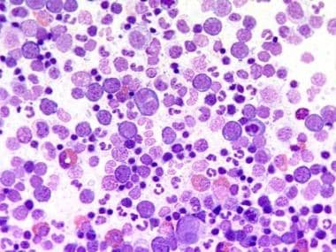
 In the plethoric phase, the goal of treatment is controlling thrombotic episodes by restraining monoclonal proliferation rather than restoring polyclonal growth and maturation of cells. Interferon (INF)-A is an exception; a few case reports have described the restoration of polyclonality.

Phlebotomy

In the plethoric phase, polycythemia vera is first treated by performing phlebotomy until the hematocrit is reasonably controlled. Most patients can tolerate the removal of 450-500 mL of blood every 2-4 days. As blood is removed and as the patient becomes iron deficient, control of the patient's hematocrit becomes progressively easier, and the phlebotomy schedule should be adjusted accordingly. Although phlebotomy is effective for controlling erythrocytosis, it does not affect the variable leukocytosis and thrombocytosis observed in polycythemia vera.

For many years, the mainstay of therapy of polycythemia vera was phlebotomy with a goal hematocrit less than 45% in men and less than 42% in women. This recommendation was based on retrospective data that are now almost 30 years old and, in the authors' opinion, potentially inaccurate.

Di Nisio and colleagues (2007) recently reported that 40-55% differences in hematocrit were not associated with a risk of thrombosis or mortality among patients with polycythemia vera.5

Landolfi and colleagues (2007) performed an extensive retrospective review of 1638 patients polycythemia vera who were examined in part of the European Collaboration on Low-Dose Aspirin in Polycythemia Vera (ECLAP) trial.6 In this trial, the hematocrit was not correlated with a risk of thrombosis. Limitations of this study were its retrospective nature and its relatively short follow-up (median, 2.8 y).

A retrospective review of the ECLAP data demonstrated that older age and previous thrombosis were risk factors for thrombotic events. The investigators found no correlation between the risk of thrombosis and the hematocrit. However, a WBC count of more than 15,000/μL (when compared with a WBC of <10,000/μL) was an independent risk factor for major thrombosis, which primary manifested as an increased risk of myocardial infarction.

Therefore, the present authors believe that the true hematocrit goal for polycythemia vera, if one exists, is unclear. This issue remains to be determined in prospective studies. Although many experts still recommend phlebotomy, it is clearly a controversial issue.

Aspirin therapy

In most patients, low-dose aspirin is started to reduce the risk of thrombotic events, and phlebotomy is continued as necessary to control the hematocrit. This recommendation is based on results of the ECLAP study in which patients with polycythemia vera and no clear indications for aspirin therapy were randomly assigned to receive aspirin 100 mg/day or no aspirin. The study showed a minor but statistically significant decrease in the risk of cardiovascular death, nonfatal myocardial infarction, nonfatal stroke, pulmonary embolism, and major venous thrombosis with aspirin therapy. Low-dose aspirin did not significantly increase rates of hemorrhage.

Use of additional agents

Whether an additional agent should be given depends on the patient's risk of thrombosis. The decision is a trade off between a reduction in thrombotic events and a increase in the incidence of malignancy.

The initial PVSG investigators compared phlebotomy, phlebotomy with 32P, and phlebotomy with chlorambucil; median survival durations were 13, 11, and 9 years, respectively. The incidence of thrombosis was 23% in the phlebotomy-only group versus 16% in the 32P-and-phlebotomy group.

Rates of AML were 1.5%, 10%, and 13% for phlebotomy, phlebotomy with 32P, and phlebotomy with chlorambucil, respectively. Because of the increase in the rate of AML when polycythemia vera was treated with chlorambucil, this drug is no longer used for myelosuppression.

In addition, rates of GI and skin cancers increased by 4%, 9%, or 12% when patients were treated with phlebotomy, phlebotomy with 32P, or phlebotomy with chlorambucil, respectively. Clearly, myelosuppression reduces the incidence of thrombotic events but increases the risk of malignancy.

Because of these results, a phase II efficacy trial was performed by using hydroxyurea instead of chlorambucil or 32P to see if this less-leukemogenic agent could control thrombosis. Fifty-one patients with polycythemia vera were given hydroxyurea 30 mg/kg/day for 1 week then 15 mg/kg/day, with the goal of maintaining a platelet count of <600,000/cm3 and a hematocrit of <50% with minimal phlebotomy.

The incidence of thrombosis in the first 2 years of treatment (when most thromboses occur) was 9%, a rate significantly lower than the historical control of 23% for phlebotomy alone in the PVSG trial. At a median follow-up of 8.6 years, the incidence for AML was 6% for hydroxyurea compared with 1.5% for phlebotomy alone. At the time of analysis, this difference was not statistically significant, but a later addition of 2 cases of myelodysplasia in the hydroxyurea arm increased the incidence to a significant 8%.

Several other studies have not fully addressed this issue. Many clinicians and investigators believe the hydroxyurea is clearly leukemogenic. However, the present authors believe that this risk has not been definitively proven and that, if a leukemogenic effect occurs, it is small in a population. However, this issue does need to be considered, especially in the treatment of young patients. Remember that clinical trials of hydroxyurea have not proven a statistically increased risk of leukemogenicity.

Because a leukemogenic risk is theoretically possible with hydroxyurea, anagrelide has been used to control increased platelet counts with the aim to reduce thrombotic events. In the PT1 trial from the United Kingdom, patients with essential thrombocythemia were randomly assigned to receive hydroxyurea or anagrelide. The study demonstrated an increased risk of thrombosis with anagrelide. The implication for polycythemia vera is unclear, but a reduction in the platelet count does not affect rate of thrombosis in essential thrombocytopenia, and one would expect a similar result in polycythemia vera. the present authors usually consider converting anagrelide to hydroxyurea unless the patient's condition is well controlled with anagrelide.

Risk stratification

Risk stratification is important in deciding whether chemotherapeutic cytoreduction is indicated. Most agree the risk of thrombosis is high when in patients older than 70 years old and/or those with a previous history of thrombosis. (Note that this age criterion differs from the age used for risk stratification in essential thrombocythemia.)

A platelet count of more than 1.5 X 10⁶/μL is a risk factor for bleeding and is frequently considered a high-risk indication favoring chemotherapeutic cytoreduction.

Most experts consider that uncontrolled traditional cardiac risk factors, especially smoking, place a patient in a high risk category. However, when these factors are well controlled, high-risk therapy may not be justified.

Summary of the authors' approach to treatment

Given the aforementioned data, the present authors recommend chemotherapeutic cytoreduction in all patients who are high risk. In general, the drug of choice is hydroxyurea. An attempt is made to titrate the drug to achieve normal WBC counts; this practice is based on the data from Landolfi et al (2007), as discussed above.6

The authors honestly tell patients that this strategy is based on retrospective data that still need to be proven in prospective studies. In addition, the authors monitor the hematocrit. Although phlebotomy is performed to manage symptoms or high values, fully achieving a goal of 45% in men and 42% in women is probably not required.

Patients who are low risk generally do not require chemotherapeutic cytoreduction. However, the concern about an increased risk of thrombosis (primarily myocardial infarction) due to leukocytosis raises the issue of whether low-risk patients with a WBC of more than 15,000/μL should receive cytoreductive therapy. This concern remains to be addressed in prospective fashion. However, for the time being, cytoreductive therapy cannot be firmly recommended in this situation.

As stated above, the authors believe that the current recommendations to conduct phlebotomy to achieve a goal hematocrit of 45% in men and 42% in women may be inaccurate. Of course, patients with symptomatic hyperviscosity should receive phlebotomy sufficient to relieve their symptoms. Phlebotomy is also considered in patients with a hematocrit more than 55%. However, current guidelines may not be binding given the data presented above.

All patients receive low-dose aspirin, usually 81 mg, unless a contraindication is present.

Special treatment situations

On occasion, special treatment situations arise. These are detailed below.

Nonadherence or limited life spans

32P is a reasonable option in the patient who is unreliable or who has a limited expected life span. 32P offers the convenience of 1 injection that results in long-term control. The principle drawback is an increased risk of malignancy.

Pregnancy

In pregnant women, INF can be used to treat polycythemia vera. The mechanism is unclear, the adverse effects are moderate and often severe, and the drug is expensive. However, INF is not teratogenic, and it can reasonably control symptoms. A few case describe a restoration of polyclonality.

INF is also a reasonable option in a young patient because of possible concerns of leukemogenicity of hydroxyurea.

Recent data suggest that pegylated INF alfa-2a (Pegasys; Hoffman-La Roche, Inc, Nutley, NJ) is likely to be as effective as INF. INF alfa-2a is given once a week, a frequency that likely improves compliance. In addition, some suggest that INF alfa-2a may be better tolerated than standard INF.

Erythromelalgia

Erythromelalgia responds to low-dose aspirin or reduction of the platelet count to normal with low-dose myelosuppressants.

Pruritus

Pruritus can be disabling and life-altering to patients with polycythemia vera. Exposure to hot water and vigorous rubbing of the skin can incite itching. Taking cool baths and patting instead of rubbing the skin dry can provide symptomatic relief. Also, starch baths in which half a box of Linit starch (Malco Products, Inc, Barberton, OH) is dissolved in a tub of water can be effective.

Pharmacologic treatment options include antihistamines (cyproheptadine 4 mg by mouth 3 times/day), H2 receptor blockers (cimetidine 300 mg by mouth 4 times/day), photochemistry, danazol, and INF-alpha. Selective serotonin reuptake inhibitors (paroxetine 20 mg by mouth once a day or fluoxetine 10 mg by mouth once a day) can also be used. In severe refractory cases, myelosuppression may be required.

Patients who have hyperuricemia with symptoms (eg, gout, urate kidney stones) are given allopurinol. The authors also monitor uric acid levels and treat asymptomatic hyperuricemia if they are notably elevated.

Surgical Care

Polycythemia vera is treated medically except in the spent phase, when splenectomy may be performed to relieve symptoms related to mass effect and pancytopenia. Patients with polycythemia vera who are undergoing surgery have a very high risk of postoperative thrombosis.

Consultations

All patients in whom polycythemia vera is suspected should be referred to a hematologist.

Diet

Patients should avoid drinking beer with cobalt-containing foam stabilizers. Other than this, a normal healthy diet is recommended.

Activity

Patients with polycythemia vera can have a normal, active life.


Drug Category: Biologic response modifiers

Biologic response modifiers elicit antiproliferative, antiviral, and immunomodulating effects. They inhibit cellular growth and alter cellular differentiation.

Drug NameINF alfa-2a and INF alfa-2b (Roferon-A, Intron A)
DescriptionProtein produced in response to viral infection and other inflammatory stimuli. Exact mechanism unknown, but believed to exert antiproliferative effect. Produced by means of recombinant DNA techniques in Escherichia coli. Controls erythrocytosis and reduces size of spleen. Unlike hydroxyurea or anagrelide, can ameliorate intractable pruritus often found in polycythemia vera. Sporadic case reports describe cytogenetic remissions, suggesting possible biologic effect.
Adult Dose3 million U SC 3 times/wk; dosage range 4.5-27 million U qwk
Pediatric Dose2.5-5 million U IM 3 times/wk; adjust dosage according to therapeutic response and toxicity
ContraindicationsDocumented hypersensitivity; history of severe mental depression (relative contraindication); decompensated liver disease, autoimmune hepatitis, history of autoimmune disease, or rapidly progressing or life-threatening visceral AIDS-related Kaposi sarcoma
InteractionsWeak cytochrome P450 (CYP) 1A2 inhibitor; may increase adverse effects of ACE inhibitors (especially granulocytopenia) and anticoagulant effects of warfarin; agranulocytosis reported when coadministered with clozapine; theophylline, zidovudine, and vinblastine may increase toxicity; corticosteroids may decrease therapeutic effects; coadministration with interleukin-2 may increase risk of renal failure; may decrease serum concentrations of melphalan; may decrease hematopoietic effects of Epo
PregnancyC - Fetal risk revealed in studies in animals but not established or not studied in humans; may use if benefits outweigh risk to fetus
PrecautionsCaution in liver or kidney dysfunction, brain metastases, seizure disorder, multiple sclerosis, compromised CNS, or history of mental depression or suicidality; common adverse effects include fever, malaise, nausea, peripheral neuropathy, and vomiting

Drug Category: Antiplatelet agents

Antiplatelet agents prevent the formation of thrombi associated with polycythemia.

Drug NameAspirin (Anacin, Ascriptin, Bayer)
DescriptionIrreversibly acetylates platelet cyclooxygenase, decreasing thromboxane A2 (prostaglandin responsible for changes in platelet shape, granule release, and aggregation). Prescribed for most patients with polycythemia vera.
Adult Dose81 mg PO qd; doses higher than this not shown to decrease rates of thrombosis and have been associated with increased rates of hemorrhage
Pediatric DoseNot established
ContraindicationsDocumented hypersensitivity; liver damage, hypoprothrombinemia, vitamin K deficiency, bleeding disorders, asthma; children (<16 y) with viral infections because of association with Reye syndrome
InteractionsMinor CYP2C8/9 substrate; may increase risk of bleeding when given with heparin, low-molecular-weight heparins, NSAIDS, platelet inhibitors, warfarin, or other oral anticoagulants; may increase bleeding when coadministered with herbal supplements (eg, cat's claw, dong quai, evening primrose, feverfew, garlic, ginger, ginkgo, red clover, horse chestnut, green tea, ginseng); certain foods contain salicylate (eg, curry powder, paprika, licorice) and may result in accumulation if eaten with aspirin (approximately 6 mg salicylate/100 g food); antacids and urinary alkalinizers may decrease effects; corticosteroids decrease serum levels of salicylate; may antagonize uricosuric effects of probenecid and increase toxicity of phenytoin and valproic acid; doses >2 g/d may potentiate glucose-lowering effect of sulfonylureas; simultaneous administration of other NSAIDs may decrease cardioprotection and stroke-preventive effects
PregnancyC - Fetal risk revealed in studies in animals but not established or not studied in humans; may use if benefits outweigh risk to fetus
PrecautionsPregnancy category D in third trimester; caution during breastfeeding; may cause excessive hemorrhage and GI ulceration; watch for transient decrease in renal function and aggravated chronic kidney disease; avoid in severe anemia or history of blood coagulation defects

Drug NameAnagrelide (Agrylin)
DescriptionInhibits postmitotic megakaryocyte maturation. Unlike hydroxyurea, selectively inhibits platelet proliferation. In polycythemia vera used only to control platelet counts, but slightly decreases mean hemoglobin and hematocrit while maintaining WBC counts. Inhibits cyclic nucleotide phosphodiesterase and release of arachidonic acid from phospholipase, possibly by inhibiting phospholipase A2. Can be used in addition to hydroxyurea for particularly difficult to control thrombocytosis in polycythemia vera.
Adult Dose1 mg/d PO initially; adjust to lowest effective dose that maintains platelet count; not to exceed 10 mg/d
Pediatric DoseNot established; limited data suggest dose similar to adult dose
ContraindicationsDocumented hypersensitivity
InteractionsIn 1 case report, sucralfate may have decreased absorption; may increase effects of drotrecogin alpha, NSAIDs, salicylates, treprostinil, and other antiplatelet agents; use herbal medicines with caution (can have anticoagulant or antiplatelet effects)
PregnancyC - Fetal risk revealed in studies in animals but not established or not studied in humans; may use if benefits outweigh risk to fetus
PrecautionsCommon adverse effects include thrombocytopenia, headaches, palpitations, fluid retention, and diarrhea; caution in renal or hepatic impairment

Drug Category: Antineoplastic agents

These agents are used off label to treat polycythemia. Pediatric doses are extrapolated from pediatric treatment regimens, including those for leukemia or myelodysplastic syndrome.

Drug NameHydroxyurea (Droxia, Hydrea)
DescriptionNonalkylating, myelosuppressive, S-phase agent. Inhibits ribonucleotide reductase (enzyme that converts ribonucleotides into deoxynucleotides), depleting deoxynucleotide and inhibiting DNA synthesis. Cellular proliferation ultimately inhibited. Leukocyte, erythrocyte, and platelet counts decrease. Mechanism of action probably differs from its action in treating sickle cell disease.
Misconception is leukemogenicity. No studies conclusively demonstrated leukemogenicity over baseline in myeloproliferative disease.
Adult DoseRanges from 500 mg/d PO to as high as needed to control erythrocytosis and as tolerated (on basis of leukocyte and platelet counts)
Pediatric Dose15 mg/kg/d PO initially, adjust dose to response; dosage based on patient's actual or ideal weight, whichever is less
ContraindicationsDocumented hypersensitivity; severe pancytopenia (WBC count <2.5 X 10⁹/L, platelet count <100 X 109/L, severe anemia)
InteractionsCoadministration with fluorouracil can increase neurotoxicity; infections possible in immunosuppressed patients immunized with live viral vaccines (eg, MMR); coadministration with didanosine has caused fatal and nonfatal pancreatitis; coadministration with other myelosuppressive agents may increase toxicity
PregnancyD - Fetal risk shown in humans; use only if benefits outweigh risk to fetus
PrecautionsCaution in renal impairment; dose-limiting myelosuppression with variable anemia and thrombocytopenia may occur; may cause nausea and GI upset; reliable birth control required for both men and women during treatment; avoid during breastfeeding (secreted in human milk)


Further Inpatient Care

  • Most patients with polycythemia vera can be treated on an outpatient basis.
  • On occasion, a symptomatic patient who has an extremely high hematocrit may need hospitalization for emergency phlebotomy. These patients should receive aggressive volume replacement with saline. Their blood counts should be monitored closely.

Further Outpatient Care

  • In the plethoric phase, patients initially require close follow-up to monitor their blood counts. The dose of hydroxyurea must be monitored closely until a steady state is achieved. Phlebotomy is occasionally required in symptomatic patients.
  • In the spent phase, therapy is switched from the removal of cells to the transfusion of cells to relieve anemia. Leukemic transformation must be managed expectantly, but patients have a poor prognosis.

Complications

  • Potential complications of polycythemia vera are primarily thromboembolic and hematologic.
  • Increases in cardiovascular morbidity and mortality can be notable, though appropriate treatment is thought to substantially reduce these risks.
  • Whether any of the current therapies can reduce the risk of hematologic transformation is unclear. Emerging treatments such as INF and possibly JAK2 inhibitors, may reduce the mutant clone population and decrease this risk.

Prognosis

  • For patients can anticipate having a normal or near-normal life span. However, polycythemia vera carries a significant potential to cause morbidity and mortality, even when it is treated correctly.
  • As Marchiol et al (2005) reported, cardiovascular events are more common in patients with polycythemia than in others and pose an ever-present risk.2
  • Transformation to myelofibrosis decreases the anticipated length of survival and, though uncommon, transformation to acute leukemia portends a poor prognosis.
  • See Mortality/Morbidity for further details.


Medical/Legal Pitfalls

  • Before hydroxyurea therapy is started, female patients must be tested for pregnancy.
  • Patients should be made aware of the theoretical risk of hydroxyurea-induced leukemia. Emphasize the fact that no risk has been conclusively proven in clinical trials.
  • The present authors are candid with our patients. We explain that many recommendations are based on insufficient data and that we expect treatment recommendations to change over the next several years as additional information is gathered.

Special Concerns

Tests for clonality and assays for Epo-independent erythroid-colony growth can be ordered as special research tests.


  1. Berk PD, Wasserman LR, Fruchtman SM. Treatment of polycythemia vera: a summary of clinical trials conducted by the polycythemia vera study group. In: Wasserman LR, Berk PD, Berlin NI, eds. Polycythemia vera and the myeloproliferate disorders. Philadelphia, Pa: WB Saunders; 1995: 166.
  2. Marchioli R, Finazzi G, Landolfi R, et al. Vascular and neoplastic risk in a large cohort of patients with polycythemia vera. J Clin Oncol. Apr 1 2005;23(10):2224-32. [Medline].
  3. Patel RK, Lea NC, Heneghan MA, et al. Prevalence of the activating JAK2 tyrosine kinase mutation V617F in the Budd-Chiari syndrome. Gastroenterology. Jun 2006;130(7):2031-8. [Medline].
  4. Thurmes PJ, Steensma DP. Elevated serum erythropoietin levels in patients with Budd-Chiari syndrome secondary to polycythemia vera: clinical implications for the role of JAK2 mutation analysis. Eur J Haematol. Jul 2006;77(1):57-60. [Medline].
  5. Di Nisio M, Barbui T, Di Gennaro L, et al. The haematocrit and platelet target in polycythemia vera. Br J Haematol. Jan 2007;136(2):249-59. [Medline].
  6. Landolfi R, Di Gennaro L, Barbui T, et al. Leukocytosis as a major thrombotic risk factor in patients with polycythemia vera. Blood. Mar 15 2007;109(6):2446-52. [Medline].
  7. Adamson JW, Fialkow PJ, Murphy S, et al. Polycythemia vera: stem-cell and probable clonal origin of the disease. N Engl J Med. Oct 21 1976;295(17):913-6. [Medline].
  8. Ang SO, Prchal JT. The polycythemia vera and other polycythemic disorders: biological aspects. In: Melo JV, Goldman JM, eds. Myeloproliferative Diseases. New York, NY: Springer-Verlag; 2006:297-319.
  9. Baxter EJ, Scott LM, Campbell PJ, et al. Acquired mutation of the tyrosine kinase JAK2 in human myeloproliferative disorders. Lancet. Mar 19-25 2005;365(9464):1054-61. [Medline].
  10. Finazzi G, Barbui T. How I treat patients with polycythemia vera. Blood. Jun 15 2007;109(12):5104-11. [Medline].
  11. Fisher MJ, Prchal JF, Prchal JT, D'Andrea AD. Anti-erythropoietin (EPO) receptor monoclonal antibodies distinguish EPO-dependent and EPO-independent erythroid progenitors in polycythemia vera. Blood. Sep 15 1994;84(6):1982-91. [Medline].
  12. Fruchtman SM, Mack K, Kaplan ME, et al. From efficacy to safety: a Polycythemia Vera Study group report on hydroxyurea in patients with polycythemia vera. Semin Hematol. Jan 1997;34(1):17-23. [Medline].
  13. Hino M, Futami E, Okuno S, et al. Possible selective effects of interferon alpha-2b on a malignant clone in a case of polycythemia vera. Ann Hematol. Mar 1993;66(3):161-2. [Medline].
  14. Hoffman R, Prchal JT, Samuelson S, et al. Philadelphia chromosome-negative myeloproliferative disorders: biology and treatment. Biol Blood Marrow Transplant. Jan 2007;13(1 Suppl 1):64-72. [Medline].
  15. Kiladjian JJ, Cassinat B, Turlure P, et al. High molecular response rate of polycythemia vera patients treated with pegylated interferon alpha-2a. Blood. Sep 15 2006;108(6):2037-40. [Medline].
  16. Kralovics R, Guan Y, Prchal JT. Acquired uniparental disomy of chromosome 9p is a frequent stem cell defect in polycythemia vera. Exp Hematol. Mar 2002;30(3):229-36. [Medline].
  17. Kralovics R, Passamonti F, Buser AS, et al. A gain-of-function mutation of JAK2 in myeloproliferative disorders. N Engl J Med. Apr 28 2005;352(17):1779-90. [Medline].
  18. Landolfi R, Marchioli R, Kutti J, et al. Efficacy and safety of low-dose aspirin in polycythemia vera. N Engl J Med. Jan 8 2004;350(2):114-24. [Medline].
  19. Liu E, Jelinek J, Pastore YD, et al. Discrimination of polycythemias and thrombocytoses by novel, simple, accurate clonality assays and comparison with PRV-1 expression and BFU-E response to erythropoietin. Blood. Apr 15 2003;101(8):3294-301. [Medline].
  20. Messora C, Bensi L, Vecchi A, et al. Cytogenetic conversion in a case of polycythaemia vera treated with interferon-alpha. Br J Haematol. Feb 1994;86(2):402-4. [Medline].
  21. Nussenzveig RH, Swierczek SI, Jelinek J, et al. Polycythemia vera is not initiated by JAK2V617F mutation. Exp Hematol. Jan 2007;35(1):32-8. [Medline].
  22. Prchal JF, Axelrad AA. Bone-marrow responses in polycythemia vera [letter]. N Engl J Med. Jun 13 1974;290(24):1382. [Medline].
  23. Prchal JT, Beutler E. Primary and secondary polycythemias (erythrocytosis). In: Lichtman MA, Beutler E, Kaushansky K, Kipps TJ, Seligsohn U, Prchal JT, eds. Williams Hematology. New York, NY: McGraw-Hill; 2005.
  24. Silver RT. Interferon alfa: effects of long-term treatment for polycythemia vera. Semin Hematol. Jan 1997;34(1):40-50. [Medline].
  25. Silver RT. Recombinant interferon-alpha for treatment of polycythaemia vera. Lancet. Aug 13 1988;2(8607):403. [Medline].
  26. Tefferi A, Thiele J, Orazi A, et al. Proposals and rationale for revision of the World Health Organization diagnostic criteria for polycythemia vera, essential thrombocythemia, and primary myelofibrosis: recommendations from an ad hoc international expert panel. Blood. Aug 15 2007;110(4):1092-7. [Medline].
  27. Ugo V, James C, Vainchenker W. A unique clonal JAK2 mutation leading to constitutive signalling causes polycythaemia vera [in French]. Med Sci (Paris). Jun-Jul 2005;21(6-7):669-70. [Medline].
  28. Ugo V, Marzac C, Teyssandier I, et al. Multiple signaling pathways are involved in erythropoietin-independent differentiation of erythroid progenitors in polycythemia vera. Exp Hematol. Feb 2004;32(2):179-87. [Medline].
  29. Vannucchi AM, Antoniolim E, Guglielmelli P, et al. Influence of the Jak2V617F Mutational load at diagnosis on major clinical aspects in patients with polycythemia vera [abstract]. Blood. 2006;108:5. [Full Text].

Polycythemia Vera excerpt

Article Last Updated: Sep 20, 2007