Practice Essentials
Pelger-Huët anomaly (PHA) is a benign, dominantly inherited defect of terminal neutrophil differentiation secondary to mutations in the lamin B receptor (LBR) gene. [1] The characteristic neutrophil appearance was first reported in 1928 by Pelger, a Dutch hematologist, who described neutrophils with dumbbell-shaped, bilobed nuclei; a reduced number of nuclear segments; and coarse clumping of the nuclear chromatin. In 1931, Huët, a Dutch pediatrician, identified it as an inherited disorder. [2] No treatment is needed.
It is important to distinguish this benign, autosomal dominant disorder from acquired, or pseudo–, Pelger-Huët anomaly (PPHA), which can be observed in individuals with myeloid leukemia, myelodysplasia, bilineage acute lymphocytic leukemia, and radiation exposure. [3, 4]
Signs and symptoms of Pelger-Huët anomaly
Patients with PHA are healthy, with no excessive predisposition to infection. Homozygous individuals are extremely rare and are diagnosed after investigation for a skeletal anomaly such as postaxial polydactyly, short metacarpals, short upper limbs, short stature, or hyperkyphosis. [5, 6]
Workup in Pelger-Huët anomaly
The diagnosis of PHA is based on the morphologic characteristics of the neutrophils observed on peripheral blood film examination. Examination of a peripheral blood smear in an individual heterozygous for PHA is remarkable for neutrophils with a predominance of bilobed, spectacle-shaped nuclei, an appearance often described as pince-nez.
Although extremely rare, the homozygous state results in neutrophils that contain a single, round, eccentric nucleus with clumped chromatin and little or no nuclear segmentation, and basophils, eosinophils, and megakaryocytes also show dense nuclear chromatin and rounded nuclear lobes.
PPHA may be predictive of the clinical onset of myelodysplastic disorders, myeloid leukemias, or myelofibrosis, and bone-marrow aspiration and biopsy may be warranted. A molecular technique that extracts and analyzes the nuclear skeleton can also be used to differentiate PHA from PPHA with a sensitivity and specificity of over 80% but is not in routine use. [7]
Management of Pelger-Huët anomaly
No treatment is needed in individuals with PHA.
Background
The practical importance of identifying PHA lies in distinguishing this defect from a bandemia with a left-shifted peripheral blood smear that can be observed in association with infection. In addition, PPHA often develops in the course of acute or chronic myelogenous leukemia and in myelodysplastic syndromes. Because PHA is autosomal dominant, when the condition is encountered on a blood smear, family studies can often relieve anxiety about PPHA and allow unnecessary investigations to be avoided.
Pathophysiology
Genome-wide analysis of individuals with PHA from the Gelenau region of Germany was used to identify the affected gene in humans as the LBR gene, located on subband 1q42.1. [8, 9] The LBR gene product is essential for maintaining nuclear membrane structure, and heterozygotes have at least half of their neutrophils with bilobed, dumbbell-shaped nuclei, also described as pince-nez (ie, looking like pinched-nose spectacles). [10, 11] The image below demonstrates neutrophils in a patient with PHA.
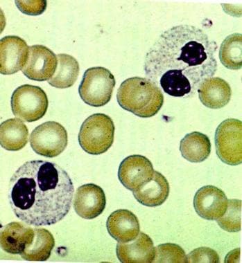 Neutrophils in this blood smear (original magnification X1500) have the 2 characteristics of the Pelger-Huët anomaly: the pince nez appearance of the bilobate nuclei and excessively coarse clumping of chromatin. Used with permission from Little, Brown.
LBR also interacts with HP-1 heterochromatin proteins; this is hypothesized to account for the excessive coarse clumping of nuclear chromatin that is observed. [12, 13] LBR abnormalities do not affect neutrophil function, and Pelger-Huët cells survive normally in circulation and can phagocytize and kill microorganisms. [14, 15]
Research also suggests that PHA and HEM skeletal dysplasia are related to a defect in cholesterol synthesis, resulting from LBR point mutations that cause a loss of sterol C14 reductase activity associated with the LBR protein. This loss occurs when the point mutations lower LBR’s affinity for the reducing agent nicotinamide adenine dinucleotide phosphate (NADPH). [16, 17]
Homozygous LBR mutations are rare, with only 11 individuals described; neutrophils have a single, round nucleus with clumped chromatin, and basophils, eosinophils, and megakaryocytes also show rounded nuclear lobes and dense nuclear chromatin. [5] Co-inherited LBR gene nonsense mutations can also result in lethal hydrops, ectopic calcification, moth-eaten (HEM) skeletal dysplasia/Greenberg skeletal dysplasia, and there continues to be debate as to how PHA and HEM skeletal dysplasia overlap. Some patients with HEM skeletal dysplasia have neutrophils with features of PHA, and some patients with PHA have mild skeletal anomalies. [10, 18, 6]
The Yakut tribe of northeastern Asia have rare cases of SOPH (short stature, optic atrophy, and PHA) syndrome, attributed to a "founder effect" and due to a mutation in neuroblastoma-associated sequences (NBASs). The wide clinical spectrum of disease associated with NBAS mutations has come to be more clearly described. [19, 20]
Epidemiology
Pelger-Huët anomaly was originally observed in individuals from Switzerland, Germany, or Holland. The anomaly has been described in all ethnic groups, including White, Black, and Asian persons of all ages, with no gender predisposition. The prevalence rate of heterozygous PHA in the United States is estimated as 1 case in 5000 population, and in the United Kingdom, 1 case in 6000 population. The highest described incidence is in the Gelenau region of Germany (1.01%) and the Vasterbotten region of Sweden (0.6%). [8] In the aforementioned Yakut tribe, PHA is seen in association with a syndrome of growth retardation.
Prognosis
Individuals with PHA are in good health, and their natural resistance to infection is unimpaired, although persons with the rare homozygous LBR mutations may have skeletal anomalies, and those with rare NBAS mutations suffer from growth retardation and progeria.
The practical importance of identifying PHA lies in distinguishing this benign entity from a bandemia with a left-shifted peripheral blood smear that can be observed in association with infection, as well as from PPHA.
-
Neutrophils in this blood smear (original magnification X1500) have the 2 characteristics of the Pelger-Huët anomaly: the pince nez appearance of the bilobate nuclei and excessively coarse clumping of chromatin. Used with permission from Little, Brown.


