Practice Essentials
Otitis media (OM) is any inflammation of the middle ear (see the images below), without reference to etiology or pathogenesis. It is very common in children.
There are several subtypes of OM, as follows:
-
Acute OM (AOM)
-
OM with effusion (OME)
-
Chronic suppurative OM
-
Adhesive OM
Signs and symptoms
AOM implies rapid onset of disease associated with one or more of the following symptoms:
-
Otalgia
-
Otorrhea
-
Headache
-
Fever
-
Irritability
-
Loss of appetite
-
Vomiting
-
Diarrhea
OME often follows an episode of AOM. Symptoms that may be indicative of OME include the following:
-
Hearing loss
-
Tinnitus
-
Vertigo
-
Otalgia
Chronic suppurative otitis media is a persistent ear infection that results in tearing or perforation of the eardrum.
Adhesive otitis media occurs when a thin retracted ear drum becomes sucked into the middle ear space and stuck.
See Clinical Presentation for more detail.
Diagnosis
OME does not benefit from antibiotic treatment. Therefore, it is critical for clinicians to be able to distinguish normal middle ear status from OME or AOM. Doing so will avoid unnecessary use of antibiotics, which leads to increased adverse effects of medication and facilitates the development of antimicrobial resistance.
Examination
Pneumatic otoscopy remains the standard examination technique for patients with suspected OM. In addition to a carefully documented examination of the external ear and tympanic membrane (TM), examining the entire head and neck region of patients with suspected OM is important.
Every examination should include an evaluation and description of the following four TM characteristics:
-
Color – A normal TM is a translucent pale gray; an opaque yellow or blue TM is consistent with middle-ear effusion (MEE)
-
Position – In AOM, the TM is usually bulging; in OME, the TM is typically retracted or in the neutral position
-
Mobility – Impaired mobility is the most consistent finding in patients with OME
-
Perforation – Single perforations are most common
Adjunctive screening techniques for OM include tympanometry, which measures changes in acoustic impedance of the TM/middle ear system with air pressure changes in the external auditory canal, and acoustic reflectometry, which measures reflected sound from the TM; the louder the reflected sound, the greater the likelihood of an MEE.
See Workup for more detail.
Management
Most cases of AOM improve spontaneously. Cases that require treatment may be managed with antibiotics and analgesics or with observation alone.
Guidelines from American Academy of Pediatrics
In February 2013, the American Academy of Pediatrics (AAP) and the American Academy of Family Physicians (AAFP) released updated guidelines for the diagnosis and management of AOM, including recurrent AOM, in children aged 6 months through 12 years. The recommendations offer more rigorous diagnostic criteria to reduce unnecessary antibiotic use.
According to the guidelines, management of AOM should include an assessment of pain. Analgesics, particularly acetaminophen and ibuprofen, should be used to treat pain whether antibiotic therapy is or is not prescribed.
Recommendations for prescribing antibiotics include the following:
-
Antibiotics should be prescribed for bilateral or unilateral AOM in children aged at least 6 months with severe signs or symptoms (moderate or severe otalgia, otalgia for 48 hours or longer, or temperature 39°C or higher) and for nonsevere, bilateral AOM in children aged 6 to 23 months
-
On the basis of joint decision-making with the parents, unilateral, nonsevere AOM in children aged 6-23 months or nonsevere AOM in older children may be managed either with antibiotics or with close follow-up and withholding antibiotics unless the child worsens or does not improve within 48-72 hours of symptom onset
-
Amoxicillin is the antibiotic of choice unless the child received it within 30 days, has concurrent purulent conjunctivitis, or is allergic to penicillin; in these cases, clinicians should prescribe an antibiotic with additional beta-lactamase coverage
In February 2016, the American Academy of Otolaryngology–Head and Neck Surgery Foundation, the AAP, and the AAFP issued updated guidelines for the assessment and management of OME.
See Treatment and Medication for more detail.
Background
Otitis media (OM) is the second most common disease of childhood, after upper respiratory infection (URI). OM is also the most common cause for childhood visits to a physician's office. Annually, an estimated 16 million office visits are attributed to OM; this does not include visits to the emergency department.
OM is any inflammation of the middle ear, without reference to etiology or pathogenesis. It can be classified into many variants on the basis of etiology, duration, symptomatology, and physical findings.
Acute OM (AOM) implies rapid onset of disease associated with one or more of the following symptoms:
-
Otalgia
-
Fever
-
Otorrhea
-
Recent onset of anorexia
-
Irritability
-
Vomiting
These symptoms are accompanied by abnormal otoscopic findings of the tympanic membrane (TM), which may include the following:
-
Opacity
-
Bulging
-
Erythema
-
Middle-ear effusion (MEE)
-
Decreased mobility with pneumatic otoscopy
AOM is a recurrent disease. More than one third of children experience six or more episodes of AOM by age 7 years.
OM with effusion (OME), formerly termed serous OM or secretory OM, is MEE of any duration that lacks the associated signs and symptoms of infection (eg, fever, otalgia, and irritability). OME usually follows an episode of AOM.
Chronic suppurative OM is a chronic inflammation of the middle ear that persists for at least 6 weeks and is associated with otorrhea through a perforated TM, an indwelling tympanostomy tube (TT; see the image below), or a surgical myringotomy.
Pathophysiology
The most important factor in middle ear disease is eustachian tube (ET) dysfunction (ETD), in which the mucosa at the pharyngeal end of the ET is part of the mucociliary system of the middle ear. Interference with this mucosa by edema, tumor, or negative intratympanic pressure facilitates direct extension of infectious processes from the nasopharynx to the middle ear, causing OM. Esophageal contents regurgitated into the nasopharynx and middle ear through the ET can create a direct mechanical disturbance of the middle ear mucosa and cause middle ear inflammation.
In children, developmental alterations of the ET, an immature immune system, and frequent infections of the upper respiratory mucosa all play major roles in AOM development. Studies have demonstrated how viral infection of the upper respiratory epithelium leads to increased ETD and increased bacterial colonization and adherence in the nasopharynx. [1]
Certain viral infections cause abnormal host immune and inflammatory responses in the ET mucosa and subsequent microbial invasion of the middle ear. The host immune and inflammatory response to bacterial invasion of the middle ear produces fluid in the middle ear and the signs and symptoms of AOM.
Although interactions between the common pathogenic bacteria in AOM and certain viruses are not fully understood, strong evidence indicates that these interactions often lead to more severe disease, lowered response to antimicrobial therapy, and OME development following AOM.
Etiology
A multitude of host, infectious, allergic, and environmental factors contribute to the development of OM.
Host factors
Immune system
The immature immune systems of infants or the impaired immune systems of patients with congenital immune deficiencies, HIV infection, or diabetes may be involved in the development of OM. [2] OM is an infectious disease that prospers in an environment of decreased immune defenses. The interplay between pathogens and host immune defense plays a role in disease progression.
Patel et al found higher interleukin (IL)-6 levels in patients with OM who also had influenza and adenoviral infections, whereas IL-1β levels were higher in patients who developed OM following URI. [3] In another study, Skovbjerg et al found that middle ear effusions with culturable pathogenic bacteria were associated with higher levels of IL-1β, IL-8, and IL-10 than sterile effusions. [4]
Familial (genetic) predisposition
Although familial clustering of OM has been demonstrated in studies that examined genetic associations of OM, separating genetic factors from environmental influences has been difficult. No specific genes have been linked to OM susceptibility. As with most disease processes, effects of environmental exposures on genetic expression probably play an important role in OM pathogenesis.
Mucins
The role of mucins in OME has been described. Mucins are responsible for gel-like properties of mucus secretions. The middle ear mucin gene expression is unique compared with the nasopharynx. Abnormalities of this gene expression, especially upregulation of MUC5B in the ear, may have a predominant role in OME.
Anatomic abnormality
Children with anatomic abnormalities of the palate and associated musculature, especially the tensor veli palantini, exhibit marked ETD and have higher risk for OM. Specific anomalies that correlate with high prevalence of OM include cleft palate, Crouzon syndrome or Apert syndrome, Down syndrome, and Treacher Collins syndrome.
Physiologic dysfunction
Abnormalities in the physiologic function of the ET mucosa, including ciliary dysfunction and edema, increase the risk of bacterial invasion of the middle ear and the resultant OME. Children with cochlear implants have a high incidence of OM, especially chronic OM and cholesteatoma formation. One study described a relation between laryngopharyngeal reflux and chronic OM (COM); the authors concluded that reflux workup should be performed as part of COM investigations and that if reflux is confirmed, reflux treatment should be initiated in addition to treatment of primary disease. [5]
Other host factors
Vitamin A deficiency is associated with pediatric upper respiratory infections and AOM.
Obesity has been linked to an increased incidence of OM, although the causal factor is unknown. Speculations include alteration of intrinsic cytokine profile, increased gastroesophageal reflux with alterations of the oral flora, and/or fat accumulation; all of these have been linked with an increased incidence of OM. Conversely, OM may increase the risk of obesity by altering the taste buds. [6]
Infectious factors
Bacterial pathogens
The most common bacterial pathogen in AOM is Streptococcus pneumoniae, followed by nontypeable Haemophilus influenzae and Moraxella (Branhamella) catarrhalis. These three organisms are responsible for more than 95% of all AOM cases with a bacterial etiology. [7]
In infants younger than 6 weeks, gram-negative bacilli (eg, Escherichia coli, Klebsiella species, and Pseudomonas aeruginosa) play a much larger role in AOM, causing 20% of cases. S pneumoniae and H influenzae are also the most common pathogens in this age group. Some studies also found Staphylococcus aureus as a pathogen in this age group, but subsequent studies suggested that the flora in these young infants may be that of usual AOM in children older than 6 weeks.
Many experts had proposed that the MEE associated with OME was sterile because cultures of middle ear fluid obtained by tympanocentesis often did not grow bacteria. This view is changing as newer studies show 30-50% incidence of positive results in middle ear bacterial cultures in patients with chronic MEE. These cultures grow a wide range of aerobic and anaerobic bacteria, of which S pneumoniae, H influenzae, M catarrhalis, and group A streptococci are the most common.
M catarrhalis–induced AOM differs from AOM caused by other bacterial pathogens in several ways. It is characterized by higher a proportion of mixed infections, younger age at the time of diagnosis, lower risk of spontaneous perforation of the tympanic membrane, and an absence of mastoiditis. [8]
Further evidence for the presence of bacteria in the MEE of patients with OME was provided by studies using polymerase chain reaction (PCR) assay to detect bacterial DNA in MEE samples that were determined to be sterile with standard bacterial culture techniques. In one such study using PCR assay, 77.3% of the MEE samples had positive results for one or more common AOM pathogens (eg, S pneumoniae, H influenzae, M catarrhalis).
In chronic suppurative OM, the most frequently isolated organisms include P aeruginosa, S aureus, Corynebacterium species, and Klebsiella pneumoniae. An unanswered question is whether these pathogens invade the middle ear from the nasopharynx via the ET (as do the bacteria responsible for AOM) or whether they enter through the perforated TM or a TT from the EAC.
The role of Helicobacter pylori in children with OME has been increasingly recognized. [9] Evidence that this agent might be responsible for OME comes from its isolation from middle ear and tonsillar and adenoidal tissue in patients with OME.
Alloiococcus otitidis is a species of gram-positive bacterium that has been discovered as a pathogen associated with OME. [10, 11] This organism is the most frequent bacterium in AOM, as well as in OME. It has also been detected in patients who had been treated with antibiotics, such as beta-lactams or erythromycin, suggesting that these agents may not be sufficiently effective to eliminate this organism. Further investigation is needed to reveal the clinical role of the organism in OM.
Viral pathogens
Because acute viral URI is a prominent risk factor for AOM development, most investigators have suspected a role for respiratory viruses in AOM pathogenesis.
Many studies have substantiated this suspicion by showing how certain respiratory viruses can cause inflammatory changes to the respiratory mucosa that lead to ETD, increased bacterial colonization and adherence, and, eventually, AOM. Studies have also shown that viruses can alter the host-immune response to AOM, thereby contributing to prolonged middle ear fluid production and development of chronic OME.
The viruses most commonly associated with AOM are respiratory syncytial virus (RSV), influenza viruses, parainfluenza viruses, rhinovirus, and adenovirus. Human parechovirus 1 (HPeV1) infection is associated with OM and cough in pediatric patients. [12] OM developed in 50% of 3-month follow-up periods that yielded evidence of HPeV1 infection but in only 14% of the HPeV1-negative periods; in recurring OM, the middle ear fluid samples were positive for HPeV in 15% of episodes.
Factors related to allergies
The relation between allergies and OM remains unclear. In children younger than 4 years, the immune system is still developing, and allergies are unlikely to play a role in recurrent AOM in this age group. Although much evidence suggests that allergies contribute to the pathogenesis of OM in older children, extensive evidence refutes the role of allergies in the etiology of middle ear disease.
The following is a brief list of evidence for and against the etiologic role of allergy in OM:
-
Many patients with OM have concomitant allergic respiratory disease (eg, allergic rhinitis, asthma)
-
Many patients with OM have positive results to skin testing or radioallergosorbent testing (RAST)
-
Although mast cells are found in the middle ear mucosa, most studies fail to show significant levels of immunoglobulin E (IgE) or eosinophils in the MEE of patients with OM
-
OM is most common in the winter and early spring, yet most major allergens (eg, tree and grass pollens) peak in the late spring and early fall
-
Most patients with concomitant OM and allergy show no marked improvement in middle ear disease with aggressive allergy management, despite marked improvements to nasal and other allergy-related symptoms
Environmental factors
Infant feeding methods
Many studies report that breastfeeding protects infants against OM. The best of these studies indicates that this benefit is evident only in children who are breastfed exclusively for the first 3-6 months of life. Breastfeeding of this duration reduces the incidence of OM by 13%. The protective effects of breastfeeding for the first 3-6 months persist for 4-12 months after breastfeeding ceases, possibly because delaying onset of the first OM episode reduces recurrence of OM in these children.
Passive smoke exposure
Many studies have shown a direct relation between passive smoke exposure and risk of middle ear disease. [13] A systematic review of 45 publications dealing with OM and parental smoking showed pooled odds ratios of 1.48 (95% confidence interval [CI], 1.08-2.04) for recurrent OM, 1.38 (95% CI, 1.23-1.55) for MEE, and 1.3 (95% CI, 1.3-1.6) for AOM. [14]
Group daycare attendance
Daycare centers create close contact among many children, which increases the risks of respiratory infection, nasopharyngeal colonization with pathogenic microbes, and OM.
Many researchers have used meta-analysis to confirm that exposure to other young children (including siblings) in group daycare settings is a major risk factor for OM. [15] A meta-analysis reported that care outside the home conferred a 2.5-fold risk for OM. Other critical reviews of studies on OM and group childcare show heightened odds ratios of 1.6-4.0:1 for center care versus home care.
Children who attend daycare centers frequently acquire antibacterial-resistant organisms in their nasopharynx, leading to AOM that may be refractory to antibacterial treatment. American Academy of Pediatrics (AAP) and American Academy of Family Physicians (AAFP) guidelines recommend high-dose amoxicillin-clavulanate as the antibiotic of choice in the treatment of AOM in children who attend daycare.
Socioeconomic status
Socioeconomic status encompasses many independent factors that affect both the risk of OM and the likelihood that OM will be diagnosed. [16]
In general, lower socioeconomic status confers higher risk for environmental exposure to parental smoking, bottle-feeding, crowded group daycare, crowded living conditions, and viruses and bacterial pathogens. Compared with children from middle-income and high-income families, children from lower socioeconomic groups use health care resources less frequently, which decreases the likelihood that OM cases will be diagnosed.
Epidemiology
United States statistics
OM, the most common specifically treated childhood disease, accounts for approximately 20 million annual physician visits. Various epidemiologic studies report the prevalence rate of AOM to be 17-20% within the first 2 years of life, and 90% of children have at least one documented MEE by age 2 years. OM is a recurrent disease. One third of children experience six or more episodes of AOM by age 7 years.
International statistics
Incidence and prevalence in other industrialized nations are similar to US rates. In less developed nations, OM is extremely common and remains a major contributor to childhood mortality resulting from late-presenting intracranial complications. International studies show increased prevalence of AOM and chronic OM (COM) among Micronesian and Australian aboriginal children.
Age-related demographics
Peak prevalence of OM in both sexes occurs in children aged 6-18 months. Some studies show bimodal prevalence peaks; a second, lower peak occurs at age 4-5 years and corresponds with school entry. Although OM can occur at any age, 80-90% of cases occur in children younger than 6 years. Children who are diagnosed with AOM during the first year of life are much more likely to develop recurrent OM and chronic OME than children in whom the first middle ear infection occurs after age 1 year.
Sex-related demographics
Several studies have now shown equal AOM prevalence in males and females; many previous studies had shown increased incidence in boys.
Race-related demographics
For some time, the prevalence of OM in the United States was reported to be higher in black and Hispanic children than in white children. However, a study that controlled for socioeconomic and other confounding factors showed equal incidence in blacks and whites. Hispanic children and Alaskan Inuit and other American Indian children have higher prevalence of AOM than white and black children in the United States.
Prognosis
US mortality is extremely low in this era of antimicrobial therapy (< 1 death per 100,000 cases). In developing nations with limited access to primary medical care and modern antibiotics, mortality figures are similar to those reported in the United States before antibiotic therapy. A study that examined the causes of death in Los Angeles County Hospital from 1928-1933, years before the advent of sulfa, showed that 1 in 40 deaths was caused by intracranial complications of OM.
Morbidity from this disease remains significant, despite frequent use of systemic antibiotics to treat the illness and its complications. Intratemporal and intracranial complications of OM are the two major types.
Intratemporal complications include the following:
-
Hearing loss (conductive and sensorineural)
-
TM perforation (acute and chronic)
-
Chronic suppurative OM (with or without cholesteatoma)
-
Tympanosclerosis
-
Petrositis
-
Labyrinthitis
-
Facial paralysis
-
Cholesterol granuloma
-
Infectious eczematoid dermatitis
Intracranial complications include the following [17] :
-
Meningitis
-
Subdural empyema
-
Brain abscess
-
Extradural abscess
-
Lateral sinus thrombosis
-
Otitic hydrocephalus
The prognosis for almost all patients with OM is excellent [18] ; the exceptions are patients in whom OM involves intratemporal and intracranial complications (< 1%).
Data on cognitive and educational outcomes of OM in the literature are limited. [19] The impact of OM on child development depends on numerous factors. OM in infants younger than 12 months predisposes to long-term speech and language problems. OM has also been reported to negatively affect preexisting cognitive or language problems. Careful follow-up and early referral are key to management.
Patient Education
Patient education topics should include the following:
-
Avoiding risk factors
-
Appropriate use of antibiotics
-
Understanding the implications of antibiotic-resistant bacteria in OM
Education for health care providers should focus on the following topics:
-
Antibiotic-resistant bacteria and the need to avoid overprescribing antibiotics
-
Importance of pneumatic otoscope examination to distinguish AOM from OME
-
Treatment differences between AOM and OME
For patient education resources, see the Ear, Nose, and Throat Center, as well as Earache.
-
Diagram of the normal tympanic membrane anatomy.
-
Healthy tympanic membrane.
-
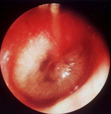
Acute otitis media with purulent effusion behind a bulging tympanic membrane.
-
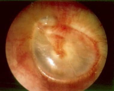
Chronic otitis media with a retraction pocket of the pars flaccida.
-
Cholesteatoma of the pars flaccida.
-
Central/pars tensa tympanic membrane perforation with a healthy middle ear membrane.
-
Central/pars tensa tympanic membrane perforation with a tympanostomy tube in place.
-
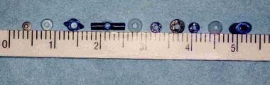
Various tympanostomy tube styles and sizes.
-
Initial presentation of a young girl with chronic right ear pain and multiple untreated middle ear infections.
-
Acute coalescent mastoiditis with a Bezold abscess in a young girl who presented with chronic right ear pain and multiple untreated middle ear infections.
-
A young girl who presented with chronic right ear pain and multiple untreated middle ear infections on the operating table for mastoidectomy and drainage of Bezold abscess.
-
Aspirating pus from the Bezold abscess for Gram staining, culturing, and sensitivity testing in a young girl who presented with chronic right ear pain and multiple untreated middle ear infections.
-
Surgical incision to aspirate pus in a young girl who presented with chronic right ear pain and multiple untreated middle ear infections.
-
Freer elevator demonstrating extension of an abscess cavity from the mastoid into the neck in a young girl who presented with chronic right ear pain and multiple untreated middle ear infections.
-
Incision is closed and a drain is placed in the abscess cavity in a young girl who presented with chronic right ear pain and multiple untreated middle ear infections.
-
Postoperative bandage in a young girl who presented with chronic right ear pain and multiple untreated middle ear infections.
-
The wound now appears clean and dry on postoperative day 4. This young girl initially presented with chronic right ear pain and multiple untreated middle ear infections.
-
Postoperative day 4: Mom is smiling. This young girl initially presented with chronic right ear pain and multiple untreated middle ear infections.


