Continually Updated Clinical Reference
 
 
  All Sources     eMedicine     Medscape     Drug Reference     MEDLINE
 
eMedicine - Nail-Patella Syndrome : Article by

Quick Find
Authors & Editors
Introduction
Clinical
Differentials
Workup
Treatment
Medication
Follow-up
Miscellaneous
References


Patient Education
Click here for patient education.


Author: Suzanne M Carter, MS, Senior Genetic Counselor, Associate, Department of Obstetrics and Gynecology, Division of Reproductive Genetics, Montefiore Medical Center, Albert Einstein College of Medicine

Suzanne M Carter is a member of the following medical societies: American Bar Association

Coauthor(s): Susan J Gross, MD, FRCS(C), FACOG, FACMG, Codirector, Division of Reproduction Genetics, Associate Professor, Department of Obstetrics and Gynecology, Albert Einstein College of Medicine

Editors: Christian J Renner, MD, Consulting Staff, Department of Pediatrics, University Hospital for Children and Adolescents, Erlangen, Germany; Mary L Windle, PharmD, Adjunct Assistant Professor, University of Nebraska Medical Center College of Pharmacy, Pharmacy Editor, eMedicine.com, Inc; Leonard G Feld, MD, PhD, MMM, Chairman of Pediatrics, Carolinas Medical Center; Chief Medical Officer, Levine Children's Hospital, Carolinas Healthcare System; Daniel Rauch, MD, FAAP, Director, Pediatric Hospitalist Program, Associate Professor, Department of Pediatrics, New York University School of Medicine; Bruce Buehler, MD, Professor, Department of Pathology and Microbiology, Director, Hattie B Munroe Center for Human Genetics, Chairman, Department of Pediatrics, University of Nebraska Medical Center

Author and Editor Disclosure

Synonyms and related keywords: nail-patella syndrome, NPS, Fong disease, NPS 1, onycho-osteodysplasia, Turner-Kieser syndrome, arthro-onychodysplasia, nephrotic syndrome, end-stage renal disease, ESRD, end-stage renal failure, LMX1B, NPS1, proteinuria

Background

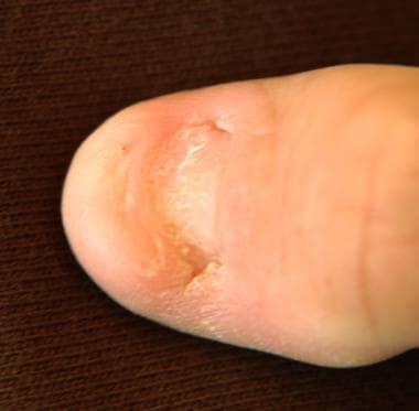
Nail-patella syndrome (NPS; OMIM 161200) is a well-known autosomal dominant condition characterized by nail dysplasia, patellar aplasia-hypoplasia, arthrodysplasia of the elbows, iliac horns, and nephropathy. Recent reports indicate that open-angle glaucoma (OAG) may also cosegregate with NPS. One of the first chromosomal linkages identified in humans is between the NPS locus and the ABO blood group on chromosome 9. LMX1B, located on band 9q34.1, is an LIM-homeodomain transcription factor required for the normal development of dorsal limb structures, the glomerular basement membrane, the anterior segment of the eye, and dopaminergic and serotonergic neurons. Heterozygous loss-of-function mutations in LMX1B cause NPS.

Pathophysiology

Although the joint anomalies in NPS may limit range of motion (ROM), the associated nephropathy may be the most serious complication. Asymptomatic proteinuria may persist for years; however, end-stage renal failure has been reported.

Frequency

United States

NPS has been studied for more than 100 years. It has a prevalence of 1 per 50,000 live births.

Mortality/Morbidity

The age of onset and degree of severity cannot be predicted. Patients are at risk for nephropathy that resembles glomerulonephritis. This occurs in 30-55% of cases, and renal failure is the leading cause of death. Proteinuria is the first clinical symptom, and some cases progress to end-stage renal disease (ESRD), which requires transplantation. Although many patients are asymptomatic until the early adult years, proteinuria may develop before age 2 years.

Research has revealed that the location of the LMXB1 mutation may play a role in the frequency and severity of proteinuria. If the mutation is located in the homeodomain, proteinuria is more frequent and more severe.

Race

No race predilection has been reported.

Sex

Males and females are equally affected.

Age

Prenatal diagnosis based on ultrasonography findings is reported; however, the associated clinical findings span all ages.


History

  • Proteinuria: This is the first sign of renal involvement. The patient may remain asymptomatic or eventually develop ESRD.
  • Hypoplastic nails: Absence of the thumbnail is the most common nail pattern. Other nails may be less fragile.
  • Knee abnormalities: Some patients report recurrent dislocation or arthritis, which may necessitate patellar replacement.
  • Limited ROM in the elbows
  • Decreased pronation or supination of the elbows, appearing unilaterally or bilaterally
  • Hypoplastic radial head

Physical

  • Renal symptoms: These symptoms range from proteinuria to nephrotic syndrome.
  • Nephropathy (resembles glomerulonephritis)
    • Prevalence ranges from 30-55%, and renal failure is the leading cause of death.
    • Patients present with proteinuria as the first clinical symptom.
    • Some patients eventually develop ESRD and require transplantation.
  • Open-angle glaucoma
    • Strong evidence supports that OAG is a pleiotropic effect of the NPS1 gene.
    • Age at diagnosis ranges from birth to late adulthood.
    • Optic nerve and visual field damage can be prevented with early diagnosis and treatment of elevated intraocular pressure.
  • Patellar abnormalities
    • Skeletal deformities include patellar absence or hypoplasia, which may decrease flexion.
    • Osteoarthritis, osteoarthrosis, and knee effusions are associated complications; however, disability is not a major concern.
  • Dysplastic nails
    • Although absent or dysplastic nails are the most common nail findings, nonspecific changes include discoloration, longitudinal ridging, and poorly formed lunulae.
    • Nails are progressively less affected toward the fifth digit.
  • Iliac horns: These symmetrical, bilateral, central-posterior, iliac processes are asymptomatic and vary from a small dimple to a well-marked spur.
  • Elbow arthrodysplasia
    • Clinical manifestations include an increased carrying angle and limited supination and pronation.
    • On radiographs, the head of the radius is underdeveloped and posteriorly displaced.

Causes

  • The LMX1B gene plays a central role in limb dorsoventral patterning and patterning of the nails, patella, and long bones during development.
  • Human fetal and adult kidneys show a high level of LMX1B expression, thereby suggesting a critical function for this gene during kidney development.
  • The loss of function in one allele of this gene is the most likely cause of nephropathy, dysplastic nails, and hypoplastic patella manifesting in nail-patella syndrome.


Other Problems to be Considered

Familial patella aplasia-hypoplasia syndrome
Genitopatellar syndrome
Small patella syndrome


Lab Studies

  • Conduct urinalysis, along with a microscopic analysis, to check for proteinuria and hematuria.
  • Obtain a 24-hour urine collection to quantitate protein and creatinine excretion. A urine protein-to-creatinine ratio on a random sample is also acceptable (normal ratio <0.2).
  • Evaluate the blood chemistry for plasma urea, BUN, and creatinine concentrations.

Imaging Studies

  • Radiography findings reveal iliac horns and hypoplastic patella in adults.
  • Conduct MRI to identify abnormal muscle insertions, which cannot be observed using radiography.
  • Ultrasonography is indicated in young children for a better evaluation of the unossified patella.

Other Tests

  • Some families have glaucoma that cosegregates with nail-patella syndrome (NPS).
  • The following annual vision checks are important:
    • Visual acuity testing
    • Slit-lamp biomicroscopic examination
    • Intraocular pressure determination
    • Applanation
    • Gonioscopy
    • Ophthalmoscopy

Procedures

  • Renal biopsy: Light microscopy reveals glomerulonephritis and basement membrane thickening; however, negative results do not rule out pathological processes in the kidney. Electron microscopy can reveal ultrastructural changes, such as irregularities and thickening of the basement membrane, which cannot be seen with light microscopy. This moth-eaten appearance is pathognomonic.

Histologic Findings

Increased lucency of the glomerular basement membrane resembles a moth-eaten appearance. A typical lesion consists of irregular basement membrane thickening, epithelial foot process fusion, and the presence of fibrillar collagenlike material within the basement.


Medical Care

  • Unless the patient presents with acute findings, perform the evaluation on an outpatient basis.
  • Suggest knee replacement if the patient develops osteoarthritis in the knees.
  • Dialysis and transplantation are necessary in patients who develop ESRD. Approximately 30-55% of patients have renal involvement.
  • The use of diuretics in patients with nephrotic syndrome depends on the glomerular filtration rate. Any therapy for edema requires an assessment of intravascular volume status.

Surgical Care

  • Renal transplantation has proven successful in patients with nail-patella syndrome (NPS) who develop ESRD.
  • Avoid excision of the radial head to correct elbow arthrodysplasia. This excision forces the ulna into the wrist, creating a painful condition.
  • MRI is necessary to reveal the abnormal muscle and nerve insertions that may complicate orthopedic procedures.

Consultations

  • Dentist
  • Geneticist
  • Nephrologist
  • Ophthalmologist
  • Orthopedist
  • Rheumatologist

Diet

  • No dietary restrictions are necessary unless hypertension or nephrotic syndrome develop. At this time, a salt-free diet is appropriate.

Activity

  • Joint abnormalities generally do not limit physical activity.


Vitamin D analogs, thiazides, and prednisone are effective in alleviating the symptoms of nephrotic syndrome and end-stage renal failure.

Drug Category: Vitamin D analog

Vitamin D is necessary to maintain the correct amount of calcium needed for strong bones and teeth and is needed throughout the body.

Drug NameCalcitriol (Rocaltrol)
DescriptionIncreases calcium levels by promoting absorption of calcium in intestines and retention in kidneys. The beneficial effects of vitamin D replacement in renal osteodystrophy appear to result from correction of hypocalcemia and secondary hyperparathyroidism.
Adult Dose0.25 mcg/d PO initially
If the response in the biochemical parameters and clinical manifestations of the disease state is not satisfactory, dosage may be increased by 0.25 mcg/d q4-8wk; typical dosage range is 0.5-1 mcg/d
Pediatric Dose<3 years: 10-15 ng/kg/d PO
>3 years: Administer as in adults
ContraindicationsDocumented hypersensitivity; hypercalcemia; malabsorption syndrome
InteractionsCholestyramine and colestipol decrease absorption of calcitriol; magnesium-containing antacids and thiazide diuretics can increase calcitriol effects
PregnancyC - Safety for use during pregnancy has not been established.
PrecautionsHypercalcemia, hypercalciuria, and hyperphosphatemia

Drug Category: Thiazide diuretics

These agents are used to treat edema caused by renal dysfunction (eg, nephrotic syndrome, chronic renal failure).

Drug NameHydrochlorothiazide (Hydro-Diuril, Microzide)
DescriptionInhibits reabsorption of sodium in distal tubules, increasing excretion of sodium, water, and potassium and hydrogen ions.
Adult Dose25-100 mg PO qd or divided bid
Pediatric Dose1-2 mg/kg PO qd or divided bid; not to exceed 37.5 mg/d (age <2 y) or 100 mg/d (age 2-12 y)
ContraindicationsDocumented hypersensitivity; anuria or renal decompensation
InteractionsMay decrease effects of anticoagulants, antigout agents, or sulfonylureas; may increase toxicity of allopurinol, anesthetics, antineoplastics, calcium salts, loop diuretics, lithium, diazoxide, digitalis, amphotericin B, or nondepolarizing muscle relaxants
PregnancyC - Safety for use during pregnancy has not been established.
PrecautionsCaution in renal or hepatic disease, gout, diabetes mellitus, or erythematosus

Drug Category: Corticosteroids

These agents induce diuresis or remission of proteinuria in nephrotic syndrome.

Drug NamePrednisone (Deltasone, Orasone)
DescriptionActs as an anti-inflammatory agent and immunosuppressant.
Dose depends on specific disease entity being treated; in situations of less severity, lower doses generally suffice, whereas, in selected patients, higher initial doses may be required; initial dosage should be maintained or adjusted prn.
Alternate day therapy (ADT) PO is a corticosteroid-dosing regimen in which twice the usual daily dose of corticoid is administered every other morning.
The purpose of this mode of therapy is to provide the patient who requires long-term pharmacologic dose treatment with the beneficial effects of corticoids while minimizing certain undesirable effects, including pituitary-adrenal suppression, the Cushingoid state, corticoid withdrawal symptoms, and growth suppression in children.
Adult Dose5-60 mg/d PO
Pediatric Dose1-2 mg/kg/d PO
ContraindicationsDocumented hypersensitivity; viral infection; peptic ulcer disease; hepatic dysfunction; connective tissue infections; fungal or tubercular skin infections; GI disease
InteractionsCoadministration with estrogens may decrease clearance; concurrent use with digoxin may cause digitalis toxicity secondary to hypokalemia; phenobarbital, phenytoin, and rifampin may increase metabolism of glucocorticoids (consider increasing maintenance dose); monitor for hypokalemia with coadministration of diuretics
PregnancyB - Usually safe but benefits must outweigh the risks.
PrecautionsAbrupt discontinuation of glucocorticoids may cause adrenal crisis; hyperglycemia, edema, osteonecrosis, myopathy, peptic ulcer disease, hypokalemia, osteoporosis, euphoria, psychosis, myasthenia gravis, growth suppression, and infections may occur with glucocorticoid use


Further Inpatient Care

  • Admit the patient for testing and renal transplantation.
  • If the patient has severe osteoarthritis, knee replacement may be required.

Further Outpatient Care

  • Monitor renal function.
  • An ophthalmological evaluation is suggested for detection of glaucoma.
  • Consider a dermatologic consultation.

In/Out Patient Meds

  • Prednisone, vitamin D replacement, and thiazides are appropriate to manage nephrotic syndrome and end-stage renal failure.

Transfer

  • Transfer may be required for further evaluation and renal transplantation.

Complications

  • Glaucoma
  • Osteoarthritis
  • Nephrotic syndrome
  • End-stage renal failure

Prognosis

  • Approximately 30-55% of patients with nail-patella syndrome (NPS) develop nephropathy, which may lead to end-stage renal failure. However, the presence of nephropathy or renal failure does not increase the risk of the same complications in their children.
  • A normal lifestyle, including procreation, is possible after renal transplantation; however, genetic counseling is recommended in patients with NPS.

Patient Education

  • Genetic counseling is recommended because the risk of having affected offspring is 50%. Prenatal diagnosis using ultrasonography and molecular analysis is possible.
  • Stress the importance of regular assessment of renal function to the patient.
  • The patient should receive annual glaucoma checks.


Medical/Legal Pitfalls

  • Failure to recognize associated conditions such as glaucoma
  • Failure to recommend genetic counseling

Special Concerns

  • Nephropathy occurs in children as young as 2 years.


  • Azouz EM, Kozlowski K. Small patella syndrome: a bone dysplasia to recognize and differentiate from the nail-patella syndrome. Pediatr Radiol. May 1997;27(5):432-5. [Medline].
  • Bennett WM, Musgrave JE, Campbell RA, et al. The nephropathy of the nail-patella syndrome. Clinicopathologic analysis of 11 kindred. Am J Med. Mar 1973;54(3):304-19. [Medline].
  • Bongers EM, Huysmans FT, Levtchenko E, et al. Genotype-phenotype studies in nail-patella syndrome show that LMX1B mutation location is involved in the risk of developing nephropathy. Eur J Hum Genet. Aug 2005;13(8):935-46. [Medline].
  • Browning MC, Weidner N, Lorentz WB Jr. Renal histopathology of the nail-patella syndrome in a two-year-old boy. Clin Nephrol. Apr 1988;29(4):210-3. [Medline].
  • Chen H, Lun Y, Ovchinnikov D, et al. Limb and kidney defects in Lmx1b mutant mice suggest an involvement of LMX1B in human nail patella syndrome. Nat Genet. May 1998;19(1):51-5. [Medline].
  • Cormier-Daire V, Chauvet ML, Lyonnet S, et al. Genitopatellar syndrome: a new condition comprising absent patellae, scrotal hypoplasia, renal anomalies, facial dysmorphism, and mental retardation. J Med Genet. Jul 2000;37(7):520-4. [Medline].
  • Cottereill CP, Jacobs P. Hereditary Arthro-osteo-onchyodysplasia associated with iliac horns. Br J Clin Pract. 1961;15(11):933-941.
  • Curtis JJ, Bhathena D, Leach RP, et al. Goodpasture''s syndrome in a patient with the Nail-Patella syndrome. Am J Med. Sep 1976;61(3):401-6. [Medline].
  • Darlington D, Hawkins, CF. Nail patella syndrome with iliachorns and hereditary nephropathy: necropsy report and anatomical dissection. J Bone Joint Surg [Am]. Jan 1967;49B:164-74. [Medline].
  • Dreyer SD, Zhou G, Baldini A, et al. Mutations in LMX1B cause abnormal skeletal patterning and renal dysplasia in nail patella syndrome. Nat Genet. May 1998;19(1):47-50. [Medline].
  • Dunston JA, Hamlington JD, Zaveri J, et al. The human LMX1B gene: transcription unit, promoter, and pathogenic mutations. Genomics. Sep 2004;84(3):565-76. [Medline].
  • Eisenberg KS, Potter DE, Bovill EG Jr. Osteo-onychodystrophy with nephropathy and renal osteodystrophy. A case report. J Bone Joint Surg [Am]. Sep 1972;54(6):1301-5. [Medline].
  • Feingold M, Itzchak Y, Goodman RM. Ultrasound prenatal diagnosis of the Nail-Patella syndrome. Prenat Diagn. Aug 1998;18(8):854-6. [Medline].
  • Fong EE. 'Iliac horns' (symmetrical bilateral central posterior iliac processes):a case report. Radiology. 1946;47:517-518.
  • Galloway G, Vivian A. An ophthalmic screening protocol for nail-patella syndrome. J Pediatr Ophthalmol Strabismus. Jan-Feb 2003;40(1):51-3. [Medline].
  • Leahy MS. The hereditary nephropathy of osteo-onychodysplasia. Nail-patella syndrome. Am J Dis Child. Sep 1966;112(3):237-41. [Medline].
  • Looij BJ Jr, te Slaa RL, Hogewind BL, van de Kamp JJ. Genetic counselling in hereditary osteo-onychodysplasia (HOOD, nail- patella syndrome) with nephropathy. J Med Genet. Oct 1988;25(10):682-6. [Medline].
  • McIntosh I, Clough MV, Gak E. Prenatal diagnosis of nail-patella syndrome [letter]. Prenat Diagn. Mar 1999;19(3):287-8. [Medline].
  • Morita T, Laughlin LO, Kawano K, et al. Nail-Patella syndrome. Light and electron microscopic studies of the kidney. Arch Intern Med. Feb 1973;131(2):271-7. [Medline].
  • Sabnis SG, Antonovych TT, Argy WP, et al. Nail-patella syndrome. Clinical Nephrology. Sep 1980;14(3):148-53. [Medline].
  • Schulz-Butulis BA, Welch MD, Norton SA. Nail-patella syndrome. J Am Acad Dermatol. Dec 2003;49(6):1086-7. [Medline].

Nail-Patella Syndrome excerpt

Article Last Updated: Feb 13, 2007