Practice Essentials
Milia are benign, keratinous cysts that commonly manifest as tiny white bumps on the face of the newborn (see the image below). When present on the gum margin and midline palate they are referred to as Bohn nodules and Epstein pearls, respectively. Milia can be broadly categorized into primary and secondary types. Congenital milia in newborns account for the vast majority of primary milia. Primary milia may also occur in association with one of several genodermatoses or sporadically without associated findings. Secondary milia may be associated with an underlying skin disease, medications, or trauma.
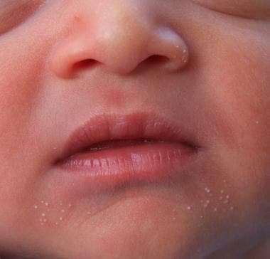 Milia in a week old infant.
Signs and symptoms
The clinical history varies somewhat between subtypes. Milia are usually asymptomatic.
Congenital milia are typically present at birth, although their onset may be delayed in premature neonates. Lesions usually resolve spontaneously.
Acquired milia have a tendency to persist without treatment. When acquired, a history of preceding trauma or associated bullous skin disease may be noted.
The typical lesion is a 1-3 mm white to yellow, dome shaped and smooth papule. Lesions may be associated with a faint blue hue in darkly pigmented skin. [1]
Lesions may be solitary or multiple.
Congenital milia predominate on the face, and the nose is commonly affected.
Benign acquired milia of children and adults favor the forehead, cheeks, eyelids, and genitalia.
Milia may be grouped.
Milia en plaque presents as an erythematous plaque studded with multiple milia. Lesions may be several centimeters in size.
Multiple eruptive milia favor the face, upper trunk, proximal extremities, and groin.
Diagnostics
Milia are easily diagnosed on clinical findings alone. Histologic examination reveals small cysts lined by stratified squamous epithelium and central keratinous material. [2] Persistent and widely distributed milia may warrant investigation for an underlying genodermatosis, particularly when other anomalies are present.
Management
Treatment of congenital milia is not necessary as these lesions have a tendency to spontaneously resolve. When multiple lesions are present in older children, topical retinoids may be effective.
Milia are easily treated with simple surgical interventions. These procedures are not painless; however, anesthesia is not needed for most patients. A small incision is made with a No. 11 blade or hypodermic needle. A comedone extractor is used to apply tangential pressure and express the contents. Other instruments may also be used, including the blunt side of a curette.
Background
In 2008, Berk and Bayliss published an updated classification of milia, as follows [3] :
Primary milia is as follows:
-
Congenital
-
Benign primary milia of children and adults
-
Milia en plaque
-
Nodular grouped milia
-
Multiple eruptive milia
-
Nevus depigmentosus with milia
-
Genodermatosis-associated
Secondary milia is as follows:
-
Disease-associated
-
Medication-associated
-
Trauma-associated
Congenital milia occur in nearly half of healthy newborns and are typically present at birth, although their onset may be delayed in premature neonates. [4] Lesions typically spontaneously resolve within weeks. Congenital milia predominate on the face, and the nose is frequently affected.
Benign acquired milia of children and adults also occur spontaneously; however, like other acquired milia, they have a tendency to persist without treatment. Benign acquired milia of children and adults favor the eyelids, cheeks, forehead and genitalia.
Multiple eruptive milia describes acquired and widespread milia that appear rather abruptly over weeks to months. Multiple eruptive milia may be associated with a genodermatosis or inherited in an autosomal dominant fashion without other apparent anomalies; however, in most cases they occur sporadically. [5]
Genodermatosis-associated milia have been reported in association with basal cell nevus syndrome, [6] Rombo syndrome, [7] Brooke-Spiegler syndrome, [8] pachyonychia congenita type 2, [9] and atrichia with papular lesions. [10]
In children, traumatic milia most commonly manifest following abrasions or burns. Milia have also been reported following skin grafting. [10] Milia may occur in association with blistering skin diseases. Epidermolysis bullosa and porphyria cutanea tarda are the classic examples. Milia associated with topical corticosteroid use is rarely reported. [11]
Pathophysiology
Histopathologic studies support the notion that primary milia arise from the lower infundibular sebaceous collar of the vellus hair, whereas secondary milia are more commonly derived from eccrine ducts. [12, 13]
Epidemiology
Frequency
Congenital milia are common, affecting 40-50% of healthy newborns. Infants born prematurely are less commonly affected, although onset may be delayed.
Race-, sex-, and age-related information
No racial predilection is observed.
In general, milia occur equally in males and females. Milia en plaque is more common in females.
Milia can affect persons of any age, but are most commonly seen in the neonatal period. Onset can be delayed for days to weeks in neonates born prematurely. Milia en plaque is most common in middle-aged adult females.
Prognosis
Congenital milia are benign cysts with a tendency for spontaneous resolution without scarring.
Acquired milia may persist without treatment.
Patient Education
Educate the family about the benign course of milia and tendency towards spontaneous resolution without scarring.
-
Milia in a week old infant.