Practice Essentials
Keratosis pilaris is a very common, benign, heritable disorder of keratinized hair follicles. [1] It is characterized by grouped, horny, keratotic, follicular papules, predominantly located over the extensor surfaces of the proximal extremities, most commonly the posterolateral upper arms and anterior thighs. Though the papules are typically asymptomatic, affected individuals may be bothered by the cosmetic appearance. There can be significant individual variation in the prominence and severity of keratosis pilaris. It is often described in children with underlying xerosis, atopy, or ichthyosis vulgaris. Treatment is marginally effective and only provides temporary relief. [2, 3]
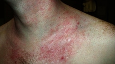
Images of keratosis pilaris are below (also see Physical Examination).
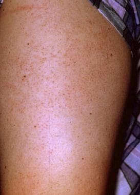 Keratosis pilaris occurs most commonly on the lateral upper arms and upper thighs.
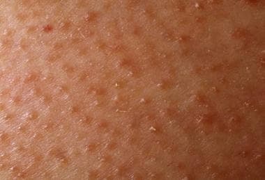 Close examination of keratosis pilaris shows keratotic papules associated with hair follicles. Keratinocytes at the follicular orifice are retained, producing keratin plugs.
Pathophysiology
Keratosis pilaris is theorized to result from defective keratinization of the follicular epithelium, resulting in the lack of proper desquamation of keratinocytes and the formation of a keratotic plug at the follicular orifice. Alternatively, some experts suggest that keratosis pilaris may not be a primary disorder of keratinization, but rather a disorder of the hair shaft. Circular hair shafts rupture the follicular epithelium, leading to inflammation and abnormal follicular keratinization. [4] More recently, atrophy or absence of sebaceous glands has been identified as an early feature in keratosis pilaris pathogenesis. [5]
Autosomal dominant inheritance has been described.
Etiology
The etiology of keratosis pilaris is not completely known. Keratosis pilaris may be due to a disorder of corneocyte adhesion that prevents normal desquamation in the area around the hair follicle. It is often associated with xerosis, ichthyosis vulgaris, and atopy. Other conditions reportedly associated with keratosis pilaris include ichthyosis follicularis, ectodermal dysplasia, keratitis ichthyosis deafness (KID) syndrome, Down syndrome, Noonan syndrome, [6] and cardiofaciocutaneous syndrome.
Al-Maawali et al propose that the gene BTG1 is critical for the development of the distinctive keratosis pilaris observed in patients with interstitial deletion of 12q21-q22. [7]
Keratosis pilaris–like eruptions have been described in patients receiving targeted cancer therapies such as RAF inhibitors (ie, vemurafenib) and the Bcr-Abl tyrosine kinase inhibitor nilotinib. [8, 9, 10, 11]
Epidemiology
Keratosis pilaris is a very common disorder observed worldwide in both children and adults.
The frequency of keratosis pilaris is increased in individuals with ichthyosis vulgaris, estimated at 74%. [12] Many older reports claim an increased incidence in patients with atopic dermatitis, but more recent studies have not demonstrated this association. Given a high prevalence and intensity of keratosis pilaris noted during puberty and in women with hyperandrogenism, some experts postulate that keratosis pilaris may be influenced by hormonal changes.
Keratosis pilaris has no known racial predilection.
Keratosis pilaris affects both males and females. The inflammatory form of keratosis pilaris may be more prevalent in females.
Keratosis pilaris most commonly develops during the first decade of life. It is estimated that 51% of cases are diagnosed during this period, while 35% are diagnosed during the second decade, 12% in the third decade, and 2% in the fourth decade.
Prognosis
Keratosis pilaris is a benign disorder and is not associated with increased mortality or long-term health consequences. Many patients find the lesions cosmetically unappealing and therefore seek treatment. A significant inflammatory component may be present, resulting in pruritus. Occasionally, keratosis pilaris may become secondarily infected due to scratching or abrasive self-therapy, in which case treatment of the infection is necessary.
The overall prognosis for patients is very good. Keratosis pilaris tends to improve over time, though it can persist with a waxing and waning course in some. Keratosis pilaris does not tend to leave scars.
A study performed by Poskitt demonstrated the following course [2] :
-
The condition dramatically improves in approximately 35% of patients, usually by late adolescence (mean age of improvement is 16 yrs).
-
The condition remains unchanged from the time of diagnosis in approximately 43% of patients.
-
Approximately 20% of patients experience a worsening of symptoms over time.
-
Approximately 50% experience a worsening of symptoms during wintertime, but only 60% of those who worsen improve over summertime.
Patient Education
Reassurance and gentle skin care are the most important recommendations the clinician can offer.
-
Keratosis pilaris occurs most commonly on the lateral upper arms and upper thighs.
-
The lesions of keratosis pilaris are evenly spaced, consistent with the follicular origin of this disorder.
-
Close examination of keratosis pilaris shows keratotic papules associated with hair follicles. Keratinocytes at the follicular orifice are retained, producing keratin plugs.
-
Bacteria associated with the follicular papules of keratosis pilaris may cause some lesions to become erythematous or pustular.