Continually Updated Clinical Reference
 
 
  All Sources     eMedicine     Medscape     Drug Reference     MEDLINE
 
eMedicine - Hypomelanosis of Ito : Article by

Quick Find
Authors & Editors
Introduction
Clinical
Differentials
Workup
Treatment
MEDICATION
Follow-up
Miscellaneous
References

Related Articles
Tuberous Sclerosis


Patient Education
Click here for patient education.


Author: Camila K Janniger, MD, Clinical Professor, Dermatology and Chief, Pediatric Dermatology, Clinical Associate Professor, Pediatrics, University Medicine and Dentistry of New Jersey, New Jersey Medical School

Camila K Janniger is a member of the following medical societies: American Academy of Dermatology

Coauthor(s): Marcio Sotero de Menezes, MD, Associate Professor, Department of Neurology, Division of Pediatric Neurology, Children's Hospital of Seattle, University of Washington

Editors: Kevin P Connelly, DO, Clinical Assistant Professor, Department of Pediatrics, Division of General Pediatrics and Emergency Care, Virginia Commonwealth University; Medical Director, Paws for Health Pet Visitation Program; Mary L Windle, PharmD, Adjunct Assistant Professor, University of Nebraska Medical Center College of Pharmacy, Pharmacy Editor, eMedicine.com, Inc; Robert A Schwartz, MD, MPH, Professor and Head of Dermatology, Professor of Medicine, Professor of Pediatrics, Professor of Pathology, Professor of Preventive Medicine and Community Health, UMDNJ-New Jersey Medical School; Merrily P M Poth, MD, Professor, Department of Pediatrics and Neuroscience, Uniformed Services University of the Health Sciences; William D James, MD, Paul R Gross Professor of Dermatology, University of Pennsylvania School of Medicine; Vice-Chair, Program Director, Department of Dermatology, University of Pennsylvania Health System

Author and Editor Disclosure

Synonyms and related keywords: hypomelanosis of Ito, HI, hypomelanosis of Ito syndrome, HI syndrome, incontinentia pigmenti achromians, whirled hypochromic skin lesions, hypopigmented skin, hyperpigmented skin, neurocutaneous disease, seizures, mental retardation, whirled-marble lesions, hemimegalencephaly, lines of Blaschko, whorled nevoid hypermelanosis, nevus depigmentosus, persistent mongolian blue spots, nevus of Ota, nevus marmoratus, angiomatous nevi, soft fibroma, pilomatrixoma, aplasia cutis, atopic dermatitis, slow hair growth, diffuse alopecia, trichorrhexis, widow's peak, generalized hirsutism, facial hypertrichosis, low hairline, ungual hypoplasia, abnormal, generalized tonic-clonic seizures, Lennox-Gastaut syndrome, autism, hypotonia, pes valgus, genu valgus, brain tumors, macrocephaly, microcephaly, medulloblastoma, choroid plexus papilloma, ataxic gait, sensory neuropathy, chronic distal spinal muscular atrophy, torticollis, auditory conduction defect, hyperactivity, spina bifida occulta, visual field defects, polymicrogyria, disarray of cortical lamination, heterotopic neurons in the white matter, tessellated fundus, radial hypopigmented streaks, unilateral heterochromic iris, myopia, hyperopia, astigmatism, megalocornea, opaque corneas, scleral melanosis, strabismus, slow pupillary response, pupillary atrophy, pupillary irregularity, nonclosure of the upper eyelid, ptosis, symblepharon, optic atrophy, choroidal atrophy, microphthalmia, macrophthalmia, epicanthal folds, dacryostenosis, nystagmus, musculoskeletal abnormalities, arm and leg length discrepancy, scoliosis, finger atrophy, syndactyly, polydactyly, clinodactyly, bifid thumb, luxatio coxae, hypertelorism, coarse facies, cleft lip and palate, delayed fontanelle closure, asymmetry of the head, imperfect teeth implantation, partial anodontia, dental dysplasia, defective enamel, hamartomatous cuspids, ventricular septum defect, atrial septum defect, pulmonary artery stenosis, Tetralogy of Fallot, incomplete right bundle branch block, cardiomegaly, hypospadias, micropenis, single kidney, urethral duplication, cryptorchidism, precocious puberty, gynecomastia, asymmetrical breasts, nephritis, segmental dilation of the colon, diaphragmatic hernia, umbilical hernia, inguinal hernia, proteinuria, focal segmental glomerulosclerosis, end-stage renal disease


Background

Hypomelanosis of Ito (HI) syndrome is characterized by the presence of whirled hypochromic skin lesions often associated with systemic manifestations.
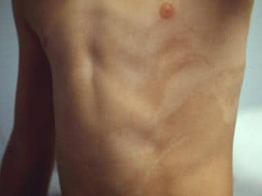
 Ito first introduced the syndrome 19511. In 1967, Hamada et al confirmed the association between the skin lesions and systemic abnormalities, including mental retardation2. Finally, Pascual-Castroviejo et al delineated the full spectrum of associated neurological abnormalities in a systematic study of the largest series published3.

Incontinentia pigmenti (IP) achromians is another term for this syndrome; however, because no true IP (melanin absent in the epidermis and present in the dermis) is present in the skin specimens, HI syndrome has become the preferred name.

In 1992, Ruiz-Maldonado and associates established some diagnostic criteria for HI syndrome4. Nonetheless, Ruiz-Maldonado et al's criteria link the diagnosis to the presence of systemic nondermatological (eg, CNS, skeletal) or chromosomal abnormalities. These criteria exclude patients with only dermatological manifestations. Patients with skin manifestations suggestive of HI with and without systemic alterations have been described in the same family, demonstrating that HI syndrome’s systemic involvement can vary. Studies that do not include systemic manifestations as diagnostic criteria for HI reported that approximately 30-74% of patients with typical HI skin lesions do not have nondermatological pathology.

Pathophysiology

The pathogenesis of HI syndrome is strongly linked to its genetics. A karyotype analysis survey was performed on 115 patients and revealed chromosomal anomalies in 60 (52%)5. Many patients have a chromosomal mosaic pattern, often leading to the generation of 2 cell lineages, which produce patterns of hypopigmented and hyperpigmented skin. X-chromosome alterations are not unusual in HI syndrome, and recent evidence points to X-chromosome inactivation, activation, and mosaicism as the main causes of these different patterns of cell behavior in the skin. Perhaps this can also be found in other tissues, such as the fundus (tessellated or radial pigmentation of the fundi), iris (hypopigmentation), and the brain (areas with abnormal cell morphology and neuroblast migration side by side with normal patterns). Karyotyping the blood cells may not be diagnostic; a skin biopsy for fibroblasts may be necessary to detect the HI-related chromosomal anomalies.

Despite recent advances, the genetic substrate for HI syndrome is far from homogenous and is not completely understood. A wide range of chromosomal abnormalities may be observed, including balanced X-autosome translocations, supernumerary X-chromosome ring fragment, ring chromosome 10, mosaic triploidy, mosaic trisomies (8, 13, 14, 18, 22), mosaic translocations, and mosaic deletions. Autosomal deletions and duplications may involve chromosomes 7, 12, 13, 14, 15, and 18. The pattern of chromosomal aberrations and the polymorphic nature of this disease have led some to believe that HI syndrome is a descriptive term rather than a true syndrome.

A familial form of HI syndrome exists; however, less than 3% of the patients have a family history of HI-type skin lesions. Although HI syndrome is most commonly a de novo occurrence, familial cases appear to be transmitted as an autosomal dominant trait. Approximately 10% of the patients report a family history of seizures or epilepsy, but the phenotypic expression varies; therefore, pigmentary changes may be the only clue to the genetic basis.

A cutaneous ultrastructural study that shows abnormal nerve termination in close proximity to basal keratinocytes, degenerated melanocytes, premelanosomes, and Langerhans cells has suggested that this finding may be important in the pathogenesis of HI6.

Frequency

International

To date, epidemiological data on this syndrome are limited. It appears to be the third most common neurocutaneous disease, second only to neurofibromatosis and tuberous sclerosis. In a pediatric neurology service in Spain, 1 in 600-700 patients referred was diagnosed with HI syndrome. It is diagnosed in 1 of every 7805 general pediatric outpatient visits, 1 of every 790 pediatric dermatology clinic visits, and 1 of every 2983 children in a general pediatric service3. Approximately three fourths of the patients with typical skin lesions have systemic manifestations.

Sex

Male-to-female ratios vary. In earlier series, the male-to-female ratio was reported to be 1:2.5. However, in larger and more recent series, the male-to-female ratio was 1:1.2. The severity of systemic manifestations appears to be similar in both sexes.

Age

Data in relation to age of diagnosis are usually reported in regard to the skin manifestations of HI syndrome. Typical skin lesions are initially demonstrated during the first year of life in as many as 70% of patients; they are noticeable at birth in 54% of patients. Rarely, lesions are not visible until mid childhood.


History

  • Hypomelanosis of Ito (HI) syndrome may involve many organs. Several manifestations, such as seizures and mental retardation, can be elicited in the history. For the sake of better understanding the subject, data from the history and physical examination are fused into a discussion of the signs and symptoms of each organ system’s involvement. The dermatological alterations are discussed first, followed by the description of systemic involvement.

Physical

Skin and neurological examinations are crucial for diagnosis; a thorough examination is extremely important.

  • Dermatological manifestations

    • Hypochromic lesions in distinctive patterns (eg, whirls, patches, streaks) characterize this syndrome; they often resemble whirled marble. The lesions may be unilateral (46%) or bilateral, and usually show a midline cutoff.

    • Patients with HI and hemimegalencephaly often have unilateral skin lesions. They are contralateral to the side of brain malformation and may show a zigzag pattern of lines. The hypochromic lesions are readily visualized in patients with pigmented skin, such as blacks, Asians, Spaniards, and Hispanic persons (eg, Mexicans). In children with fair skin, the use of a Wood lamp (ultraviolet light) is helpful in demonstrating these hypochromic lesions, which develop on the trunk, legs, arms, and face. They are more prominent in the ventral surface of the trunk and flexor surface of the limbs. The lesions are located on both the torso and the extremities in 59% of patients, on just the torso in 23% of patients, and exclusively on the extremities in 6% of patients. Some advocate stricter criteria for diagnosis, requiring cutaneous involvement of 2 or more segments for a diagnosis of HI.

    • Skin lesions usually appear during the first year of life (70%) and may be noticeable at birth (54%). Lesions are not visible until mid childhood in rare cases.

    • Hypopigmented or depigmented lesions may appear in swirls or patches with irregular borders. They tend to run parallel to one another, following the lines of Blaschko; however, HI is not the only syndrome associated with pigmentary anomalies along the lines of Blaschko. In 22% of the patients, lesions are patchy (square or rounded) rather than linear.

    • In linear or whorled nevoid hypermelanosis and nevus depigmentosus, lesions also follow these lines. The lines of Blaschko are relatively consistent and distinct from dermatomal lines. They represent orderly migration of mesodermal and ectodermal precursors during embryogenesis, occurring after X inactivation or activation. Genetic alterations (see Pathophysiology) or other problems that perturb morula cells and their daughter cells generate the swirl pattern of hypopigmentation along the lines of Blaschko. The color changes observed in skin lesions of HI may be present at birth but are frequently more obvious later in infancy or early childhood.

    • Hypohidrosis of hypopigmented skin can be detected in HI by application of iodine and starch in the skin, followed by a subcutaneous injection of pilocarpine hydrochloride.

    • Nonspecific skin lesions are reported in 20-40% of individuals with HI. These lesions are most frequently café-au-lait spots, but the following lesions have also been described:

      • Persistent mongolian blue spots

      • Nevus of Ota

      • Nevus marmoratus and angiomatous nevi

      • Soft fibroma

      • Pilomatrixoma

      • Aplasia cutis

      • Atopic dermatitis

    • Hair abnormalities include the following:

      • Slow growth

      • Diffuse alopecia

      • Trichorrhexis

      • Widow’s peak

      • Generalized hirsutism

      • Facial hypertrichosis

      • Coarse and curly hair

      • Low hairline (7% of patients)

      • Appearance of a zone of alopecia or white hair in the scalp (may precede hypopigmented lesions)

    • Fingernails may show some alterations, especially ungual hypoplasia.

    • Abnormal sweat glands are reported.

  • Neurological manifestations

    • Neurological involvement is found in 76% of patients during the first decade of life. Mental retardation and seizures are the most common presenting symptoms.

    • In one series of 19 patients, 10 displayed neurological symptoms before cutaneous symptoms arose; however, the proportion of patients with neurological involvement is clearly biased7. The pattern of referral is as high as 84% in a pediatric neurology clinic–generated series but somewhat lower in a dermatologist-generated series. When systemic involvement was used as a key diagnostic criterion, as many as 90% of the patients were found to have neurological involvement. Approximately 50% of the patients presented with seizures, although a lower frequency (37%) was reported in the pediatric dermatology clinic–based series. Generalized tonic-clonic seizures were most common (25%), whereas partial seizures were noted in 12% of patients, infantile spasms were reported in 8%, and myoclonic seizures were observed in 4%. Patients with Lennox-Gastaut syndrome have been reported.

    • Seizure control was achieved in 40-70% of patients. Some patients with partial seizures had very localized dysplastic lesions on neuroimaging that showed the typical localized, almost continuous, spike activity using electroencephalography (EEG).

    • Approximately one half to two thirds of patients have mental retardation (ie, intelligence quotient [IQ] <70). Chromosomal anomalies do not appear to increase the risk of mental retardation. Approximately 40% of patients with HI have an IQ of less than 50, and fewer than one fourth have an IQ of more than 85.

    • Autistic behavior has been found in approximately 11% of patients with HI. It may be associated with infantile spasms and other severe seizures. Similarly, the presence of mental retardation is linked with seizures (65%). Although the causal association of seizures and mental retardation or autism is attractive and present outside of HI, a common mechanism for both can be easily demonstrated (eg, patients with neuroblast migration and neuronal dysplasias).

    • HI is occasionally associated with hemimegalencephaly; however, more focal dysplastic lesions are also observed. Approximately one fourth of patients have a motor development delay. Hypotonia, which is usually accompanied by pes and genu valgus, is also a common finding.

    • Somatic hemihypertrophy, macrocephaly, and microcephaly may also be present (see below).

    • Brain tumors, including medulloblastoma and choroid plexus papilloma, are associated with HI.

    • Other neurological problems include ataxic gait, sensory neuropathy, chronic distal spinal muscular atrophy, torticollis, auditory conduction defect, hyperactivity, and spina bifida occulta. 

    • Patients with occipital lesions may have visual field defects but could represent sporadic associations.

    • Neuropathological studies demonstrate polymicrogyria, disarray of cortical lamination, heterotopic neurons in the white matter, and giant cells.

  • Ophthalmologic abnormalities

    • Approximately one fifth of patients present with ocular abnormalities.

    • Retinal pigment abnormalities are described as tessellated fundus, radial hypopigmented streaks, or geographic areas of hypopigmentation. Unilateral heterochromic iris with hypopigmentation of the cornea has also been described.

    • Cataracts and retinal detachment may produce vision loss.

    • Other ophthalmologic changes include myopia, hyperopia, astigmatism, megalocornea, opaque corneas, scleral melanosis, strabismus, slow pupillary response, pupillary atrophy or irregularity, nonclosure of the upper eyelid, ptosis, symblepharon, optic atrophy, choroidal atrophy, microphthalmia, macrophthalmia, epicanthal folds, dacryostenosis, and nystagmus.

  • Musculoskeletal abnormalities

    • Musculoskeletal abnormalities are common. One fifth of patients have hemihypertrophy, usually ipsilateral-to-hypomelanotic lesions.

    • Arm and leg length discrepancy and scoliosis are reported.

    • Fingers may be abnormal, showing atrophy, syndactyly, polydactyly, clinodactyly, or bifid thumb.

    • Other limb anomalies include luxatio coxae (caused by hypoplastic femoral heads) and genu valgus. 

  • Head and face anomalies

    • Facial malformations include hypertelorism, coarse facies, cleft lip and palate, bifid uvula, and nose and ear anomalies.

    • Macrocephaly, often associated with coarse facies, develops in 3-23% of patients.

    • In a study by Pascual-Castroviejo et al, microcephaly was described in 8% of patients8

    • Delayed fontanelle closure and asymmetry of the head can be present.

    • Dental problems include imperfect teeth implantation, partial anodontia and dental dysplasia, defective enamel, and hamartomatous cuspids.

  • Cardiac anomalies

    • Ventricular septum defect

    • Atrial septum defect

    • Pulmonary artery stenosis

    • Tetralogy of Fallot

    • Incomplete right bundle branch block and cardiomegaly of unknown etiology (occasionally noted)

  • Genital and reproductive organ anomalies

    • Hypospadias

    • Micropenis

    • Single kidney

    • Urethral duplication

    • Cryptorchidism

    • Precocious puberty

    • Gynecomastia

    • Asymmetrical breasts

    • Nephritis

  • Other types of abnormalities

    • Hepatomegaly

    • Segmental dilation of the colon

    • Diaphragmatic, umbilical, and inguinal hernia

  • HI is considered a cutaneous disease with multisystem involvement. It may be associated with the above findings and may also be associated with proteinuria, focal segmental glomerulosclerosis and end-stage renal disease9

  • Malignant and benign tumors are associated with HI. One patient study reported a mature cystic teratoma in the posterior mediastinum and a diploic epidermoid cyst of the parietal bone. Brain tumors may be present (see above); however, the benign tumors in patients with HI are often associated with chromosomal anomalies.

Causes

  • Several chromosomal abnormalities have been reported in HI (see Pathophysiology), but the etiology remains elusive.


Tuberous Sclerosis

Other Problems to be Considered

Many other skin pigmentary abnormalities may be associated with systemic and neurological abnormalities. In tuberous sclerosis, the lesions are round, oval, or in the shape of an ash leaf and do not follow the lines of Blaschko10.

Linear and whorled nevoid hypermelanosis

These conditions are characterized by brown pigmentation and hypopigmentation in streaks and whorls, which follow the lines of Blaschko. Because determining whether the darker or lighter skin color is the normal one is difficult, Sybert suggests that differentiating the dermatological features of linear and whorled nevoid hypermelanosis from the ones found in hypomelanosis of Ito (HI) is virtually impossible11. In patients with linear and whorled nevoid hypermelanosis, the lesions appear in infancy and gradually spread though the body. This feature is not helpful in the differentiation from HI, in which skin lesions are present at birth in more than one half of patients. Linear and whorled nevoid hypermelanosis are not associated with systemic abnormalities, unlike in HI.

Nevus depigmentosus

Nevus depigmentosus is characterized by hypochromic lesions in streaks and whorls, which also follow the lines of Blaschko. The hypochromic lesions tend to be circumscribed and are present at birth, changing little thereafter. Systemic abnormalities are rare in nevus depigmentosus.

Incontinentia pigmenti

IP is a condition seen mostly in girls; thus, X-linked dominant transmission is postulated. Patients with IP frequently have systemic involvement, similar to the involvement in patients with HI, including CNS manifestations. In patients with IP, cutaneous lesions undergo 3 stages, which may overlap.

The first phase is characterized by vesiculobullous lesions that appear in a linear array (with no dermatomal distribution) and are present from birth or develop in the first 2 weeks of life in 90% of patients. Vesicles are proximal in the limbs, are located in flexor surfaces, and contain eosinophils. This first phase may last days to months. Between the second and sixth week of life, the vesicles become pustular, verrucous, or keratotic, marking the second phase. In this phase, the lesions tend to be more distally and dorsally prominent in the limbs.

The second phase, during which hypopigmentation and skin atrophy develop, usually lasts for months.

During the third phase, hyperpigmentation of lesions is observed, peaking from 12-26 weeks of life. The dermatological appearance involves streaks, whorls, macules, and flecks. The color of the lesions is chocolate-brown or tan. Some patients are born with lesions already in the third phase. This phenomenon is thought to be caused by in utero onset of the inflammatory process.

The term incontinentia pigmenti is used because melanin is not observed in the epidermis but is present in the dermis, as if it had leaked or dropped into the deeper layer of the skin; thus, the epidermis is incontinent of melanin. This incontinence of melanin is not observed in the skin of patients with HI. Alopecia may be observed in one third of patients with IP.

Neuropathologically, patients with IP may show neuroblast migration disturbances, such as polymicrogyria. Inflammatory and destructive alterations often accompany IP lesions, which is a pattern often missing in HI. The reader is referred to Rosman, 1987, for a review of the clinical picture of incontinentia pigmenti12.

Vitiligo

Segmental vitiligo may be a consideration13, 14.


Lab Studies

  • Chromosomal analysis

    • Blood karyotyping is indicated, especially when systemic abnormalities are present.

    • Fibroblast karyotyping (sampling the dark and light skin) can reveal mosaicism but is not mandatory for diagnosis.

Imaging Studies

  • Neuroradiological abnormalities are reported in at least one third of the patients. Because many of the lesions are dysplastic or neuroblastic migration abnormalities, MRI is more useful than CT scanning.

    • Increase in the T2 signal of white matter is one of the most common findings. White matter abnormalities are somewhat predictive of a poor neurological outcome.

    • Neuroblast migration includes heterotopia, pachygyria, and polymicrogyria. Heterotopia may be observed at the level of the basal ganglia or as a periventricular band. Some of the dysplastic lesions may be localized, and hemimegalencephaly is also visualized.

    • Cerebral atrophy can be unilateral or generalized. Cases of cerebral hemiatrophy and porencephaly are often associated with a history of perinatal hypoxia or low birth weight.

  • Other rare imaging associations include the following:

    • Noncommunicating hydrocephalus

    • Megacisterna magna

    • Arteriovenous malformation

    • Cerebellar hypoplasia (hemispheres and vermis)

    • Brainstem hypoplasia

    • Brain tumors: MRI rarely reveals brain tumors; thus, patients occasionally have abnormal MRI findings but are neurologically healthy.

  • Brain imaging in patients with hypomelanosis of Ito (HI) and medically refractory epilepsy: The area that generates seizures (zone of ictal onset) should be found using a prolonged video-EEG, single-photon emission computed tomography (SPECT) or positron emission tomography (PET), and high-resolution MRI. If resective epilepsy surgery is a serious consideration after the preliminary tests are done (video-EEG, SPECT or PET) and the zone of ictal onset could not be determined, the patient may need to undergo invasive EEG monitoring with subdural grids or strips.

  • Other imaging tests

    • Musculoskeletal abnormalities often require radiography for proper quantification. A CT scan of the chest may be necessary when mediastinal tumors are investigated.

    • Abdominal ultrasonography may be required for diagnosis of genitourinary anomalies such as single kidney and urethral duplication.

Other Tests

  • In patients with seizures, an EEG is indicated to show focal discharges and slowing.

  • Most patients with cardiac anomalies require an ECG.


  • Perform a slit lamp examination in patients with ophthalmologic abnormalities.

Procedures

  • Biopsies of affected and nonaffected skin are sometimes indicated.

  • In select patients with cardiac anomalies, cardiac catheterization is recommended for proper diagnosis.

Histologic Findings

Dihydroxyphenylalanine (DOPA) staining of the skin may reveal decreased size and number of melanosomes in hypopigmented areas. The melanocytes may be smaller and fewer, and their dendrites are short and sparse. Melanin incontinence (ie, melanin is absent in the epidermis but present in the deeper dermis) is not observed in patients with HI. Histopathological alterations are not always typical; normal histology findings have been described in some cases. Neuropathological studies demonstrate polymicrogyria, disarray of cortical lamination, and heterotopic neurons in the white matter and giant cells.

Ultrastructural cutaneous studies may reveal normal-appearing basal and malpighian keratinocytes but a lack of melanosomes in the malpighian cells6. Melanosomes are dramatically reduced in the basal keratinocytes and appear small, single, or clustered and surrounded by a membrane. Melanocytic degeneration may be evident, and dendritic melanocytes contain various stages of nonmelanized premelanosome (stage II), partially melanized premelanosome (stage III), and, rarely, stage IV melanosomes. Unmyelinated axons of nerve-containing melanosomes may be seen at the dermoepidermal junction. Abnormal nerve termination has been observed in close relationship with basal keratinocytes, degenerated melanocytes, premelanosomes, and Langerhans cells.


Medical Care

  • No specific treatment is available for hypomelanosis of Ito (HI).

  • Treat seizures depending on the specific seizure type and epileptic syndrome (see Status Epilepticus, Complex Partial Seizures).

  • Partial seizures may respond to the usual anticonvulsant medications, such as carbamazepine, phenytoin, lamotrigine, gabapentin, and topiramate.

  • Infantile spasms should be treated accordingly with adrenocorticotropic hormone (ACTH), vigabatrin, valproic acid, or topiramate.

  • Approximately 30% of patients with seizures do not respond to anticonvulsant medications; therefore, these patients may need an appropriate evaluation to verify if they are good candidates for resective epilepsy surgery, ketogenic diet, or vagal nerve stimulation. In these patients, perform a prolonged video-EEG to document the zone of ictal onset.

  • Always offer the patient and parents genetic consultation.

Surgical Care

  • Approximately 30% of patients with seizures do not respond to anticonvulsant medications. In these patients, conduct an appropriate evaluation to verify if they are good candidates for resective epilepsy surgery or vagal nerve stimulation.

  • The patient may have large lesions and require a hemispherectomy for the treatment of their refractory epilepsy; however, in other cases, the removal of a more focal lesion may stop the seizures.

  • Cataracts and retinal detachment may produce loss of vision and can be successfully treated with surgery.

  • In patients with craniofacial malformation, such as cleft lip and palate, repair is done in the same fashion as in patients without HI.

Consultations

  • Consultation with an orthopedic specialist is indicated for patients with skeletal abnormalities.

  • Suggest consultation with an ophthalmologist for patients with ophthalmologic abnormalities.

  • Consultation with a nephrologist is recommended for patients with renal abnormalities.

  • Suggest a consultation with an endocrinologist for patients with associated abnormalities.

  • Patients with HI who were initially seen by a dermatologist may benefit from a consultation with a neurologist; conversely, patients initially referred to a neurologist may benefit from a consultation with a dermatologist and geneticist. Always offer the patient and parents the option of a consultation with a geneticist.

  • Refer patients who have seizures that are not completely controlled by anticonvulsant medications to an appropriate tertiary center with a comprehensive epilepsy program for proper evaluation (video-EEG, SPECT or PET, high-resolution MRI).

Diet

  • No dietary restrictions are indicated.

  • Patients with seizures who are unresponsive to anticonvulsant medication may benefit from a high-fat, low-carbohydrate diet (ie, the ketogenic diet).

Activity

  • No restriction in activity is recommended.


Currently, no drug therapy is specifically indicated for this disease. Treat comorbidities (eg, seizures) according to standard treatment. See Treatment.


Deterrence/Prevention

  • Hypomelanosis of Ito (HI) cannot be prevented, except in rare cases of familial HI. Because familial HI is autosomal dominant, genetic counseling is indicated as a way to prevent new cases in the same family. Nonetheless, most cases are a de novo occurrence.

Complications

  • Seizures are due directly to cerebral malformations. Patients with seizures, especially those with infantile spasms, are at risk for mental retardation and autistic behavior. Careful screening and follow-up with cognitive testing are suggested. Because seizures are often difficult to control using anticonvulsant medications, patients who have intractable partial seizures with secondary generalization or infantile spasms are at risk for neurocognitive deterioration; therefore, a workup should be completed at an epilepsy center.

  • Because of the possibility of ophthalmologic complications or manifestations, refer the patient to an ophthalmologist (see Consultations).

  • Similarly, patients with the following urologic (cryptorchidism), endocrinic, and renal complications need specialized follow-up:


    • Hypospadias

    • Micropenis

    • Single kidney

    • Urethral duplication

    • Precocious puberty

    • Gynecomastia

    • Asymmetrical breasts

    • Nephritis

  • Various benign and malignant tumors may complicate the course of HI because their manifestations may be protean. Remain attentive for any change in clinical status and consider the possibility of a tumor. Similarly, any significant and unexplained change in neurological status should raise suspicion for a brain tumor; therefore, consider MRI in these cases.

  • Craniofacial malformations, such as cleft lip and palate, may significantly affect the well-being of patients by interfering with their feeding and speech. The management of these complications requires consultation with a team of craniofacial specialists, including a plastic surgeon, craniofacial surgeon, or both and a speech therapy specialist.

  • Many of the dental malformations (eg, dental dysplasia, defective enamel, hamartomatous cuspids) seen in patients with HI may lead to secondary dental problems, which, in turn, may lead to increased decay. These require careful attention by a dentist.

Prognosis

  • Prognosis depends on the patient’s manifestations and complications of the disease.

  • About three fourths of patients with typical HI skin lesions have systemic manifestations of the disease.

  • As many as three fourths of patients have neurological symptoms in the first decade of life. Cognitive deficit is defined as an IQ of less than 70. Association of mental retardation and seizures suggests HI, although the cause-effect link is difficult to prove, except in the intractable cases.

  • Patients with chromosomal anomalies are at risk for tumors.


Medical/Legal Pitfalls

  • The main medicolegal concerns of hypomelanosis of Ito (HI) are related to improper diagnosis and genetic counseling. Failure to warn the family about the potential of systemic complications in a patient with dermatological signs of HI may lead to litigation if the family believes that unnecessary suffering resulted from the lack of diagnosis and information.

  • Educate patients with typical HI skin lesions about the potential for systemic complications, especially neurocognitive (eg, seizures, mental retardation) and tumor-related complications.

  • Proper documentation of the discussion with the parents and the patient is strongly suggested. Provide printed educational material to the families. Always discuss genetic counseling, although HI is only rarely familial.


  1. Ito M. A singular case of naevus depigmentosus systematicus bilateralis. Jpn J Dermatol. 1951;61:31-2.
  2. Hamada T, Saito T, Sugai T, Morita Y. "Incontinentia pigmenti achromians (Ito)". Arch Dermatol. Dec 1967;96(6):673-6. [Medline].
  3. Pascual-Castroviejo I, Lopez-Rodriguez L, de la Cruz Medina M, Salamanca-Maesso C, Roche Herrero C. Hypomelanosis of Ito. Neurological complications in 34 cases. Can J Neurol Sci. May 1988;15(2):124-9. [Medline].
  4. Ruiz-Maldonado R, Toussaint S, Tamayo L, Laterza A, del Castillo V. Hypomelanosis of Ito: diagnostic criteria and report of 41 cases. Pediatr Dermatol. Mar 1992;9(1):1-10. [Medline].
  5. Sybert VP. Hypomelanosis of Ito: a description, not a diagnosis. J Invest Dermatol. Nov 1994;103(5 Suppl):141S-143S. [Medline].
  6. Palungwachira P, Palungwachira P. Incontinentia pigmenti achromians of Ito: an ultrastructural study. J Med Assoc Thai. Feb 2006;89(2):253-7. [Medline].
  7. Glover MT, Brett EM, Atherton DJ. Hypomelanosis of Ito: spectrum of the disease. J Pediatr. Jul 1989;115(1):75-80. [Medline].
  8. Pascual-Castroviejo I, Roche C, Martinez-Bermejo A, et al. Hypomelanosis of ITO. A study of 76 infantile cases. Brain Dev. Jan 1998;20(1):36-43. [Medline].
  9. Gatter N, Hoppe B, Nutzenadel F, Waldherr R, Querfeld U. A cutaneous disease with multisystem involvement: hypomelanosis of Ito may be associated with proteinuria, focal segmental glomerulosclerosis and end-stage renal disease. Nephrol Dial Transplant. Mar 8 2007;[Medline].
  10. Jozwiak S, Schwartz RA, Janniger CK, Bielicka-Cymerman J. Usefulness of diagnostic criteria of tuberous sclerosis complex in pediatric patients. J Child Neurol. Oct 2000;15(10):652-9. [Medline].
  11. Sybert VP, Pagon RA, Donlan M, Bradley CM. Pigmentary abnormalities and mosaicism for chromosomal aberration: association with clinical features similar to hypomelanosis of Ito. J Pediatr. Apr 1990;116(4):581-6. [Medline].
  12. Rosman NP. Incontinentia Pigmenti. In: Gomez MR, ed. Neurocutaneous Diseases: A Practical Approach. Boston: Butterworths; 1987:294-300.
  13. Huggins RH, Schwartz RA, Janniger CK. Vitiligo. Acta Dermatovenerol Alp Panonica Adriat. Dec 2005;14(4):137-42, 144-5. [Medline].
  14. Huggins RH, Janusz CA, Schwartz RA. Vitiligo: a sign of systemic disease. Indian J Dermatol Venereol Leprol. Jan-Feb 2006;72(1):68-71. [Medline].
  15. Ardinger HH, Bell WE. Hypomelanosis of Ito. Wood's light and magnetic resonance imaging as diagnostic measures. Arch Neurol. Aug 1986;43(8):848-50. [Medline].
  16. Battistella PA, Peserico A, Bertoli P, et al. Hypomelanosis of Ito and hemimegalencephaly. Childs Nerv Syst. Nov 1990;6(7):421-3. [Medline].
  17. Berg B. Neurocutaneous syndromes: Phakomatoses and allied conditions. In: Swaiman K, ed. Pediatric Neurology: Principles and Practice. 2nd ed. 1994:1045-1069.
  18. Berg BO. Unusual neurocutaneous syndromes. Neurol Clin. Feb 1985;3(1):165-78. [Medline].
  19. Chevalier C, Colon S, Faraj G, et al. [Glomerulopathy and hypomelanosis of Ito]. Nephrologie. 1997;18(4):125-7. [Medline].
  20. Neurocutaneous Disorders. In: Hurwitz S, ed. Clinical Pediatric Dermatology. 2nd ed. Philadelphia, Penn: WB Saunders; 1993:624-640.
  21. Fonseca E, Contreras F, Soto J. Marcadores cutos de los sromes neuroectodermicos. In: Pascual-Castroviejo I, Barcelona JR, eds. Transtornos Neuroectodermicos. 1989:73-84.
  22. Fryburg JS, Lin KY, Matsumoto J. Abnormal head MRI in a neurologically normal boy with hypomelanosis of Ito. Am J Med Genet. Dec 11 1996;66(2):200-3. [Medline].
  23. Hypomelanosis of Ito. In: Sybert VP, ed. Genetic Skin Disorders. New York, NY: Oxford University Press; 1997:318-22.
  24. Glab E, Barg E, Wikiera B. Hypomelanosis of Ito and precocious adrenarche in a girl. J Pediatr Endocrinol Metab. Mar 2006;19(3):283-4. [Medline].
  25. Hara M, Kozasa M, Mituisi Y. Ito Syndrome (Hypomelanosis of Ito) as a cause of intractable epilepsy. In: Pascual-Castroviejo I, Barcelona JR eds. Transtornos Neuroectodermicos. 1989:221-5.
  26. Ishikawa T, Kanayama M, Sugiyama K, Katoh T, Wada Y. Hypomelanosis of Ito associated with benign tumors and chromosomal abnormalities: a neurocutaneous syndrome. Brain Dev. 1985;7(1):45-9. [Medline].
  27. Keng WT, Harewood L, Grace E, et al. A balanced reciprocal translocation in a case of hypomelanosis of Ito with confirmation of mosaicism using buccal cell interphase FISH. Am J Med Genet A. May 15 2006;140(10):1111-3. [Medline].
  28. Khandpur S, Minz A, Sharma VK. An unusual association of pigmentary mosaicism (hypomelanosis of Ito) with generalized hypertrichosis. Clin Exp Dermatol. May 2006;31(3):467-8. [Medline].
  29. Nehal KS, PeBenito R, Orlow SJ. Analysis of 54 cases of hypopigmentation and hyperpigmentation along the lines of Blaschko. Arch Dermatol. Oct 1996;132(10):1167-70. [Medline].
  30. Ono J, Harada K, Kodaka R, Ishida M, Okada S. Regional cortical dysplasia associated with suspected hypomelanosis of Ito. Pediatr Neurol. Oct 1997;17(3):252-4. [Medline].
  31. Pascual-Castroviejo I. Hypomelanosis of Ito. In: Gomez MR, ed. Neurocutaneous Diseases: A Practical Approach. Boston, MA: Butterworths; 1987:85-90.
  32. Pascual-Castroviejo I. Nevus Acro de Ito. In: Castro-Viejo I, ed. Neurologia Infantil. Madrid, Spain: Editorial Cientifico; 1983:678-680.
  33. Rafay MF, Al-Futaisi A, Weiss S, Armstrong D. Hypomelanosis of Ito and Moyamoya disease. J Child Neurol. Nov 2005;20(11):924-6. [Medline].
  34. Reese PD, Judisch GF. Hypomelanosis of Ito. Arch Ophthalmol. Aug 1986;104(8):1136-7. [Medline].
  35. Ross DL, Liwnicz BH, Chun RW, Gilbert E. Hypomelanosis of Ito (incontinentia pigmenti achromians)--a clinicopathologic study: macrocephaly and gray matter heterotopias. Neurology. Sep 1982;32(9):1013-6. [Medline].
  36. Rubin MB. Incontinentia pigmenti achromians. Multiple cases within a family. Arch Dermatol. Mar 1972;105(3):424-5. [Medline].
  37. Ruggieri M, Pavone L. Hypomelanosis of Ito: clinical syndrome or just phenotype?. J Child Neurol. Oct 2000;15(10):635-44. [Medline].
  38. Ruggieri M, Tigano G, Mazzone D, Tine A, Pavone L. Involvement of the white matter in hypomelanosis of Ito (incontinentia pigmenti achromiens). Neurology. Feb 1996;46(2):485-92. [Medline].
  39. Steiner J, Adamsbaum C, Desguerres I, et al. Hypomelanosis of Ito and brain abnormalities: MRI findings and literature review. Pediatr Radiol. Nov 1996;26(11):763-8. [Medline].
  40. Sybert VP, Pagon RA. Hypomelanosis of Ito in a girl with plexus papilloma and translocation (X;17) [letter]. Hum Genet. Feb 1994;93(2):227. [Medline].
  41. Tagawa T, Futagi Y, Arai H, Mushiake S, Nakayama M. Hypomelanosis of Ito associated with hemimegalencephaly: a clinicopathological study. Pediatr Neurol. Sep 1997;17(2):180-4. [Medline].
  42. Urban J, Toruniowa B, Janniger CK, Czelej D, Schwartz RA. Incontinentia pigmenti (Bloch-Sulzberger syndrome): multisystem disease observed in two generations. Cutis. Nov 1996;58(5):329-36. [Medline].
  43. Urgelles E, Pascual-Castroviejo I, Roche C, et al. Arteriovenous malformation in hypomelanosis of Ito. Brain Dev. Jan-Feb 1996;18(1):78-80. [Medline].
  44. Weaver RG Jr, Martin T, Zanolli MD. The ocular changes of incontinentia pigmenti achromians (hypomelanosis of Ito). J Pediatr Ophthalmol Strabismus. May-Jun 1991;28(3):160-3. [Medline].
  45. Zajac V, Kirchhoff T, Levy ER, et al. Characterisation of X;17(q12;p13) translocation breakpoints in a female patient with hypomelanosis of Ito and choroid plexus papilloma. Eur J Hum Genet. Mar-Apr 1997;5(2):61-8. [Medline].

Hypomelanosis of Ito excerpt

Article Last Updated: May 25, 2007