Practice Essentials
A dentoalveolar abscess is an acute lesion characterized by localization of pus in the structures that surround the teeth. Most patients are treated easily with analgesia, antibiotics, drainage, and/or referral to a dentist or oral-maxillofacial surgeon. However, the physician should be aware of potential complications of simple dentoalveolar abscess. [1] (See the image below.)
Signs and symptoms of dental abscess
The following may be reported in patients with dental abscess:
-
Localized pain and swelling (may progress over a few hours to days)
-
Thermal sensitivity (periapical abscess)
-
Fever
-
Gingival bleeding (on occasion with periodontal abscess)
-
Decreased intake of fluid, food, or both
See Presentation for more detail.
Diagnosis of dental abscess
Laboratory studies
For uncomplicated dental abscess, no laboratory studies are needed.
For complicated abscess (accompanying cellulitis), the following are recommended:
-
Complete blood cell (CBC) count
-
Blood culture (aerobic and anaerobic) before starting parenteral antibiotic therapy
-
Needle aspirate for Gram staining and aerobic and anaerobic cultures
Imaging studies
Depending on the severity of the abscess based on clinical presentation, the following is recommended:
-
Periapical radiography is the first level of investigation
-
Panoramic radiography (pantomography) is most helpful in emergency situations
If cellulitis swelling extends beyond the local area, the following is indicated:
-
Lateral and anteroposterior neck views
-
CT scanning with intravenous contrast
See Workup for more detail.
Management of dental abscess
The primary therapeutic modality is surgical drainage of any pus collection. A pulpectomy or incision and drainage is the recommended management of a localized acute apical abscess in the permanent dentition. Most dental abscesses respond to surgical treatment (incision and drainage, root canal, or extraction) and elimination of the source of infection. The addition of antibiotics is not recommended for a localized dental abscess.
See Treatment and Medication for more detail.
Pathophysiology
The term dentoalveolar abscess comprises 3 distinct processes, as follows:
-
A periapical abscess that originates in the dental pulp and is usually secondary to dental caries is the most common dental abscess in children. Dental caries erode the protective layers of the tooth (ie, enamel, dentin) and allow bacteria to invade the pulp, producing a pulpitis. Pulpitis can progress to necrosis, with bacterial invasion of the alveolar bone, causing an abscess.
-
A periodontal abscess involves the supporting structures of the teeth (periodontal ligaments, alveolar bone). [2] This is the most common dental abscess in adults, but may occur in children with impaction of a foreign body in the gingiva.
-
Pericoronitis describes the infection of the gum flap (operculum) that overlies a partially erupted tooth.
Developmental and acquired conditions are associated with dental abscesses in childhood. Developmental conditions include abnormal morphology of the crown (eg, dens invaginatus, dens evaginatus) and abnormal structure of the dentine (eg, dentine dysplasia, dentinogenesis imperfecta, osteogenesis imperfecta, familial hypophosphatemia). Acquired conditions include pre-eruptive intracoronal resorption and mandibular infected buccal cyst. [3]
Odontogenic infections are polymicrobial, with an average of 4-6 different causative bacteria. The dominant isolates are strictly anaerobic gram-negative rods and gram-positive cocci, in addition to facultative and microaerophilic streptococci. Anaerobic bacteria outnumber aerobes 2-3:1. [4] In general, strictly anaerobic gram-negative rods are more pathogenic than facultative or strictly anaerobic gram-positive cocci.
Generally, a nonpathologic resident bacterium gains entry when the host's defenses are breached, rather than when a nontypical microorganism is introduced. The predominant species associated with dental abscess include Bacteroides, Fusobacterium, Actinomyces, Peptococcus,Peptostreptococcus, and Porphyromonas as well as Prevotella oralis, Prevotella melaninogenica, and Streptococcus viridans. Beta-lactamase producing organisms occur in approximately one third of dental abscesses. [5]
The use of molecular techniques such as 16S rRNA gene sequencing and polymerase chain reaction (PCR) have identified difficult-to-culture organisms and expanded knowledge of the microflora associated with dental abscess. Examples include Treponema, Atopobium, Bulleidia extructa, and Mogibacterium species, as well as Cryptobacterium curtum. [6] A recent Brazilian study using 16S rRNA PCR and sequencing performed on cultivable bacteria from acute apical abscesses revealed the most common identified bacteria were Prevotella sp, Pseudoramibacter alactolyticus, Parvimonas micra, Dialister invisus, Filifactor alocis and Peptostreptococcus stomatis. Recently, a study using 16S rRNA sequencing in 15 patients with primary endodontic infections with and without a sinus tract to the oral cavity revealed Propionibacterium acnes as the most prevalent isolate recovered from lesions with an intraoral communication. Additionally, the authors found no difference in the number of species identified from lesions with or without intraoral communication. [7]
Etiology
Dental caries are caused by the following:
-
"Infant-bottle" tooth decay or "nursing" caries: The term "early childhood" caries is replacing these terms because the description also includes dental caries in breastfed babies. The American Academy of Pediatrics (AAP) along with the American Academy of Pediatric Dentistry issued a clinical report entitled "Oral and Dental Aspects of Child Abuse and Neglect," which states that the caregivers of children who present for dental care with severe early childhood caries must be carefully interviewed to differentiate caregivers with adequate knowledge and willful failure to seek dental care from caregivers without knowledge or awareness of a child's dental needs. Failure to seek dental care may result from many socioeconomic factors, and clinicians should determine if dental care is readily available and accessible when considering the possibility of negligence. Physicians and dentists are required by law to report suspected cases of child negligence and abuse. [8]
-
Plaque: This is a noncalcified precipitate of microorganisms and their byproducts that adheres to the enamel of teeth.
In immunocompromised patients, bacteria may hematogenously spread to invade the pulp of the tooth.
Gingivitis is an inflammation of the gingiva without attachment loss or with nonprogressing attachment loss.
Posttraumatic infection or postsurgical infection may also cause dental abscess.
Epidemiology
Race-, sex-, and age-related demographics
No race predilection is observed.
No sex predilection is noted.
Dental abscess is rare in infants because abscesses do not form until teeth erupt. In children, periapical abscess is the most common type of dental abscess. This is because of the combination of poor hygiene, thinner enamel, and the primary dentition having more abundant blood supply, which allows for an increased inflammatory response. In adults, periodontal abscess is more common than periapical abscess.
Prognosis
The prognosis is excellent with proper incision, drainage, antibiotic therapy, tooth extraction, root canal therapy, and follow-up care.
Morbidity/mortality
Mortality is rare and is usually due to airway compromise. Morbidity relates to pain, probable tooth loss, and dehydration.
Complications
Complications include the following:
-
Dentocutaneous fistulae arise from chronic dental infections. The fistulous pathway develops as the chronic inflammation erodes through the alveolar bone, perforates the periosteum, and spreads into the surrounding soft tissues. The diagnosis is often missed because a chronic asymptomatic dental infection is usually present and the skin lesion is mistakenly thought to arise locally.
-
Acute suppurative osteomyelitis was common before the era of antibiotic therapy. Osteomyelitis is an inflammation of the medullary cavity and adjacent cortex of bone. The mandible is more commonly involved than the maxilla because the maxilla has a better blood supply. Garr é osteomyelitis is a chronic nonsuppurative sclerosing osteomyelitis that is characterized by a localized, hard, nontender swelling of the mandible and is usually associated with dental caries of the lower first molar. Radiography may reveal a focal area of bone proliferation with a periosteal reaction that has an onion-peel or laminated appearance. A study by Moratin et al found that more severe infection, a history of diabetes, and the use of clindamycin were associated with an increased risk for the development of osteomyelitis in patients with dental abscess. [9]
-
Cavernous sinus thrombosis (CST) may be a complication. Approximately 10% of patients with CST have an odontogenic focus. Spread of infection from dental abscesses to the cavernous sinus is believed to occur via the valveless pterygoid venous plexus by way of the retromandibular vein. Patients often present with headache, unilateral retro-orbital pain, periorbital edema, fever, proptosis, chemosis, and ptosis. Treatment consists of antibiotics, anticoagulants, and, occasionally, surgery. [10]
-
Ludwig angina is rapidly spreading cellulitis of the bilateral sublingual, submandibular, and submental spaces. Abscesses of the second and third mandibular molars account for 75% of cases. Ludwig angina manifests as swelling of the floor of the mouth with elevation and posterior displacement of the tongue. A rapidly spreading gangrenous cellulitis produces a brawny edema of the suprahyoid region of the neck. The infection begins unilaterally but quickly spreads to include the entire neck. The most common presenting symptoms include oral, neck, and dental pain; neck swelling; odynophagia; drooling; dysphagia; dysphonia; trismus; and tongue swelling. The patient may lean forward in order to maintain airway patency. The infection can extend into the retropharyngeal space and the mediastinum. This is a life-threatening infection. Ludwig angina is unusual in children.
-
Maxillary sinusitis may occur from direct extension of an odontogenic infection or from perforation of the floor of the sinus during extraction. Case reports exist of odontogenic orbital abscess secondary to spread of infection to the maxillary sinus. These patients can present with facial edema, ocular pain, ophthalmoplegia and proptosis. [11]
-
Facial-space swelling secondary to spread of the infection most often involves the following areas:
Submandibular swelling is caused by dental abscesses from the second or third molars whose roots lie below the attachment of the mylohyoid bone. This space contains the submandibular gland, Wharton duct (the opening of the submandibular salivary gland), lingual and hypoglossal nerves, facial artery, and vein. A firm, ill-defined, and often significant-sized swelling is present below the mandible. The inferior border and angle of the mandible are difficult to palpate. The patient may have mild trismus.
Sublingual swelling is caused by any lower tooth whose apex is above the mylohyoid muscle attachment (ie, incisors, canines, premolars, mesial roots of the first molar). Infections produce a unilateral elevation of the floor of the mouth near the offending tooth but can spread across the midline, causing pain, dysphagia, and an elevation of the base of the tongue, leading to potential airway compromise.
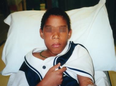
Buccal swelling originates from infected maxillary or mandibular molars. Clinically, infection produces a large tender swelling of the cheek without trismus. Boundaries for this type of infection may extend from the philtrum of the lip, to the border of the parotid, and up to the eye.
Less frequently involved facial-space swellings include submental, masticator, canine, lateral pharyngeal, and retropharyngeal. Retropharyngeal space infections are serious infections with the potential to cause airway obstruction and infection of the organs in the mediastinum.
-
Necrotizing fasciitis of the face or neck that results from an odontogenic abscess is very rare.
Patient Education
Most dentoalveolar abscesses are preventable.
-
Inquire if drinking water is fluorinated. If not, counsel parents about fluoride supplementation (see Prevention).
-
Instruct patients about proper dental hygiene, including brushing teeth after meals, flossing, and regular dental check-ups.
-
Obvious swelling of the right cheek due to dental abscess.
-
Side view. Fluctuant mass extending toward the buccal side of the gum end to the gingival-buccal reflection.


