Continually Updated Clinical Reference
 
 
  All Sources     eMedicine     Medscape     Drug Reference     MEDLINE
 
eMedicine - Slipped Capital Femoral Epiphysis : Article by

Quick Find
Authors & Editors
Introduction
Indications
RELEVANT ANATOMY
Contraindications
Workup
Treatment
Complications
Outcome and Prognosis
Future and Controversies
Multimedia
References


Patient Education
Click here for patient education.


Author: Mihir M Thacker, MBBS, MS(Orth), DNB(Orth), FCPS(Orth), D'Ortho, Assistant Professor of Orthopedic Surgery and Pediatrics, Thomas Jefferson University; Consulting Staff, Department of Pediatric Orthopedic Surgery, Alfred I duPont Hospital for Children; Orthopedic Oncologist, Helen F Graham Cancer Center and Christiana Care Health Services

Mihir M Thacker is a member of the following medical societies: Limb Lengthening and Reconstruction Society ASAMI-North America, Medical Council of India, and Musculoskeletal Tumor Society

Coauthor(s): Michael S Clarke, MD, Clinical Associate Professor, Department of Orthopedic Surgery, University of Missouri-Columbia School of Medicine

Editors: Steven I Rabin, MD, Clinical Associate Professor, Loyola University Medical Center; Chair, Department of Orthopedic Surgery, Dreyer Medical Clinic; Francisco Talavera, PharmD, PhD, Senior Pharmacy Editor, eMedicine; James McCarthy, MD, FAAOS, Associate Professor of Orthopedic Surgery, Temple University School of Medicine; Assistant Chief of Staff, Medical Director of Gait Laboratory, Shriners Hospital for Children of Philadelphia; Dinesh Patel, MD, FACS, Associate Clinical Professor of Orthopedic Surgery, Harvard Medical School; Chief of Arthroscopic Surgery, Department of Orthopedic Surgery, Massachusetts General Hospital; William L Jaffe, MD, Clinical Professor of Orthopedic Surgery, New York University School of Medicine; Vice Chairman, Department of Orthopedic Surgery, Hospital for Joint Diseases

Author and Editor Disclosure

Synonyms and related keywords: SCFE, adolescent coxa vara, slipped upper femoral epiphysis, knee pain, hip pain, knee injury, hip injury, obesity, hypothyroidism, osteotomy

Slipped capital femoral epiphysis (SCFE) was first described by Ernst Muller (1888-1889), who called it Schenkelhalsverbiegungen im Jungesalter, meaning "bending of the femoral neck in adolescence." The term slipped capital femoral epiphysis is a misnomer because the epiphysis is held in the acetabulum by the ligamentum teres; thus, the metaphysis actually moves proximally and anteriorly while the epiphysis remains in the acetabulum.

In most patients, this appears radiographically as a varus relationship between the head and the neck. Occasionally, the slip appears to be in a valgus position, with the epiphysis displaced superiorly in relation to the neck.1, 2 In the vast majority of cases, the etiology is unknown, although atypical slips may be associated with a known endocrine disorder,3, 4, 5 with renal failure osteodystrophy,6 or with previous radiation therapy.4, 7

Related eMedicine topics:
Slipped Capital Femoral Epiphysis (Radiology)
Slipped Capital Femoral Epiphysis (Sports Medicine)

Related Medscape topics:
The Limping Child
Overweight Children and Adolescents: Recommendations for Screening, Assessment and Management

History of the Procedure

See Surgical therapy.

Problem

This condition is not life threatening. However, untreated and complicated SCFE can lead to deformity and early osteoarthrosis of the hip and, thus, can cause considerable morbidity. Factors that increase morbidity include avascular necrosis (AVN) of the hip and chondrolysis. Both of these may result in damage severe enough to warrant a salvage procedure, in the form of an arthrodesis or a total hip arthroplasty. Prompt diagnosis is critical to prevent further deformity and AVN. The diagnosis is often subtle, and symptoms, such as groin or knee pain, can be misleading.

Frequency

Prevalence varies widely even within the continental United States. Prevalence has been reported to be 2.13 cases per 100,000 population in the southwestern United States and 10.08 cases per 100,000 population in the northeastern United States.8  Prevalence is lowest in the mountain and Great Plains states. Prevalence is reported to be quite low in Asia, with just 0.2 cases per 100,000 children affected in eastern Japan.9

A race predilection exists for SCFE. The relative racial frequency of SCFE is as follows10:


  • Whites - 1.0
  • Pacific islanders - 4.5
  • Blacks - 2.2
  • American Indians and Hispanic individuals - 1.05
  • Indonesian-Malay peoples (eg, Chinese, Japanese, Thai, Vietnamese) - 0.5
  • Indo-Mediterranean peoples (those of Near East, North African, or Indian subcontinent ancestry) - 0.1

Males are affected more commonly than females. The male-to-female ratio is 2-5:1.

The mean age at diagnosis is 13.5 years in boys (age range 13-15 y) and 12 years in girls (age range 11-13 y).10 This corresponds to the period of maximum skeletal growth. Juvenile (in children <10 y) SCFE should raise the suspicion of an underlying cause, such as an endocrinopathy. Radiation-associated slips tend to occur in young children.

Etiology

Causative factors include the following:

  • Habitus
    • Obesity (At least 50% of patients are above the 95th percentile for weight.11)
    • Excessive tallness, thinness
  • Endocrinopathy (If height is <10th percentile, likelihood of an underlying endocrinopathy is high.12, 13 ])
  • Radiation therapy, especially for childhood leukemias or lymphomas
    • Tend to occur in young children
    • Affected children are also light, so tend to have mild slips
  • Renal failure
    • High incidence of simultaneous bilateral presentations (approximately 87%)
    • Highest incidence of severe slips in these patients
    • Two distinct subgroups of patients, as follows:
      • Those whose disease is controlled early (less hyperparathyroidism) tend to have mild slips.
      • Those with poorly controlled disease (significant hyperparathyroidism) tend to have severe slips.

Related Medscape Topics:
Resource Center Hypothyroidism
Resource Center Hypogonadism
Resource Center Thyroid Disease
CME Endocrine Emergencies

Related eMedicine Topics
:
Obesity
Hypothyroidism
Acromegaly
Acute Renal Failure
Chronic Renal Failure

Pathophysiology

SCFE is caused by increased stresses across a weakened physis, with a combination of both biomechanical and biochemical factors contributing to the development of the slip. Factors affecting stability of the physis include the following:

  • Perichondrium - Thick in children, but progressively thins with age
  • Perichondrial ring - Fibrous ring that spans the physeal plate and extends from the metaphysis to the epiphysis; this also thins with age
  • Transphyseal collagen - Weakens due to progressive mineralization; cross-linkage of collagen affected in osteolathyrism, leading to slippage in these patients
  • Mammillary projections - Assume increasing importance as the perichondrial ring thins
  • Contour of the growth plate - Normally convex toward the physis with undulations at the periphery, which contribute to its resistance to linear shear and torque forces
  • Growth plate thickness - Mainly affected by biochemical and endocrine factors
  • Mechanical factors - Increased stress across the physis.14 Also includes the following:
    • Relative femoral retroversion, which also increases the stress across the physis.15, 16, 17, 18
    • Obesity 8, 10 - Increases the forces across the physis; associated with femoral retroversion, which also increases the stress across the physis (The average femoral version of patients who are not obese was found to be 10.6°, compared with 0.40° in patients who are obese.15)
    • Increased physeal slope19 - Increase in inclination of the physis at the time of rapid skeletal growth around puberty (Patients with SCFE have been shown to have a higher physeal inclination even on the opposite side.)
    • Deeper acetabula - Higher mean center-edge angle of Wiberg in children with SCFE compared with control subjects20; lead to increased coverage of the femoral heads and increased stress across the physis
  • Biochemical factors - SCFE most common in the peripubertal age group21; possible contribution of hormones on the physes to the likelihood of developing a slip
    • Growth hormone - Causes widening of the physes and consequent weakening
    • Sex hormones - Increased physeal width and decreased physeal strength from testosterone22,  probably accounting for increased frequency in boys; narrowing of the physis and increased physis strength from estrogen, possibly explaining why slips seldom occur in postpubertal females23
    • Thyroid hormone - Effect not very clear, although slips can occur in patients with hypothyroidism and those receiving thyroid hormone replacements for hypothyroidism

Even though most children with SCFE do not have an overt endocrinopathy, they very well may have some subtle endocrine disorder.24, 25, 26, 27, 28 A delay in bone age of some of these children lends further credence to this theory.22, 23

Shear stresses across a physis made vulnerable by the biomechanical and biochemical factors outlined above leads to the slip. The displacement is decided by the direction of the deforming force. Posteroinferior displacement of the head (anterosuperior migration of the neck) is the most common pattern, although rarely, the head may displace posterosuperiorly, giving rise to an apparent valgus slip (see Image 1).1, 2

Clinical

SCFE must be considered in the differential diagnosis in children presenting with knee pain. Typically, knee examination and radiographic findings are normal in individuals with SCFE. All children with knee pain should have their hips evaluated.

Patients may present with the following symptoms:

  • Pain:The presenting feature is hip (groin) pain in most patients. Up to 46% of patients with chronic slips may present with thigh pain or knee pain (referred pain, obturator nerve).
  • Limp
  • Inability to bear weight in acute slips
  • History of irradiation, renal failure, endocrinopathy (hypothyroidism and treatment thereof, acromegaly, short stature [growth hormone supplementation])

Either side may be affected. The left hip is involved in 60% of unilateral cases. In most studies, 18-50% of cases are bilateral, although other studies have reported a prevalence of bilaterality of as high as 80%.29, 30, 31. Bilateral involvement may be evident at the time of initial presentation.10 Most patients who subsequently develop a contralateral slip do so within 18-24 months of initial symptoms, although slips may occur until physeal closure.10, 23, 29, 32, 33, 34, 35

The following physical signs may be present:

  • Externally rotated attitude of the affected lower limb
  • Restriction of flexion, abduction, and internal rotation of the affected hip in varus slips and restriction of flexion, adduction, and internal rotation in valgus slips
  • Obligatory external rotation on flexion (This is a diagnostic clue.)
  • Wasting of the thigh in chronic slips
  • True supratrochanteric shortening (Bryant triangle, Nélaton line)
  • Trendelenburg sign and gait possible; antalgic gait in acute or acute-in-chronic slips

Differentials

  • Knee injury
  • "Groin pull"
  • Trauma: Hemarthrosis or fracture may manifest similarly to an acute slip.
  • Infections: Septic arthritis and acute osteomyelitis must be excluded in acute cases.
  • Legg-Calvé-Perthes disease: This disease usually affects children aged 4-9 years.
  • Juvenile rheumatoid arthritis: This condition may manifest with hip involvement and minimal systemic manifestations.
  • Transient synovitis
  • Benign or malignant tumors


See Surgical therapy for indications for various procedures.


See Surgical therapy.


The treatment of a slip is stabilization. Although surgery should be performed promptly, it is elective; and therefore, severe medical conditions that would significantly increase surgical risk should be addressed.


Lab Studies

  • A renal panel in patients with renal failure–associated slips may reveal elevated BUN and creatinine levels.
  • An endocrine panel is appropriate if endocrinopathy is suspected.
    • Hypothyroidism is a common endocrinopathy associated with SCFE. In cases in which hypothyroidism is suspected, triiodothyronine, thyroxine, and thyrotropin levels must be obtained.
    • Other endocrinopathies also may be associated with SCFE (see Pathophysiology). In these cases, the appropriate investigations must be performed.

Imaging Studies

  • Diagnostic radiography
    • In the preslip stage, the only positive findings are a widening and irregularity of the physis with rarefaction of its juxtaepiphysial portion.
    • Early diagnosis is made using a Lauenstein (frog-leg) lateral view (see Image 2) or true lateral view of the hip36 because small slips may be missed on the anteroposterior (AP) view. A frog-leg lateral view should not be attempted in persons with acute or unstable slips because it may cause further displacement.
    • The Klein line, a line along the superior border of the femoral neck, intersects less of the femoral head than on the opposite side (Trethowan sign) on the AP radiograph (see Image 3).
    • Localized rarefaction of the inferior medial metaphysis of the neck may be evident.
    • The "metaphyseal blanch" of Steel37 is a crescent-shaped area of increased density overlying the metaphysis adjacent to the physis on the AP radiograph. This is an early sign of a posterior slip without significant medial slip and indicates the need for a lateral radiograph to identify the slip.
    • In moderate-to-severe slips, the overlap of the head and the metaphysis is visible.
    • Remodeling in chronic slips is seen as callus on the posteroinferior portion of the neck and rounding-off of the anterosuperior bare area of the neck, which is seen as a rounded hump.
  • Bone scanning38
    • This can be used to identify patients at high risk of AVN. A cold bone scan (demonstrating an absence of vascularity) essentially is seen only in unstable cases. When a patient has such a bone scan finding, the risk of subsequently developing AVN is 80-100%.
    • Bone scans help in the early detection of AVN (ie, decreased activity/uptake on the femoral side).
    • These scans also help in the early detection of chondrolysis (ie, increased activity/uptake on both the acetabular and the femoral sides).
  • CT scanning
    • CT scanning can be used to measure the amount of retroversion and the degree of slip. Three-dimensional imaging of the upper femur, which may be useful in the preoperative planning of osteotomies, is also possible.
    • CT scans may incidentally reveal asymptomatic, mild, contralateral slips.
  • Ultrasonography
    • The presence of an effusion indicates an unstable slip.
    • The presence of remodeling indicates a stable slip.
  • MRI: This is used for early detection of AVN.

Histologic Findings

The zone of hypertrophy is thickened, and the normal columnar arrangement of chondrocytes is lost. Chondrocyte clustering and disarray are evident.39, 40

Anatomically, the slip occurs in the layer of cartilage adjacent to the zone of provisional calcification (unlike a type 1 Salter-Harris injury, which occurs between the proliferating and the hypertrophic zones). However, the plane of cleavage may be irregular and may pass through different zones of the physis.

Ultrastructural studies show defective collagen fibrils and defects in collagen banding41 in the zone of hypertrophy and changes in the proteoglycan and glycoprotein concentrations in the zone of proliferation, with increased glycoprotein staining in the territorial matrix and increased proteoglycan staining in the extraterritorial matrix.

Staging

  • Traditional classification42, 43, 44, 45, 46: This classification system may not be accurate and is not useful for prognosis.
    • Preslip: Discomfort (pain and restricted internal rotation) is mild. The diagnosis is based primarily on radiological findings, with widening and irregularity of the physis47 and generalized osteopenia in the affected hemipelvis.
    • Acute slip (10-15% of cases)33, 44: The duration of symptoms is less than 3 weeks, with no evidence of remodeling on the radiograph (see Image 5).
    • Chronic slip (85% of cases)33: Symptoms persist for more than 3 weeks.
    • Acute-on-chronic: An acute-on-chronic SCFE is one associated with chronic symptoms initially and with subsequent development of acute symptoms, as well as a sudden increase in the degree of slip, usually with minor trauma.
  • Classifications based on stability: The focus of classification has shifted to determining the stability of the slip because this significantly affects the prognosis. The classification into stable and unstable categories is important because it is predictive of the prognosis. The traditional classification of slips as acute or chronic is misleading because it does not take into account the stability of the hip, which is highly predictive of the development of AVN.
    • Loder classification48
      • The SCFE is considered stable when the child is able to walk with or without crutches.
      • SCFE is considered unstable when the child cannot walk with or without crutches.
    • Kallio classification49, 50: This radiographic classification depends on the presence or absence of a hip effusion on ultrasound images.
      • If the ultrasound images demonstrate the absence of metaphyseal remodeling and the presence of an effusion, an acute event is likely to have occurred, and the SCFE is considered unstable.
      • If the ultrasound images demonstrate metaphyseal remodeling and the absence of an effusion, an acute event has not occurred, and the SCFE is considered stable.
  • Classifications based on severity
    • Based on epiphyseal-metaphyseal angle: On a true lateral radiograph, the capital femoral epiphysis and the femoral neck usually lie at right angles to each other (up to 87° considered normal). The physeal-neck angle decreases proportionate to the amount of slip.
      • In mild cases, the neck is displaced less than one-third the diameter of the femoral head.
      • In moderate cases, one-third to one-half of the diameter of the femoral head is displaced.
      • In severe cases, displacement is more than half the diameter of the femoral head.
    • Based on the epiphyseal-shaft angle (angular measurement method): Depending on the epiphyseal-shaft angle (slip angle of Southwick51), this classification is probably most important with regard to long-term prognosis (see Image 4). Mild and moderate slips have an excellent long-term prognosis when treated with in situ pinning, whereas severe slips are associated with a more rapid decline in hip function over time.52, 53
      • Mild is less than 30°.
      • Moderate is 30-60°.
      • Severe is more than 60°.


Medical therapy

Medical care includes treatment of any underlying conditions (eg, renal failure, endocrinopathies).

The following consultations may be useful:

  • A consultation with a pediatric endocrinologist may be useful in cases in which an underlying endocrinopathy is suspected.
  • A consultation with a pediatric nephrologist is useful for management of renal failure.
  • A dietitian may be consulted for a graduated weight loss program. At least 50% of patients are above the 95th percentile for weight.

Surgical therapy

The treatment of SCFE is essentially surgical. Principles of treatment are stabilization of the slip to prevent progression and promotion of closure of the upper femoral physis. The current treatment methods for a patient with an SCFE include the following:

  • Hip-spica cast immobilization54 
  • In situ stabilization with single or multiple pins or screws43, 46, 55, 56
  • Open epiphysiodesis with autograft57, 58 or allograft59 bone
  • Open reduction with a corrective osteotomy through the physis and internal fixation with use of multiple pins60, 61
  • Compensatory basilar neck osteotomy with in situ stabilization with multiple-pin fixation62, 63 
  • Intertrochanteric osteotomy with internal fixation64
  • The advantages and disadvantages of each method are discussed below.

Hip-spica cast

Duration is usually approximately 12 weeks.

Advantages are as follows:

  • It avoids the complications of anesthesia and surgery.
  • It also provides prophylactic treatment for the opposite hip. Bilateral slips are seen in 20-40% of cases.10, 29, 30, 31

Disadvantages are as follows:

  • The hip-spica cast does not stabilize the SCFE, and most investigators have reported progression of the slip in 5-10% of patients, despite immobilization in the cast.54, 65
  • The frequency of chondrolysis appears to be higher than with surgical treatment54 65
  • It is cumbersome, especially if the patient is obese, and restricts the mobility of the patient. It can also cause cast sores.

For all of these reasons, the authors do not recommend a hip-spica cast for treatment of SCFE. The routine use of a hip-spica cast in the treatment of SCFE is no longer recommended.

In situ stabilization with use of single or multiple pins or screws

The patient lies supine on a fracture table or a radiolucent-top table for this procedure. Excellent intraoperative imaging is absolutely essential. In acute slips, positioning of the patient's affected limb in slight flexion and internal rotation may result in an incidental reduction. An aggressive reduction maneuver should be avoided.

A screw or screws are inserted percutaneously under fluoroscopic guidance as described previously by other authors58, 66 Use of 7.3-mm cannulated screws is recommended. Use of a single screw for a stable slip is preferred. Whether to use 1 or 2 screws for unstable slips is controversial. Use of multiple (>2) screws is not recommended because it increases the risk for iatrogenic damage to the vascularity of the femoral head.

The entry point must be at or above the level of the lesser trochanter to avoid subtrochanteric fracture. It also should be anterolateral, as opposed to the lateral entry point used in fixation of fractures around the hip. Screws are directed from anterolateral to posteromedial. Care is taken to remain in the center of the capital epiphysis and perpendicular to the physis on AP and lateral views.66 Posterosuperior placement in the epiphysis is to be avoided at all costs to prevent damage to the lateral epiphyseal vessels.32, 67 At least 2.5 threads should engage the epiphysis for a good hold.

Screw position must be confirmed. Although temporary penetration of guide pins into the joint has not been shown to be associated with chondrolysis, hardware that remains in the joint must be avoided to decrease the risk of chondrolysis and joint degeneration.

Advantages to pinning are as follows:

  • It is a simple, short procedure.
  • Blood loss is minimal.
  • Success rates are high, rates of slippage and complications are low.8, 66, 68

Disadvantages are as follows:

  • It may be technically difficult in patients with severe slips.
  • It is associated with a risk of pin penetration into the joint, especially if multiple pins are used.

The results of single-screw fixation in patients with SCFE have been gratifying. In 1992, Aronson and Carlson reported excellent or good results in 36 (95%) of 38 mild slips, 10 of 11 moderate slips, and 8 of 9 severe slips. AVN developed in only 1 patient (2%) with an unstable SCFE, and chondrolysis developed in no patients.43

In 1992, Ward et al reported on 42 patients (53 hips) with an SCFE treated with single-screw fixation.46 After a mean duration of follow-up of 32 months, 92% of the patients demonstrated physeal fusion and were able to participate in full activities. Neither chondrolysis nor AVN developed in any patient.

In 1996, Samuelson and Olney, using a similar percutaneous technique, reported excellent results in 7 patients treated with 2 Knowles pins and in 17 patients treated with a single Knowles pin.27

Remodeling or adaptation may occur after pinning, and the loss of internal rotation often improves. Pinning is currently the method of choice for stabilization of all slips.

Controversies include the following:

  • Single pins are preferred in stable slips, but the number of pins to use for unstable slips is controversial.
  • Data at this time remain controversial regarding whether early reduction and stabilization in unstable slips provides more benefit than delayed surgery.48, 69, 70
  • Prophylactic pinning of the contralateral hip is recommended in noncompliant patients and when follow-up is not ensured, as well as in patients with endocrinopathy or renal disease in whom the risk of contralateral slip is very high.71, 72 The indications in other patients are less well defined.

Open epiphysiodesis with iliac crest or allogeneic bone graft

This procedure was first reported by Ferguson and Howorth in 1931.73 The hip is exposed using the iliofemoral approach. A rectangular window of bone is removed from the anterior aspect of the femoral neck. A cylindrical tunnel is created across the physis, and multiple corticocancellous strips of iliac crest bone graft are driven into the tunnel as bone pegs across the proximal femoral physis in an attempt to promote early closure of the physis. A cortical strut allograft, for structural support, also may be used.59

Advantages include the following:

  • The chance of damaging the vascularity of the femoral head is reduced because the graft is usually inserted at a proper angle.
  • The chance of the graft being inserted too deeply and causing joint penetration is also reduced.
  • It avoids the complications of internal fixation, including unrecognized pin penetration and hardware failure.
  • It provides rapid, reliable closure of the physis.

Disadvantages include the following:

  • The curettage may make the slip even more unstable, and fixation afforded by bone graft is not as solid as that provided by pins. A risk of additional slippage remains.
  • The surgery and period the patient is under anesthesia are longer.
  • Blood loss is increased.
  • The incision is larger.
  • A spica cast is needed.

In 1984, Weiner et al reported on their 30-year experience with this technique, which they used to treat 159 patients (185 hips) who had an SCFE. Additional slipping developed in 4 hips (2%), AVN developed in 1 (1%), and chondrolysis developed in none.58

In 1996, Rao et al reported on evaluations of 43 patients (64 hips) treated by open bone-peg epiphysiodesis. At the time of healing, 27 hips (42%) had additional slipping. AVN developed in 4 hips (6%), chondrolysis developed in 3 (5%), and additional complications developed in 14 (22%).57

In 1996, Schmidt et al reported on evaluations of 31 patients (38 hips) with a mean duration of follow-up of 3 years and 6 months and stated that the Harris hip rating was excellent for 35 hips, good for 1, and fair for 2. Six patients (19%) had a major complication; AVN, chondrolysis, femoral neck fracture, subtrochanteric hip fracture, bilateral coxa vara deformity, and unilateral coxa vara deformity occurred in 1 patient each.59 Despite these complications, those authors still recommend this technique, particularly for a patient with a severe SCFE.

Osteotomies

These are recommended most commonly as secondary procedures after clinically significant residual deformity develops. They are aimed at altering the arc of motion and at attempting to retard the onset of osteoarthrosis.

The osteotomies can be performed through the physis, neck, or subtrochanteric regions. As the osteotomies move from proximal to distal, the correction moves further from the point of deformity, but the risk of AVN decreases.

Open reduction with corrective osteotomy through the physis and internal fixation with use of multiple pins

This procedure is described by Fish and by Dunn and Angel (see Image 10).74, 75 The hip is exposed by the anterior Smith-Petersen60 or anterolateral61 approach. A wedge of bone is removed from the metaphysis of the femoral neck, allowing the epiphysis to be anatomically repositioned on the metaphysis without creating tension on the epiphyseal vasculature. After the femoral neck is sufficiently shortened, the epiphysis is reduced and internally fixed using 3 pins.

The advantage is that correction is performed at the site of deformity.

The disadvantage is that this procedure is associated with a high risk of AVN.

In 1994, Fish reported on cuneiform osteotomy in 61 patients (66 hips) and stated that 55 hips (83%) had an excellent result, 6 (9%) had a good result, 2 (3%) had a fair result, and 3 (5%) had a poor result.74

In 1996, DeRosa et al reported on evaluations of 23 patients (27 hips) with a severe SCFE treated by cuneiform osteotomy.60 After a mean duration of follow-up of 8 years and 5 months, no hip had an excellent result, 19 hips (70%) had a good result, 4 (15%) had a fair result, and 4 had a poor result. AVN developed in 4 hips (15%), and chondrolysis developed in 8 hips (30%). In addition, 2 patients (7%) lost fixation and required additional surgery, a skin erosion developed over 1 of the pins and required pin removal, and a buttock pressure sore developed in another patient. Despite the 15% rate of AVN in their study, DeRosa et al stated that they would still recommend the cuneiform osteotomy for patients with a severe SCFE.

In 1998, Velasco et al reported on evaluations of 65 patients (66 hips) treated with open reduction for SCFE.61 In 60 hips, the open reduction of the slip was combined with a cuneiform subcapital wedge resection of the femoral neck according to the technique described by Dunn and Angel. At a mean of 16 years, chondrolysis had developed in 8 hips (12%) and AVN in 7 (11%). Of the 48 hips that had been followed for a minimum of 10 years (mean, 20.6 y), 22 (46%) had a good result, 16 (33%) had a moderate result, and 10 (21%) had a poor result. Degenerative arthritis was seen in 19 (40%) of the 48 hips.

Compensatory basilar neck osteotomy with in situ stabilization of the slipped capital femoral epiphysis with use of multiple pin fixation

This technique is described by Barmada et al, Crawford, and Kramer et al. The anterosuperior-based wedge osteotomy is described by Kramer et al, and the extracapsular basilar neck osteotomy is described by Barmada et al.62, 63, 76

Advantages are as follows:

  • The prevalence of AVN associated with basilar neck osteotomy is less than that associated with cuneiform osteotomy.

Disadvantages are as follows:

  • It shortens the femoral neck, which may result in impingement of the greater trochanter against the lateral aspect of the acetabulum during hip abduction.
  • If the proximal femoral physis closes prematurely, as is often seen in patients with an SCFE, shortening of the femoral neck may aggravate a limb-length discrepancy.
  • Only 35-55° of correction is possible with this technique.

Kramer et al described an anterosuperior-based wedge osteotomy of the femoral neck. They stabilized both the osteotomy site and the SCFE with multiple pins.63 They reported on 55 patients (56 hips) who all had a positive preoperative Trendelenburg test result and had walked with a lurching gait. After the osteotomy, 48 patients (87%) had a negative Trendelenburg test result. Nine hips (16%) had a poor result because of pain, a limp, or a decreased range of motion. AVN developed in 2 patients (4%), and chondrolysis developed in 1 (2%).

Intertrochanteric osteotomy with internal fixation

This technique is described by Crawford, Schai et al, and Southwick64, 67, 77 It was initially described by Southwick in 1967 as the primary treatment for severe slips. It is now primarily used if restricted range of motion persists even after remodeling of the slip. This procedure is an intertrochanteric osteotomy at the level of the lesser trochanter with flexion, abduction, and internal rotation of the distal fragment. The osteotomy site is fixed with a compression hip screw, blade plate, or other fixation device.

Advantages are as follows:

  • It improves hip motion.
  • It is rarely associated with AVN.

Disadvantages are as follows:

  • It results in shortening and limb length discrepancy.
  • The correction is limited to 45° on the AP radiograph and to 60° on the lateral radiograph, as measured by the Southwick method.78

In 1996, Schai et al reported on evaluations of 51 patients who had an SCFE of 30-60° that was treated with an intertrochanteric osteotomy and followed for a mean duration of 24 years.77 Moderate osteoarthritis developed in 14 patients (27%), and severe osteoarthritis developed in 9 (18%). Thirty-five patients (69%) had a shorter limb on the affected side, and 2 patients (4%) had a limb-length equalization procedure. AVN developed in only 1 patient (2%).

Postoperative details

In stable slips, partial weightbearing with axillary crutches can be started as soon as the patient is comfortable after pinning, progressing to full weightbearing as tolerated. In unstable slips, toe touch weightbearing with axillary crutch protection is used for 6 weeks after pinning. Discharge the patient on the day after pinning and after 3-5 days for uncomplicated osteotomies.

Follow-up

In patients with unilateral slips, the patient must return for an evaluation if any pain develops on the contralateral side.


The most common complications are AVN, chondrolysis, and pin penetration into the joint.

Avascular necrosis

AVN is the most devastating complication of SCFE.79 The factors responsible for the development of AVN are as follows1, 32, 60, 67, 74, 80:

  • An acute unstable SCFE
  • Over-reduction of an acute SCFE
  • Attempts at reduction of the chronic component of an acute-on-chronic SCFE
  • Placement of pins in the superolateral quadrant of the femoral head
  • Femoral neck osteotomy, especially if performed prior to physeal closure

Clinical features include pain in the groin or knee. On physical examination, loss of motion of the hip, particularly internal rotation, is evident, and the hip is irritable upon passive internal and external rotation.

Plain radiographs are unremarkable early in the course of the disorder, but changes diagnostic of AVN (collapse of the femoral head with cyst formation and sclerosis) develop after a few months. All cases of AVN after SCFE are radiographically apparent within 1 year. Early bone scanning81 or MRI often reveals asymmetry between the femoral heads, predicting the eventual development of AVN.

Symptomatic treatment includes nonweightbearing walking with crutches, range-of-motion exercises, traction, and anti-inflammatory medication. The radiographic evaluation should reassess the position of the hardware, which can protrude into the hip joint with femoral head collapse. Clearly, this should be backed out of the joint or removed if the physis is closed. In severe cases, a hip arthrodesis or joint arthroplasty may be needed.

Chondrolysis

The factors responsible for chondrolysis are unknown. The possible role of an autoimmune phenomenon or some factor interfering with cartilage nutrition is yet to be defined. Risk factors for developing chondrolysis include the following:

  • Immobilization in a cast
  • Unrecognized permanent pin penetration
  • Severe SCFE

The prevalence of chondrolysis is not increased in the black population,36, 40, 55 as has been previously reported.10 Prevalence is 5-7%.10 Clinical features include pain in the groin or knee and a loss of hip motion, particularly internal rotation.

Radiographs demonstrate a decrease in the width of the joint space of greater than 50% compared with the uninvolved side or, in patients who have bilateral involvement, a joint space of less than 3 mm. Hips that demonstrate increased uptake with premature closure of the greater trochanter on an early bone scan have been associated with an increased risk for the development of chondrolysis.81

Symptomatic treatment is similar to that of AVN. Early and aggressive physical therapy may help regain range of motion. Some authors have recommended surgical interventions such as extensive releases or distraction with external fixation.

Device penetration into the joint

The frequency of problems related to internal fixation devices (slip progression, pin breakage, and joint penetration by the pins) is decreasing with the use of fluoroscopic guidance and cannulated single-screw fixation. The risk of fracture through an unused pinhole can be avoided with the use of fluoroscopy to position the guide-pin and by entering the bone proximal to the lesser trochanter.


Long-term follow-up, if the SCFE is mild or moderate in severity, is good if it is maintained between the femoral head and the acetabulum, and AVN and chondrolysis do not develop. Hips with a severe SCFE and those with AVN or chondrolysis undergo more rapid deterioration with degenerative changes and ultimately require reconstructive procedures.


The treatment of unstable slips is controversial. Whether to use 1 or 2 pins for stabilization is debated, and some authors are now advocating open reduction maneuvers. Prophylactic pinning of the contralateral hip, which has long been the practice in some countries, is now being considered more frequently in North America.


Media file 1:  Valgus slip (rare).
Click to see larger pictureClick to see detailView Full Size Image
Media type:  X-RAY

Media file 2:  Frog-leg (Lauenstein) lateral view, showing a mild slip that can easily be missed on an anteroposterior view.
Click to see larger pictureClick to see detailView Full Size Image
Media type:  X-RAY

Media file 3:  Klein line (line drawn along the superior border of the neck intersects less of the capital epiphysis than on the unaffected side).
Click to see larger pictureClick to see detailView Full Size Image
Media type:  X-RAY

Media file 4:  Southwick head-shaft angle (angle between the metaphyseal surface of the physis and the shaft of the femur on a frog leg lateral view). Difference from the opposite side is used to grade the severity of the slip.
Click to see larger pictureClick to see detailView Full Size Image
Media type:  X-RAY

Media file 5:  
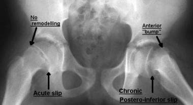
Bilateral slipped capital femoral epiphysis. One side shows evidence of remodeling of the neck and an anterior bone bump that restricts flexion. The other side demonstrates an acute slip as seen by the absence of any evidence of remodeling.
Click to see larger pictureClick to see detailView Full Size Image
Media type:  X-RAY

Media file 6:  Chronic slipped capital femoral epiphysis showing the extent of remodeling along the anterior neck (arrow).
Click to see larger pictureClick to see detailView Full Size Image
Media type:  X-RAY

Media file 7:  Pin placement (anteroposterior view). A: The entry point must be at or above the level of the lesser trochanter to avoid the risk of subtrochanteric fracture. B: The pin (screw) should be in the center of the epiphysis. C: At least 2.5 threads engaging the epiphysis should be used for a secure hold.
Click to see larger pictureClick to see detailView Full Size Image
Media type:  X-RAY

Media file 8:  Pin placement (lateral view). Arrow shows entry point in the anterolateral cortex.
Click to see larger pictureClick to see detailView Full Size Image
Media type:  X-RAY

Media file 9:  Osteotomies in slipped capital femoral epiphysis (SCFE). A: Subcapital osteotomy. B: Base of the neck compensatory osteotomy. C: Intertrochanteric compensatory osteotomy. Note that the amount of correction increases from subcapital to intertrochanteric osteotomies.
Click to see larger pictureClick to see detailView Full Size Image
Media type:  Image

Media file 10:  Principle of the Dunn osteotomy. Reduction of the slip after shortening the neck to avoid stretch on the vessels and decrease the risk of avascular necrosis.
Click to see larger pictureClick to see detailView Full Size Image
Media type:  Image


  1. Schein AJ. Acute severe slipped capital femoral epiphysis. Clin Orthop. Mar-Apr 1967;51:151-66. [Medline].
  2. Segal LS, Weitzel PP, Davidson RS. Valgus slipped capital femoral epiphysis. Fact or fiction?. Clin Orthop. Jan 1996;(322):91-8. [Medline].
  3. Loder RT, Wittenberg B, DeSilva G. Slipped capital femoral epiphysis associated with endocrine disorders. J Pediatr Orthop. May-Jun 1995;15(3):349-56. [Medline].
  4. McAfee PC, Cady RB. Endocrinologic and metabolic factors in atypical presentations of slipped capital femoral epiphysis. Report of four cases and review of the literature. Clin Orthop. Nov 1983;(180):188-97. [Medline].
  5. Wells D, King JD, Roe TF, Kaufman FR. Review of slipped capital femoral epiphysis associated with endocrine disease. J Pediatr Orthop. Sep-Oct 1993;13(5):610-4. [Medline].
  6. Loder RT, Hensinger RN. Slipped capital femoral epiphysis associated with renal failure osteodystrophy. J Pediatr Orthop. Mar-Apr 1997;17(2):205-11. [Medline].
  7. Loder RT, Hensinger RN, Alburger PD, et al. Slipped capital femoral epiphysis associated with radiation therapy. J Pediatr Orthop. Sep-Oct 1998;18(5):630-6. [Medline].
  8. Kelsey JL, Acheson RM, Keggi KJ. The body build of patients with slipped capital femoral epiphysis. Am J Dis Child. Aug 1972;124(2):276-81. [Medline].
  9. Ninomiya S, Nagasaka Y, Tagawa H. Slipped capital femoral epiphysis. A study of 68 cases in the eastern half area of Japan. Clin Orthop. Sep 1976;(119):172-6. [Medline].
  10. Loder RT. The demographics of slipped capital femoral epiphysis. An international multicenter study. Clin Orthop. Jan 1996;(322):8-27. [Medline].
  11. Murray AW, Wilson NI. Changing incidence of slipped capital femoral epiphysis: A RELATIONSHIP WITH OBESITY?. J Bone Joint Surg Br. Jan 2008;90(1):92-4. [Medline].
  12. Loder RT, Greenfield ML. Clinical characteristics of children with atypical and idiopathic slipped capital femoral epiphysis: description of the age-weight test and implications for further diagnostic investigation. J Pediatr Orthop. Jul-Aug 2001;21(4):481-7. [Medline].
  13. Burrow S, Alman B, Wright J. Relationship between short stature and endocrinopathy in slipped capital femoral epiphysis. Pediatrics. 1998;102(suppl):734.
  14. Abraham E, Gonzalez MH, Pratap S, Amirouche F, Atluri P, Simon P. Clinical implications of anatomical wear characteristics in slipped capital femoral epiphysis and primary osteoarthritis. J Pediatr Orthop. Oct-Nov 2007;27(7):788-95. [Medline].
  15. Galbraith RT, Gelberman RH, Hajek PC, et al. Obesity and decreased femoral anteversion in adolescence. J Orthop Res. 1987;5(4):523-8. [Medline].
  16. Gelberman RH, Cohen MS, Shaw BA, et al. The association of femoral retroversion with slipped capital femoral epiphysis. J Bone Joint Surg Am. Sep 1986;68(7):1000-7. [Medline].
  17. Pritchett JW, Perdue KD. Mechanical factors in slipped capital femoral epiphysis. J Pediatr Orthop. Jul-Aug 1988;8(4):385-8. [Medline].
  18. Kordelle J, Millis M, Jolesz FA, et al. Three-dimensional analysis of the proximal femur in patients with slipped capital femoral epiphysis based on computed tomography. J Pediatr Orthop. Mar-Apr 2001;21(2):179-82. [Medline].
  19. Mirkopulos N, Weiner DS, Askew M. The evolving slope of the proximal femoral growth plate relationship to slipped capital femoral epiphysis. J Pediatr Orthop. May-Jun 1988;8(3):268-73. [Medline].
  20. Wiberg G. Studies on dysplastic acetabula and congenital subluxation of the hip joint. With special reference to the complication of osteoarthrosis. Acta Chir Scandinavica. 1939;58.
  21. Exner GU. Growth and pubertal development in slipped capital femoral epiphysis: a longitudinal study. J Pediatr Orthop. Jul-Aug 1986;6(4):403-9. [Medline].
  22. Morscher E. Strength and morphology of growth cartilage under hormonal influence of puberty. Animal experiments and clinical study on the etiology of local growth disorders during puberty. Reconstr Surg Traumatol. 1968;10:3-104. [Medline].
  23. Sorensen KH. Slipped upper femoral epiphysis. Clinical study on aetiology. Acta Orthop Scand. 1968;39(4):499-517. [Medline].
  24. Brenkel IJ, Dias JJ, Iqbal SJ, Gregg PJ. Thyroid hormone levels in patients with slipped capital femoral epiphysis. J Pediatr Orthop. Jan-Feb 1988;8(1):22-5. [Medline].
  25. Eisenstein A, Rothschild S. Biochemical abnormalities in patients with slipped capital femoral epiphysis and chondrolysis. J Bone Joint Surg Am. Jun 1976;58(4):459-67. [Medline].
  26. Mann DC, Weddington J, Richton S. Hormonal studies in patients with slipped capital femoral epiphysis without evidence of endocrinopathy. J Pediatr Orthop. Sep-Oct 1988;8(5):543-5. [Medline].
  27. Samuelson T, Olney B. Percutaneous pin fixation of chronic slipped capital femoral epiphysis. Clin Orthop. May 1996;(326):225-8. [Medline].
  28. Wilcox PG, Weiner DS, Leighley B. Maturation factors in slipped capital femoral epiphysis. J Pediatr Orthop. Mar-Apr 1988;8(2):196-200. [Medline].
  29. Hagglund G, Hansson LI, Ordeberg G. Epidemiology of slipped capital femoral epiphysis in southern Sweden. Clin Orthop. Dec 1984;(191):82-94. [Medline].
  30. Jerre R, Billing L, Hansson G, Wallin J. The contralateral hip in patients primarily treated for unilateral slipped upper femoral epiphysis. Long-term follow-up of 61 hips. J Bone Joint Surg Br. Jul 1994;76(4):563-7. [Medline].
  31. Jerre R, Billing L, Hansson G, et al. Bilaterality in slipped capital femoral epiphysis: importance of a reliable radiographic method. J Pediatr Orthop B. Spring 1996;5(2):80-4. [Medline].
  32. Dreghorn CR, Knight D, Mainds CC, Blockey NJ. Slipped upper femoral epiphysis--a review of 12 years of experience in Glasgow (1972-1983). J Pediatr Orthop. May-Jun 1987;7(3):283-7. [Medline].
  33. Loder RT, Aronson DD, Greenfield ML. The epidemiology of bilateral slipped capital femoral epiphysis. A study of children in Michigan. J Bone Joint Surg Am. Aug 1993;75(8):1141-7. [Medline].
  34. Wilson PD, Jacobs B, Schecter L. Slipped capital femoral epiphysis: An end-result study. J Bone Joint Surg Am. Sep 1965;47:1128-45. [Medline].
  35. Riad J, Bajelidze G, Gabos PG. Bilateral slipped capital femoral epiphysis: predictive factors for contralateral slip. J Pediatr Orthop. Jun 2007;27(4):411-4. [Medline].
  36. Loder RT. Effect of femur position on the angular measurement of slipped capital femoral epiphysis. J Pediatr Orthop. Jul-Aug 2001;21(4):488-94. [Medline].
  37. Steel HH. The metaphyseal blanch sign of slipped capital femoral epiphysis. J Bone Joint Surg Am. Jul 1986;68(6):920-2. [Medline].
  38. Rhoad RC, Davidson RS, Heyman S, et al. Pretreatment bone scan in SCFE: a predictor of ischemia and avascular necrosis. J Pediatr Orthop. Mar-Apr 1999;19(2):164-8. [Medline].
  39. Agamanolis DP, Weiner DS, Lloyd JK. Slipped capital femoral epiphysis: a pathological study. II. An ultrastructural study of 23 cases. J Pediatr Orthop. Jan-Feb 1985;5(1):47-58. [Medline].
  40. Ponseti IV, McClintock R. The pathology of slipping of the upper femoral epiphysis. J Bone Joint Surg Am. Jan 1956;38-A(1):71-83. [Medline].
  41. Agamanolis DP, Weiner DS, Lloyd JK. Slipped capital femoral epiphysis: a pathological study. I. A light microscopic and histochemical study of 21 cases. J Pediatr Orthop. Jan-Feb 1985;5(1):40-6. [Medline].
  42. Aadalen RJ, Weiner DS, Hoyt W, Herndon CH. Acute slipped capital femoral epiphysis. J Bone Joint Surg Am. Oct 1974;56(7):1473-87. [Medline].
  43. Aronson DD, Carlson WE. Slipped capital femoral epiphysis. A prospective study of fixation with a single screw. J Bone Joint Surg Am. Jul 1992;74(6):810-9. [Medline].
  44. Boyer DW, Mickelson MR, Ponseti IV. Slipped capital femoral epiphysis. Long-term follow-up study of one hundred and twenty-one patients. J Bone Joint Surg Am. Jan 1981;63(1):85-95. [Medline].
  45. Fahey JJ, O''Brien ET. Acute slipped capital femoral epiphysis: Review of the literature and report of ten cases. J Bone Joint Surg Am. Sep 1965;47:1105-27. [Medline].
  46. Ward WT, Stefko J, Wood KB, Stanitski CL. Fixation with a single screw for slipped capital femoral epiphysis. J Bone Joint Surg Am. Jul 1992;74(6):799-809. [Medline].
  47. Ponseti I, Barta CK. The evaluation of treatment of the capital epiphysis. Surg Gynecol Obstet. 1948;86:87-97.
  48. Loder RT, Richards BS, Shapiro PS, et al. Acute slipped capital femoral epiphysis: the importance of physeal stability. J Bone Joint Surg Am. Aug 1993;75(8):1134-40. [Medline].
  49. Kallio PE, Paterson DC, Foster BK, Lequesne GW. Classification in slipped capital femoral epiphysis. Sonographic assessment of stability and remodeling. Clin Orthop. Sep 1993;(294):196-203. [Medline].
  50. Kallio PE, Mah ET, Foster BK, et al. Slipped capital femoral epiphysis. Incidence and clinical assessment of physeal instability. J Bone Joint Surg Br. Sep 1995;77(5):752-5. [Medline].
  51. Damaceno FL, Santili C, Longui CA. Normal reference values of Southwick's anteroposterior angle in prepubertal and pubertal normal adolescents. J Pediatr Orthop B. Nov 2007;16(6):389-92. [Medline].
  52. Carney BT, Weinstein SL, Noble J. Long-term follow-up of slipped capital femoral epiphysis. J Bone Joint Surg Am. Jun 1991;73(5):667-74. [Medline].
  53. Carney BT, Weinstein SL. Natural history of untreated chronic slipped capital femoral epiphysis. Clin Orthop. Jan 1996;(322):43-7. [Medline].
  54. Betz RR, Steel HH, Emper WD, et al. Treatment of slipped capital femoral epiphysis. Spica-cast immobilization. J Bone Joint Surg Am. Apr 1990;72(4):587-600. [Medline].
  55. Aronson DD,