Continually Updated Clinical Reference
 
 
  All Sources     eMedicine     Medscape     Drug Reference     MEDLINE
 
eMedicine - Sever Disease : Article by

Quick Find
Authors & Editors
Introduction
Clinical
Differentials
Workup
Treatment
Medication
Follow-up
Miscellaneous
Multimedia
References

Related Articles
Achilles Tendon Pathology

Calcaneus Fractures

Non-neoplastic Conditions Simulating Bone Tumors

Osteomyelitis

Tarsal Coalition


Patient Education
Foot, Ankle, Knee, and Hip Center


Author: Mark A Noffsinger, MD, Clinical Instructor, Department of Orthopedic Surgery, Michigan State College of Human Medicine; Medical Director, Deptartment of Orthopedic Surgery, Bronson Methodist Hospital, Consulting Staff, Kalamazoo Orthopedic Clinic

Mark A Noffsinger is a member of the following medical societies: American Academy of Orthopaedic Surgeons, American Association of Orthopaedic Medicine, American College of Physician Executives, American Fracture Association, American Medical Association, American Medical Directors Association, Christian Medical & Dental Society, Indiana State Medical Association, International Society on Thrombosis and Haemostasis, Michigan State Medical Society, Mid-America Orthopaedic Association, and Phi Beta Kappa

Editors: James K DeOrio, MD, Director of Foot and Ankle Fellowship Program, Assistant Professor of Orthopedic Surgery, Orthopedic Surgery, St. Luke's Hospital, Jacksonville, Florida; Francisco Talavera, PharmD, PhD, Senior Pharmacy Editor, eMedicine; Shepard R Hurwitz, MD, Executive Director Designate, American Board of Orthopaedic Surgery; Dinesh Patel, MD, FACS, Associate Clinical Professor of Orthopedic Surgery, Harvard Medical School; Chief of Arthroscopic Surgery, Department of Orthopedic Surgery, Massachusetts General Hospital; Jason H Calhoun, MD, FAAOS, Chairman, J Vernon Luck Distinguished Professor, Department of Orthopedic Surgery, University of Missouri

Author and Editor Disclosure

Synonyms and related keywords: Sever's disease, calcaneal apophysitis, calcaneal epiphysitis, traction apophysitis, Achilles tendon pain, heel pain

Background

Sever disease is a painful inflammation of the calcaneal apophysis. It is classified with the child and adolescent nonarticular osteochondroses. (The other disease in this group is Iselin disease, which is inflammation of the base of the fifth metatarsal.) The etiology of pain in Sever disease is believed to be repetitive trauma to the weaker structure of the apophysis, induced by the pull of the tendo Achilles on its insertion. This results in a clinical picture of heel pain in a growing active child, which worsens with activity.

In 1912, J.W. Sever, MD, first described this condition in the New York Medical Journal. He described it as an inflammation of the calcaneal apophysis resulting in the clinical symptoms of pain at the posterior heel, mild swelling, and difficulty with walking.

Pathophysiology

The calcaneal apophysis develops as an independent center of ossification (possibly multiple). It appears in boys aged 9-10 years and fuses by age 17 years; it appears in girls at slightly younger ages. During the rapid growth surrounding puberty, the apophyseal line appears to be weakened further because of increased fragile calcified cartilage. Microfractures are believed to occur because of shear stress leading to the normal progression of fracture healing. This theory explains the clinical picture and the radiographic appearance of resorption, fragmentation, and increased sclerosis leading to eventual union. The radiographs showing fragmentation of the apophysis are not diagnostic because multiple centers of ossification may exist in the normal apophysis, as noted above. However, the degree of involvement that occurs in children displaying the clinical symptoms of Sever disease appears to be more pronounced.

Frequency

International

Although no exact figures of the occurrence of Sever disease exist, it is a relatively common problem in growing active children.

Mortality/Morbidity

Although no well recognized long-term sequelae of untreated Sever disease exist, Sever disease causes pain that can limit performance and participation in sports, and, if left untreated, the pain can significantly limit even simple activities of daily life.

Sex

Incidence is higher in boys than in girls. Micheli and Ireland reported on 85 patients, 64% who were male (Micheli, 1987).

Age

Sever disease occurs most frequently in active 10- to 12-year-old boys. In Micheli and Ireland's report on 85 patients, the average age of presentation was 11 years 10 months for boys and 8 years 8 months for girls (Micheli, 1987).


History

The typical clinical presentation is the active child (aged 9-10 y) who complains of pain at the posterior heel made worse by sports, especially those involving running or jumping. The onset is usually gradual. Often, the pain has been relieved somewhat with rest and, therefore, patiently monitored by the patient, parents, coaches, trainers, and family physicians, expecting it to resolve. When the pain continues to interfere with sports performance and then with daily activities, further consultation is sought.

Physical

Physical examination varies depending on the severity and length of involvement. Bilateral involvement is present in approximately 60% of cases. Most patients experience pain with deep palpation at the Achilles insertion and pain when performing active toe raises. Forced dorsiflexion of the ankle also proves uncomfortable and is relieved with passive equinus positioning. Swelling may be present, but usually is mild. In long-standing cases, the child may have calcaneal enlargement.

Causes

Sever disease, like other similar conditions (eg, Osgood-Schlatter disease, little-leaguer's elbow, iliac apophysitis), is believed to be caused by decreased resistance to shear stress at the bone–growth plate interface. Studies have indicated that traction apophyses have a higher composition of fibrocartilage than epiphyses subjected more to axial load, which are composed of predominately hyaline cartilage. The anatomy of the calcaneal apophysis lends to significant shear stress because of its vertical orientation and the direction of pull from the strong gastroc-soleus muscle group (see Image 3).


Achilles Tendon Pathology
Calcaneus Fractures
Non-neoplastic Conditions Simulating Bone Tumors
Osteomyelitis
Tarsal Coalition


Imaging Studies

  • Radiograph findings include increased sclerosis and fragmentation of the calcaneal apophysis. However, it should be stressed that these findings are nonspecific and also are observed in asymptomatic feet (see Image 1). Radiographic evaluation is beneficial to exclude fracture or rare tumor. Remember that radiographic changes on plain x-ray films are neither diagnostic nor prognostic; they are beneficial to exclude other causes of heel pain. Explain this to patients and parents.
  • Remember that if pain continues, becomes significant at rest, awakens the patient from sleep, or is associated with significant swelling, tests should be performed to look for other causes. Tarsal coalition is another hindfoot disorder that must be distinguished from Sever disease. Thus, if reduction of subtalar motion is found on physical examination, a CT scan can be helpful in differentiating this disease from failure of the bones of the hindfoot to separate.
  • In cases of high suspicion, MRI may be of use to rule out osteomyelitis (see Image 2).

Histologic Findings

Sever disease is characterized by disorder of the normal process of enchondral ossification.


Medical Care

As children become involved in sports at younger ages and compete at higher levels, and as expectations of the participant, parents, and coach increase, incidence of overuse syndromes increases in growing athletes. When Sever disease occurs, not only does the child experience pain that can limit performance and participation, but, if left untreated, the pain can significantly limit even simple activities of daily life. This raises concern in all persons involved. Although no well-recognized long-term sequelae of untreated Sever disease exist, the physician's role is to minimize pain and allow for the child to return to normal activities as soon as possible to enhance psychosocial development. The physician also must be able to differentiate Sever disease from other causes of heel pain in the child that are potentially more serious, such as tumor or osteomyelitis.

  • Treatment initially is focused on reducing the present pain and limitations, and then on preventing recurrence. Limitation of activity (especially running and jumping) usually is necessary. In Micheli and Ireland's study, 84% of 85 patients were able to resume sports activities after 2 months.
  • In patients with symptoms that are not severe enough to warrant limiting sports activities or if the patient and parents are unwilling to miss a critical portion of the sport season, wearing a half-inch inner-shoe heel lift (at all times during ambulation), a monitored stretching program, presport and postsport icing, and judicious use of anti-inflammatory agents normally reduces the symptoms and allows continued participation. If symptoms worsen, activity modification must be included.
  • In severe cases, short-term (2-3 wk) cast treatment in mild equinus can be used.

Activity

Limitation of activity (especially running and jumping) usually is necessary (see Treatment, Medical Care).


Judicious use of anti-inflammatory agents may be helpful for patients wishing to avoid limiting their sports activities (see Treatment, Medical Care).

Drug Category: Nonsteroidal anti-inflammatory drugs

Have analgesic, anti-inflammatory, and antipyretic activities. Their mechanism of action is not known, but they may inhibit cyclooxygenase activity and prostaglandin synthesis. Other mechanisms may exist as well, such as inhibition of leukotriene synthesis, lysosomal enzyme release, lipoxygenase activity, neutrophil aggregation, and various cell-membrane functions.

Drug NameIbuprofen (Motrin, Ibuprin, Excedrin IB, Advil)
DescriptionDOC for patients with mild to moderate pain. Inhibits inflammatory reactions and pain by decreasing prostaglandin synthesis.
Adult Dose200-400 mg PO q4-6h while symptoms persist; not to exceed 3.2 g/d
Pediatric Dose6 months to 12 years: 4-10 mg/kg per dose PO tid/qid
>12 years: Administer as in adults
ContraindicationsDocumented hypersensitivity; peptic ulcer disease, recent GI bleeding or perforation, renal insufficiency, high risk of bleeding
InteractionsCoadministration with aspirin increases risk of inducing serious NSAID-related side effects; probenecid may increase concentrations and, possibly, toxicity of NSAIDs; may decrease effect of hydralazine, captopril, and beta-blockers; may decrease diuretic effects of furosemide and thiazides; may increase PT when taking anticoagulants (instruct patients to watch for signs of bleeding); may increase risk of methotrexate toxicity; phenytoin levels may be increased when administered concurrently
PregnancyB - Usually safe but benefits must outweigh the risks.
PrecautionsCategory D in third trimester of pregnancy; caution in congestive heart failure, hypertension, and decreased renal and hepatic function; caution in coagulation abnormalities or during anticoagulant therapy

Drug NameNaproxen (Naprosyn, Naprelan, Anaprox)
DescriptionFor relief of mild to moderate pain; inhibits inflammatory reactions and pain by decreasing activity of cyclo-oxygenase, which results in a decrease of prostaglandin synthesis.
Adult Dose500 mg PO followed by 250 mg q6-8h; not to exceed 1.25 g/d
Pediatric Dose<2 years: Not established
>2 years: 2.5 mg/kg/dose PO; not to exceed 10 mg/kg/d
ContraindicationsDocumented hypersensitivity; peptic ulcer disease; recent GI bleeding or perforation; renal insufficiency
InteractionsCoadministration with aspirin increases risk of inducing serious NSAID-related side effects; probenecid may increase concentrations and, possibly, toxicity of NSAIDs; may decrease effect of hydralazine, captopril, and beta-blockers; may decrease diuretic effects of furosemide and thiazides; may increase PT when taking anticoagulants (instruct patients to watch for signs of bleeding); may increase risk of methotrexate toxicity; phenytoin levels may be increased when administered concurrently
PregnancyB - Usually safe but benefits must outweigh the risks.
PrecautionsCategory D in third trimester of pregnancy; acute renal insufficiency, interstitial nephritis, hyperkalemia, hyponatremia, and renal papillary necrosis may occur; patients with preexisting renal disease or compromised renal perfusion risk acute renal failure; leukopenia occurs rarely, is transient, and usually returns to normal during therapy; persistent leukopenia, granulocytopenia, or thrombocytopenia warrants further evaluation and may require discontinuation of drug


Deterrence/Prevention:

  • To prevent recurrence, patients, parents, coaches, and trainers should be instructed regarding a good preexercise stretching program for the child. Early in the season, encouragement should be given for a preseason conditioning and stretching program. Coaches and trainers should be educated about recognition of the clinical symptoms so they are able to initiate early protective measures and seek medical referral when necessary.

Complications:

  • No known complication exists from failure to make the correct diagnosis because the disease is self-limited.

Prognosis:

  • Sever disease is a self-limited condition.

Patient Education:


Medical/Legal Pitfalls

  • Failure to instruct players, parents, coaches and trainers regarding limiting the patient's activity and proper preexercise and postexercise stretching can lead to prolonged symptoms and further limitation of performance.
  • Failure to instruct patients and parents that continual pain, significant swelling or redness, and fever are not signs of Sever disease and, therefore, require further evaluation could result in failure to diagnose a condition with much more serious long-term consequences.


Media file 1:  Sever disease. Lateral radiograph of foot in symptomatic 9-year-old male soccer player. Sclerosis is not diagnostic of Sever disease but is a characteristic radiographic finding.
Click to see larger pictureClick to see detailView Full Size Image
Media type:  X-RAY

Media file 2:  Transverse MRI of foot in symptomatic 11-year-old girl with heel pain showing osteomyelitis. Pain was increased with activity but more constant and with more associated night pain than expected with Sever disease. Treatment included surgical debridement and antibiotic therapy.
Click to see larger pictureClick to see detailView Full Size Image
Media type:  MRI

Media file 3:  
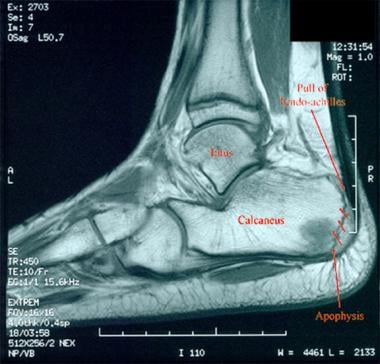
Labeled MRI depicts the anatomy and mechanical forces responsible for the development of Sever disease (shear stress at the calcaneal apophysis).
Click to see larger pictureClick to see detailView Full Size Image
Media type:  MRI


  • Katz JF. Nonarticular Osteochondroses. Clinical Orthopaedics and Related Research. 1981;158:70.
  • Micheli LJ, Ireland ML. Prevention and management of calcaneal apophysitis in children: an overuse syndrome. J Pediatr Orthop. Jan-Feb 1987;7(1):34-8. [Medline].
  • Pappas AM. The osteochondroses. Pediatr Clin North Am. Aug 1967;14(3):549-70. [Medline].
  • Pizzutillo PD, Sullivan JA, Grana WA. Osteochondroses, Chapter in The Pediatric Athlete, American Academy of Orthopaedic Surgeons Seminar, Oklahoma City Oklahoma, November 1998.
  • Roy DR. Accessory Navicular and Osteochodroses of the Foot and Ankle in the Child and Adolescent. Foot and Ankle Clinics. Philadelphia: WB Saunders;1998.
  • Sever JW. Apophysitis of the Os Calcis. New York Medical Journal. 1912;95:1025-1029.
  • Sever JW. Apophysitis of the Os Calcis. American Journal of Orthopaedics. 1917;15:659.
  • Turek SL. '. Orthopaedics, Principles and Their Application. JB Lippencott Company;1984: 1474.

Sever Disease excerpt

Article Last Updated: Jul 12, 2004